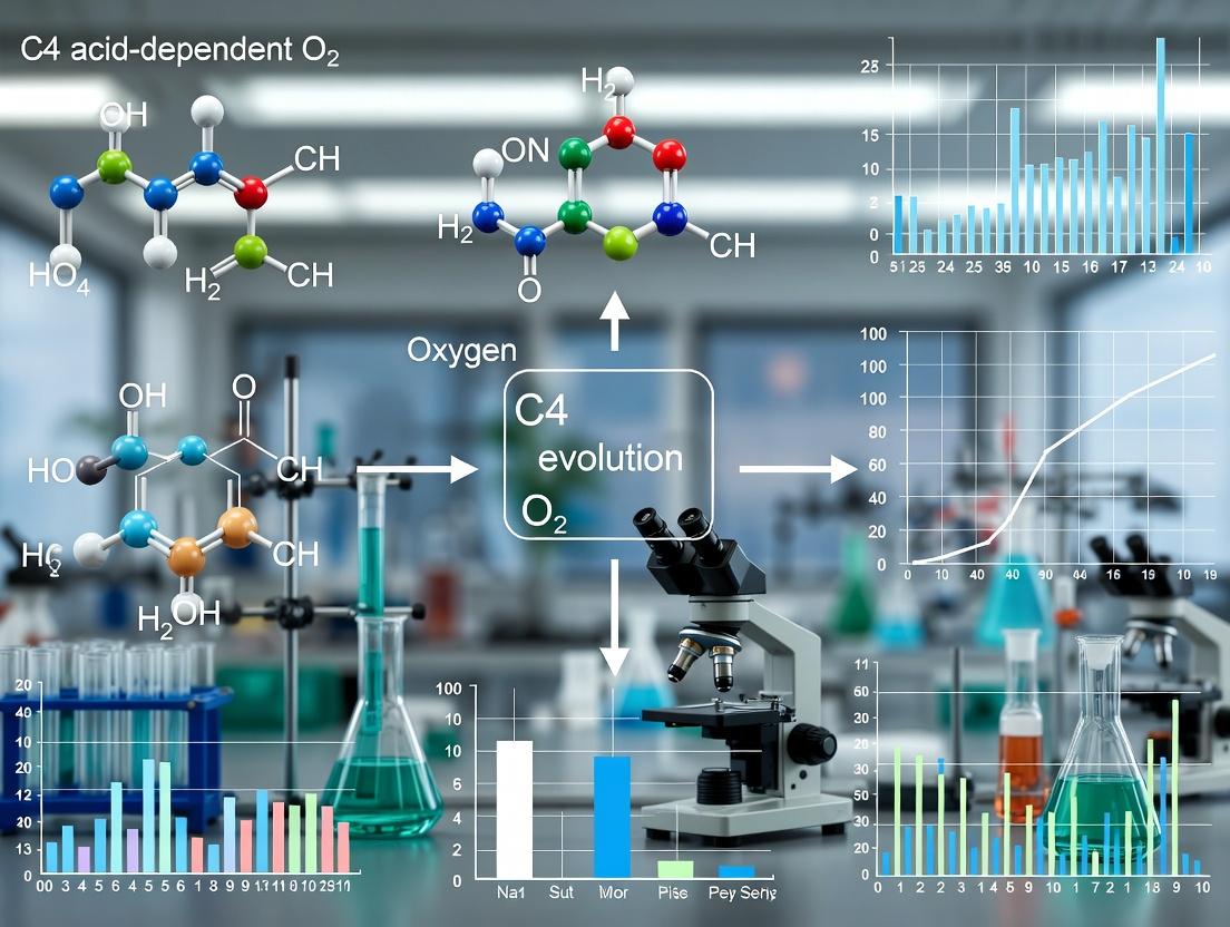
Beyond Photosynthesis: C4 Acid-Dependent O2 Evolution as a Novel Bioenergetic Assay for Drug Discovery and Metabolic Research
This article provides a comprehensive guide to the C4 acid-dependent O2 evolution experiment, a critical assay for probing the photorespiratory and metabolic functions of plant and algal systems.
Beyond Photosynthesis: C4 Acid-Dependent O2 Evolution as a Novel Bioenergetic Assay for Drug Discovery and Metabolic Research
Abstract
This article provides a comprehensive guide to the C4 acid-dependent O2 evolution experiment, a critical assay for probing the photorespiratory and metabolic functions of plant and algal systems. Targeted at researchers and drug development professionals, it covers the foundational theory of the C2 cycle and its role in photorespiration, details precise methodological protocols for measuring O2 flux, offers advanced troubleshooting and optimization strategies for reliable data, and validates the assay against contemporary techniques like chlorophyll fluorescence and mass spectrometry. By integrating these four intents, the article serves as a complete resource for applying this sensitive bioassay to screen for metabolic inhibitors, evaluate stress responses, and advance research in agricultural biotechnology and biofuel development.
Decoding Photorespiration: The Science Behind C4 Acid-Dependent O2 Evolution
Within the broader thesis on C4 acid-dependent O2 evolution, photorespiration represents a critical counter-pathway. This process is initiated when Ribulose-1,5-bisphosphate carboxylase/oxygenase (Rubisco) fixes O2 instead of CO2, leading to the production of phosphoglycolate and initiating the photorespiratory C2 cycle. Research into modulating this pathway is crucial for understanding plant metabolic efficiency and has implications for enhancing crop yields and informing bioengineering strategies in pharmaceutical contexts (e.g., for plant-based drug precursor production).
Core Mechanism: Rubisco's Dual Function and the C2 Cycle
Rubisco catalyzes two competing reactions:
- Carboxylation: RuBP + CO₂ → 2 molecules of 3-phosphoglycerate (3-PGA).
- Oxygenation: RuBP + O₂ → 1 molecule of 3-PGA + 1 molecule of 2-phosphoglycolate (2-PG).
The 2-PG is toxic and must be recycled via the photorespiratory C2 cycle (or glycolate pathway), spanning chloroplasts, peroxisomes, and mitochondria. This cycle consumes energy and releases previously fixed CO₂.
Quantitative Comparison of Rubisco Reactions:
Table 1: Kinetic Parameters of Rubisco's Dual Activity
| Parameter | Carboxylase Reaction (CO₂ fixation) | Oxygenase Reaction (O₂ fixation) | Notes |
|---|---|---|---|
| Substrate | CO₂ | O₂ | |
| Primary Product | 2 x 3-PGA | 1 x 3-PGA + 1 x 2-PG | |
| Relative Velocity (Vc/Vo) | ~3-4 : 1 | Varies with [CO₂]/[O₂] | Measured at 25°C, typical [CO₂] and [O₂] |
| Km for CO₂ (Kc) | ~10-20 µM | Not Applicable | Varies by species |
| Km for O₂ (Ko) | Not Applicable | ~200-600 µM | Varies by species |
| Specificity Factor (τ = VcKo / VoKc) | 80-100 (C3 plants) | Higher τ indicates greater CO₂/O₂ specificity |
Diagram 1: Rubisco's Dual Catalysis Initiating Competing Pathways (Max 760px)
Application Notes & Protocols
Protocol: Measuring Rubisco Oxygenase ActivityIn Vitro
Objective: To quantify the initial rate of Rubisco's oxygenase activity isolated from leaf tissue.
Thesis Context: Establishing a baseline Rubisco O₂ fixation rate is prerequisite for experiments testing the effect of exogenous C4 acids on suppressing photorespiration in isolated systems.
Materials: See "Scientist's Toolkit" (Section 4).
Method:
- Rubisco Extraction: Homogenize 1 g of fresh leaf tissue in 5 mL of ice-cold extraction buffer. Centrifuge at 12,000g for 10 min at 4°C. Desalt the supernatant using a pre-equilibrated PD-10 desalting column into assay buffer.
- Assay Setup: In a sealed, temperature-controlled (25°C) oxygen electrode chamber, add:
- 980 µL of assay buffer (containing 10 mM NaHCO₃ to suppress inherent carboxylation).
- 10 µL of 100 mM RuBP.
- 10 µL of enzyme extract.
- Initiation & Measurement: Close the chamber, ensuring no air bubbles. Monitor O₂ consumption (in µmol O₂·min⁻¹) for 2-3 minutes after injection. The initial linear slope is the oxygenase activity.
- Control: Run a blank without RuBP to account for non-specific O₂ consumption.
- Calculation: Activity = (∆[O₂]/min * Chamber Volume) / (Enzyme Protein * Time). Express as µmol O₂·min⁻¹·mg protein⁻¹.
Protocol: Monitoring Photorespiratory CO₂ Release via Gas Exchange
Objective: To measure the post-illumination CO₂ burst (PIB) as an in vivo indicator of photorespiratory flux.
Thesis Context: This protocol can be adapted to measure how feeding C4 acids (e.g., malate, aspartate) alters the magnitude of the PIB, indicating direct or indirect suppression of the C2 cycle.
Method:
- Plant Material: Use intact leaf or whole plant in a gas exchange cuvette (e.g., LI-6800).
- Stabilization: Set conditions to induce photorespiration: light intensity of 1000 µmol photons·m⁻²·s⁻¹, leaf temperature 25°C, [O₂] = 21%, low [CO₂] (e.g., 50 ppm). Allow photosynthesis to stabilize for 20-30 minutes.
- Dark Transition & Measurement: Simultaneously switch off the light and close the IRGA inlet. Rapidly record the CO₂ concentration inside the sealed cuvette every second for 2-3 minutes.
- Data Analysis: The rapid CO₂ efflux immediately following darkness is the PIB. Integrate the CO₂ burst curve over the first 60 seconds to quantify total photorespiratory CO₂ release (µmol CO₂·m⁻²).
- Experimental Arm: Repeat the process with the petiole of a detached leaf placed in a solution of a C4 acid (e.g., 10 mM malate) during the initial stabilization period.
Diagram 2: Post-Illumination Burst Assay Workflow (Max 760px)
The Scientist's Toolkit
Table 2: Key Reagents & Materials for Photorespiration Research
| Item | Function/Description | Example & Rationale |
|---|---|---|
| Oxygen Electrode | Measures real-time O₂ concentration in solution for in vitro oxygenase assays. | Clark-type electrode; essential for direct quantification of Rubisco O₂ consumption. |
| Infrared Gas Analyzer (IRGA) | Measures CO₂ and H₂O vapor fluxes for in vivo photorespiration (PIB) and gas exchange. | LI-6800 Portable Photosynthesis System; enables non-destructive, whole-leaf kinetics. |
| RuBP (Ribulose-1,5-bisphosphate) | The 5-carbon substrate for Rubisco. Must be pure and freshly prepared. | Sodium salt, ≥95% purity; unstable, prepare aliquots in neutral pH, store at -80°C. |
| Rubisco Extraction Buffer | Maintains enzyme stability and activity during isolation. | Typically contains Tris-HCl (pH 8.0), MgCl₂, EDTA, DTT, and PVP to inhibit phenolics. |
| Desalting Columns | Rapidly removes small molecules (e.g., endogenous metabolites, salts) from crude extract. | Sephadex G-25 (PD-10) columns; critical to remove interfering compounds and dissolved inorganic carbon. |
| C4 Acid Solutions | Experimental compounds to test impact on photorespiratory pathway. | Sodium malate, sodium aspartate (10-50 mM); potential donors for CO₂ concentration mechanisms. |
| ¹⁸O₂ Isotope | Radioactive tracer for precise, sensitive measurement of O₂ fixation pathways. | Used in advanced mass spectrometry-based assays to trace O₂ incorporation into metabolites. |
Within the broader thesis investigating C4 acid-dependent oxygen evolution in photosynthetic and photorespiratory contexts, the oxidation of glycolate stands out as a critical, quantifiable source of O2. This reaction is central to the photorespiratory carbon oxidation cycle, where glycolate, a two-carbon product of the oxygenase activity of Rubisco, is metabolized. Its subsequent oxidation in peroxisomes (via glycolate oxidase) and mitochondria generates measurable O2 as a direct byproduct. This application note details the protocols and mechanistic insights for capturing and quantifying this specific O2 flux, distinguishing it from the dominant O2 evolution of the light-dependent reactions of photosynthesis.
Core Biochemical Pathway and Quantitative Data
The primary pathway for glycolate-dependent O2 release involves two key enzymatic steps leading to net O2 consumption and evolution.
Diagram 1: Glycolate Oxidation Pathway in Photorespiration
Table 1: Stoichiometry of O2 Exchange in Glycolate Metabolism
| Reaction | Enzyme/Location | O2 Consumed | O2 Produced | Net O2 per 2 Glycolate |
|---|---|---|---|---|
| 2 Glycolate + 2 O₂ → 2 Glyoxylate + 2 H₂O₂ | Glycolate Oxidase (Peroxisome) | 2 | 0 | -2 |
| 2 H₂O₂ → 2 H₂O + O₂ | Catalase (Peroxisome) | 0 | 1 | +1 |
| Overall (Peroxisomal) | Glycolate → Glyoxylate | 2 | 1 | -1 |
| Further Metabolism to 3-PGA | Complete PCO Cycle (incl. Mitochondria) | 0 | 0.5* | -0.5 net |
Note: *Complete oxidation in mitochondria via glycine decarboxylase can release CO₂ and NH₃, but the net O₂ release measured in isolated peroxisomal preparations is defined by the catalase reaction.
Table 2: Typical Measured O2 Evolution Rates from Glycolate
| System | Glycolate Concentration | Buffer Conditions | Temperature | Typical O2 Release Rate (µmol O2 mg⁻¹ Chl min⁻¹) |
|---|---|---|---|---|
| Isolated Spinach Peroxisomes | 5 mM | 50 mM HEPES-KOH, pH 7.2 | 25°C | 0.8 - 1.2 |
| Intact C3 Plant Leaf Discs (High O₂, Low CO₂) | N/A | In vivo conditions | 25°C | 1.5 - 3.0* |
| Algal Cells (Chlamydomonas) | 10 mM | Minimal medium, pH 7.5 | 25°C | 2.0 - 4.0 |
Note: *In vivo rates represent net photorespiratory flux, where glycolate-derived O2 release is partially masked by concurrent O2 consumption.
Experimental Protocols
Protocol 1: Measuring Glycolate-Driven O2 Release in Isolated Peroxisomes
Objective: To isolate functional peroxisomes and directly quantify O2 evolution from glycolate oxidation.
Materials: See "The Scientist's Toolkit" below.
Method:
- Peroxisome Isolation:
- Homogenize 50g of young spinach leaves in 150 ml of ice-cold grinding buffer (0.3 M sucrose, 50 mM HEPES-KOH pH 7.5, 1 mM EDTA, 5 mM L-ascorbic acid, 0.1% BSA).
- Filter through 4 layers of Miracloth.
- Centrifuge filtrate at 1,500 x g for 10 min (4°C) to remove debris and chloroplasts.
- Centrifuge supernatant at 10,000 x g for 20 min (4°C) to pellet crude peroxisomes.
- Resuspend pellet in 2 ml of wash buffer (0.3 M sucrose, 20 mM HEPES-KOH pH 7.2). Layer onto a pre-formed Percoll density gradient (10-50% in wash buffer).
- Centrifuge at 40,000 x g for 45 min (4°C). Collect the lower, dense band (intact peroxisomes).
- Dilute 5-fold with wash buffer and pellet at 10,000 x g for 15 min. Resuspend in a small volume (~0.5 ml) of storage buffer. Keep on ice.
- O2 Evolution Assay (Clark-type Electrode):
- Calibrate the electrode with air-saturated assay buffer (50 mM HEPES-KOH pH 7.2, 0.3 M sucrose) and by adding a known amount of sodium dithionite (zero O2).
- Add 1.9 ml of assay buffer to the reaction chamber at 25°C. Stir continuously.
- Add 100 µl of isolated peroxisome suspension.
- Close chamber and record baseline O2 concentration.
- Inject 10 µl of 1 M sodium glycolate (final conc. 5 mM) through the injection port.
- Record the rate of O2 increase for 2-3 minutes. The initial linear slope represents glycolate oxidase + catalase activity.
- Control: Run a reaction without glycolate to account for any endogenous respiration.
Data Analysis: Calculate the rate using the electrode's calibration factor. Normalize to protein content (Bradford assay).
Protocol 2: Demonstrating Glycolate-Dependent O2 Evolution in Intact Leaf Discs
Objective: To induce photorespiratory glycolate production and measure associated O2 release under non-photosynthetic conditions.
Workflow Diagram:
Method:
- Cut 10 leaf discs (1 cm² each) from a dark-adapted C3 plant (e.g., tobacco, Arabidopsis).
- Infiltrate discs under vacuum for 5 minutes with assay buffer (20 mM BICINE, pH 8.5) containing 50 µM DCMU to inhibit photosynthetic O2 evolution. Rinse.
- Place discs in a gas-tight, illuminated O2 electrode chamber with the same buffer.
- Flush the chamber with N2 gas to create a low-O2, low-CO2 atmosphere (<2% O₂, ≤50 ppm CO₂).
- Illuminate with saturating light (1000 µmol photons m⁻² s⁻¹). Record the baseline (should be near zero O2 change).
- Switch the gas inflow to 21% O₂, 0% CO₂ (balance N2) to initiate photorespiration. Observe a slow increase in O2 concentration due to glycolate metabolism.
- To confirm the source, open the chamber and add sodium glycolate to a final concentration of 10 mM. Resume measurement. A marked increase in O2 evolution rate confirms the capacity for glycolate-driven O2 release.
The Scientist's Toolkit
Table 3: Key Research Reagent Solutions
| Item | Function/Benefit in Experiment | Example/Specification |
|---|---|---|
| Sodium Glycolate | The direct substrate for glycolate oxidase. High-purity grade ensures minimal side reactions. | Sigma-Aldrich, ≥98% purity. Prepare 1 M stock in assay buffer, pH to 7.2. |
| DCMU (Diuron) | A potent PSII inhibitor. Crucial for isolating non-photosynthetic O2 evolution in intact tissue experiments. | Prepare 10 mM stock in ethanol. Final working conc. 10-50 µM. |
| HEPES-KOH Buffer | Maintains stable physiological pH (7.2-7.5) during peroxisomal isolation and assays, critical for enzyme activity. | 50-100 mM concentration, pH 7.2 for assays. |
| Percoll Density Medium | Used for isopycnic centrifugation to obtain highly purified, intact peroxisomes free of chloroplasts and mitochondria. | GE Healthcare, diluted in sucrose/HEPES buffer. |
| Catalase Inhibitor (e.g., 3-AT) | Negative control. Inhibits catalase, preventing O2 release from H₂O₂, converting net O2 release to consumption. | 3-Amino-1,2,4-triazole (3-AT), 10-50 mM. |
| Clark-type Oxygen Electrode | The essential instrument for real-time, quantitative measurement of dissolved O2 concentration changes. | Hansatech Instruments OxyLab or equivalent. Requires proper membrane and electrolyte maintenance. |
Application Notes: Metabolic Context in C4 Acid-Dependent O2Evolution Research
Within the thesis framework of C4 acid-dependent oxygen evolution research, glycolate and glyoxylate are critical two-carbon (C2) substrates. They are not primary products of the C4 pathway but are intrinsically linked to its photorespiratory interactions. In C4 plants, the concentration of O2 in the bundle sheath cells can be elevated due to active decarboxylation of C4 acids, creating a microenvironment conducive to photorespiration. RuBisCO's oxygenase activity generates phosphoglycolate, which is rapidly dephosphorylated to glycolate.
Glycolate is exported from the chloroplast and metabolized in the peroxisomes via the photorespiratory C2 cycle. Here, it is oxidized to glyoxylate by glycolate oxidase (GOX). Glyoxylate is a pivotal branch-point metabolite. Its primary fate is transamination to glycine, but it can also undergo non-enzymatic decarboxylation or serve as a substrate for other enzymes, influencing the net carbon and nitrogen economy of the cell. Understanding the flux through these substrates is essential for quantifying photorespiratory losses and engineering strategies to enhance C4 photosynthetic efficiency. The interplay between C4 acid decarboxylation (releasing CO2) and glycolate metabolism (releasing CO2 and NH3) directly impacts measured O2 evolution patterns in experimental systems.
Table 1: Key Kinetic Parameters of Enzymes Involved in Glycolate/Glyoxylate Metabolism
| Enzyme (EC Number) | Substrate | Km (μM) | Vmax (μmol mg-1 min-1) | Primary Location | pH Optimum |
|---|---|---|---|---|---|
| Glycolate Oxidase (1.1.3.15) | Glycolate | 200 - 500 | 4.0 - 8.0 | Peroxisome | 8.0 - 8.5 |
| O2 | ~500 | - | - | - | |
| Glyoxylate Reductase (1.1.1.79) | Glyoxylate | 20 - 100 | 10 - 25 | Peroxisome/Cytosol | 6.5 - 7.5 |
| NADPH | 10 - 50 | - | - | - | |
| Glutamate:Glyoxylate Aminotransferase (2.6.1.4) | Glyoxylate | 1,000 - 5,000 | 15 - 40 | Peroxisome | 7.5 - 8.0 |
| RuBisCO Oxygenase (4.1.1.39) | O2 | 400 - 500 μM | - | Chloroplast Stroma | 8.0 - 8.5 |
| RuBP | ~20 μM | - | - | - |
Table 2: Typical Steady-State Metabolite Concentrations in C4 Mesophyll/Bundle Sheath Cells
| Metabolite | Approx. Concentration (μM) | Cellular Compartment | Notes |
|---|---|---|---|
| Glycolate | 10 - 50 | Chloroplast/Peroxisome | Highly variable, light-dependent |
| Glyoxylate | 1 - 10 | Peroxisome | Tightly regulated, potentially toxic |
| Glycine | 500 - 3000 | Mitochondria/Peroxisome | Photorespiration-driven accumulation |
| Serine | 200 - 1000 | Mitochondria/Peroxisome | - |
Experimental Protocols
Protocol 1: Spectrophotometric Assay of Glycolate Oxidase (GOX) Activity in Leaf Extracts
Purpose: To quantify GOX activity, a key driver of glycolate to glyoxylate conversion, relevant to photorespiratory flux in C4 research. Materials: See "Scientist's Toolkit" below. Procedure:
- Extract Preparation: Grind 100 mg of fresh leaf tissue (bundle sheath-enriched strands if possible) in 1 mL of ice-cold extraction buffer (50 mM HEPES-KOH pH 8.2, 1 mM EDTA, 5 mM DTT, 0.1% (v/v) Triton X-100, 1% (w/v) PVP-40). Centrifuge at 12,000 g for 10 min at 4°C. Use supernatant as crude extract.
- Assay Mix: Prepare 1 mL reaction mix in a UV-transparent cuvette: 50 mM HEPES-KOH (pH 8.2), 5 mM glycolate (sodium salt), 0.1 mM FMN, 20 units of catalase. Equilibrate to 25°C.
- Reaction Initiation: Add 50-100 µL of crude extract to start the reaction. Mix quickly.
- Measurement: Immediately monitor the increase in absorbance at 324 nm (A324) due to the production of glyoxylate-phenylhydrazone for 3 minutes. Use a molar extinction coefficient (ε) of 1.7 x 10⁴ M-1 cm-1.
- Calculation: Activity = (ΔA324 / min * Vtotal) / (ε * Venzyme * path length) expressed as µmol glyoxylate produced min-1 mg-1 protein.
Protocol 2: HPLC-Based Measurement of Glycolate and Glyoxylate Pools
Purpose: To accurately quantify intracellular concentrations of glycolate and glyoxylate under different O2 evolution experimental conditions. Materials: See "Scientist's Toolkit." Procedure:
- Rapid Metabolite Quenching: Flash-freeze leaf discs (50 mg) from C4 plants (pre-adapted to specific O2/CO2 conditions) in liquid N2.
- Extraction: Homogenize tissue in 500 µL of 1 M HClO4 pre-chilled to -20°C. Incubate on ice for 15 min. Neutralize with 250 µL of 2 M K2CO3 in 0.5 M triethanolamine. Centrifuge at 15,000 g for 10 min at 4°C. Filter supernatant through a 0.22 µm nylon membrane.
- Derivatization: Mix 100 µL of extract with 100 µL of 20 mM 2,4-dinitrophenylhydrazine (in 2 M HCl). Incubate at 37°C for 30 min to form hydrazone derivatives.
- HPLC Analysis:
- Column: Reverse-phase C18 column (5 µm, 250 x 4.6 mm).
- Mobile Phase: A: 0.1% (v/v) TFA in water; B: Acetonitrile. Gradient: 20% B to 60% B over 25 min.
- Flow Rate: 1.0 mL/min.
- Detection: UV-Vis detector at 360 nm.
- Quantification: Use external calibration curves of authentic glycolate and glyoxylate standards processed identically.
Visualization: Pathways and Workflows
Title: Photorespiratory Glycolate Pathway in C4 O2 Evolution
Title: Workflow for Glycolate/Glyoxylate Quantification
The Scientist's Toolkit
Table 3: Essential Research Reagents and Materials
| Item | Function/Description | Example Vendor/Catalog |
|---|---|---|
| Glycolate (Sodium Salt) | Primary substrate for GOX assays; standard for quantification. | Sigma-Aldrich, G9126 |
| Glyoxylic Acid (Monohydrate) | Standard for quantification; substrate for glyoxylate reductase assays. | Sigma-Aldrich, 128465 |
| Flavin Mononucleotide (FMN) | Essential cofactor for Glycolate Oxidase activity. | MilliporeSigma, F2253 |
| 2,4-Dinitrophenylhydrazine (DNPH) | Derivatizing agent for carbonyl groups (glyoxylate) for HPLC-UV detection. | Thermo Scientific, AC119442500 |
| Catalase (from bovine liver) | Added to GOX assays to scavenge H2O2 and prevent inhibition. | Sigma-Aldrich, C9322 |
| Perchloric Acid (HClO4) | Strong acid for rapid metabolite quenching and extraction. | VWR, 470302-568 |
| Polyvinylpolypyrrolidone (PVP-40) | Added to extraction buffers to bind phenolic compounds. | Sigma-Aldrich, P6755 |
| C18 Reverse-Phase HPLC Column | For separation of derivatized organic acids. | Agilent, ZORBAX Eclipse XDB-C18 |
| HEPES Buffer | Biological buffer for maintaining pH 8.0-8.2 in GOX assays. | Fisher Scientific, BP310 |
| Microcentrifuge Tubes (Safe-Lock) | For safe grinding and handling of perchloric acid extracts. | Eppendorf, 022363352 |
Application Notes on C4 Acid-Dependent O₂ Evolution
Within the broader thesis on C4 acid-dependent O₂ evolution, understanding the biological context of its underlying pathways is critical for designing physiologically relevant experiments. This pathway is not universally active but is induced under specific environmental and developmental conditions, primarily serving as a carbon-concentrating mechanism (CCM) to mitigate photorespiration.
Key Biological Contexts:
- Spatial Context in Plants: In C4 plants (e.g., maize, sugarcane), the pathway is anatomically compartmentalized between mesophyll and bundle sheath cells. It is active in leaf tissues under illuminated conditions.
- Spatial Context in Algae: In many green algae (e.g., Chlamydomonas reinhardtii), the CCM, which can involve C4 acid cycles, is induced in single cells, often localized to the pyrenoid, a chloroplast sub-compartment.
- Temporal Context: Pathway activity is dynamically regulated. It is induced under conditions of low CO₂ availability, high light intensity, and high O₂ concentration—conditions that promote wasteful photorespiration. Induction occurs over timescales of minutes to hours following an environmental shift.
Experimental Implication: Laboratory experiments aiming to measure C4 acid-dependent O₂ evolution must replicate these inducing conditions (e.g., low CO₂, high light) in the growth environment prior to assay to ensure the pathway is fully operational.
Table 1: Induction Conditions for CCM/C4 Pathway Activity in Model Organisms
| Organism | Inducing Condition | Typical Induction Time | Measured Increase in CCM Activity | Key Reference |
|---|---|---|---|---|
| Chlamydomonas reinhardtii (Alga) | Transfer to Low CO₂ (0.04% → 0.01%) | 2-4 hours | 3-5 fold increase in apparent CO₂ affinity | Meyer & Griffiths, 2013 |
| Arabidopsis thaliana (C3 Plant) | High Light (100 → 1000 µmol photons m⁻² s⁻¹) | 24-48 hours | Upregulation of photorespiratory genes; no true C4 cycle | Foyer et al., 2009 |
| Zea mays (C4 Plant) | Developmental: Fully expanded leaf | Constitutive in mature bundle sheath cells | N/A (constitutive) | Langdale, 2011 |
| Hydrilla verticillata (Facultative C4 Plant) | Low CO₂, High pH, High Light | 7-10 days | Shift from C3 to C4 δ¹³C isotope signature | Reiskind et al., 1997 |
Experimental Protocols
Protocol 1: Inducing CCM/C4 Pathway Activity inChlamydomonas reinhardtiifor O₂ Evolution Assays
Purpose: To precondition algal cultures to activate the Carbon Concentrating Mechanism (CCM), which may involve C4 acid metabolism, prior to measuring O₂ evolution kinetics.
Materials:
- Tris-Acetate-Phosphate (TAP) or Minimal (HSM) medium.
- CO₂-controlled incubator or air-lift bioreactors with regulated air/CO₂ mixtures.
- High-intensity growth lights (≥ 200 µmol photons m⁻² s⁻¹ PAR).
Methodology:
- Grow wild-type C. reinhardtii (e.g., strain CC-125) to mid-log phase (2-5 x 10⁶ cells/mL) in TAP medium under high CO₂ (2-5% v/v) and moderate light (50 µmol photons m⁻² s⁻¹).
- Harvest cells by gentle centrifugation (3000 x g, 5 min at 25°C).
- Resuspend the cell pellet in fresh, low-CO₂ (0.01-0.04% v/v) HSM medium to a density of 2 x 10⁶ cells/mL.
- Transfer culture to an induction apparatus bubbled with low-CO₂ air and high light (150-200 µmol photons m⁻² s⁻¹).
- Allow induction to proceed for a minimum of 4 hours. Monitor cell density and pH.
- Post-induction, harvest cells gently and resuspend in assay buffer for immediate use in O₂ evolution measurements.
Protocol 2: Isolation of Bundle Sheath Strands from C4 Leaves for Enzymatic Assay
Purpose: To isolate the compartment where decarboxylation of C4 acids occurs in C4 plants, enabling tissue-specific verification of pathway activity.
Materials:
- Fresh, mature leaves from a C4 plant (e.g., Zea mays).
- Pre-chilled mechanical blender.
- Isolation buffer: 50 mM HEPES-KOH (pH 7.3), 0.33 M sorbitol, 2 mM EDTA, 1 mM MgCl₂, 1 mM MnCl₂, 2 mM DTT.
- Nylon mesh filters (100 µm and 40 µm pore size).
Methodology:
- Remove the midrib from leaves and cut tissue into 2 cm segments.
- Blend segments in ice-cold isolation buffer using 3-5 short pulses (3 seconds each).
- Filter the homogenate sequentially through 100 µm and then 40 µm nylon mesh.
- Bundle sheath strands and chloroplasts will be retained on the 40 µm mesh. Gently wash with isolation buffer.
- Resuspend the retained material in a small volume of lysis buffer for enzymatic assay (e.g., NADP-malic enzyme activity) or RNA/protein extraction.
- Compare activity profiles to those from total leaf extracts to confirm compartmentalization.
Pathway and Workflow Diagrams
Title: Induction and Localization of CCM/C4 Pathways
Title: Experimental Workflow for Context Analysis
The Scientist's Toolkit
Table 2: Key Research Reagent Solutions for C4 Acid-Dependent O₂ Evolution Studies
| Reagent/Material | Function in Context | Example Use Case |
|---|---|---|
| CO₂-controlled Growth Chambers | Precisely maintain low-CO₂ (0.01-0.04%) or high-CO₂ (2-5%) atmospheres to induce or repress the CCM/C4 pathway. | Pre-conditioning algae or facultative plants prior to assay. |
| Clark-type Oxygen Electrode | Measure the rate of O₂ evolution from photosynthesis with high temporal resolution in response to added C4 acid substrates. | Direct measurement of O₂ evolution from malate or oxaloacetate in isolated chloroplasts/cells. |
| PEP Carboxylase (PEPC) Activity Kit | Quantify the activity of the primary CO₂-fixing enzyme in C4 plants and some algal CCMs. | Confirm tissue-specific (mesophyll) or condition-induced enzyme activity. |
| Inhibitors (e.g., DCDP, DTT) | Specific chemical inhibitors for photosynthetic enzymes (DCDP for NADP-ME, DTT for PEPC) to dissect pathway contribution. | Block specific decarboxylation steps during O₂ evolution assays to identify electron sources. |
| ¹⁴C or ¹³C-labeled C4 Acids (Malate, Aspartate) | Radiolabeled or stable isotope tracers to track carbon flux through the C4 cycle. | Pulse-chase experiments to quantify carbon flow and decarboxylation rates. |
| RNA Isolation Kit (for Polysaccharide-rich samples) | Extract high-quality RNA from algae or plant tissues with high starch/polysaccharide content (e.g., pyrenoid, bundle sheath). | Analyze gene expression changes (e.g., CAH, PEPC) upon CCM induction. |
This application note details the principle and protocol of the O2 evolution assay as a core measurement technique in the study of C4 photosynthetic mechanisms. Framed within a broader thesis on C4 acid-dependent O2 evolution, this document provides researchers with a rigorous methodological bridge between theoretical models of carbon concentration and empirical, quantitative measurement of photosynthetic electron transport.
Theoretical Foundation: C4 Acid-Dependent O2 Evolution
In C4 photosynthesis, the initial fixation of CO2 into oxaloacetate (a C4 acid) and its subsequent decarboxylation in bundle sheath cells concentrates CO2 around Rubisco. The O2 evolution assay, typically using a Clark-type oxygen electrode, measures the net oxygen produced by the photosystem II (PSII)-driven water-splitting activity. This measurement becomes specifically informative in C4 research when decarboxylation of supplied C4 acids (e.g., malate, aspartate) provides the sole internal source of CO2 for the Calvin cycle, thereby linking O2 evolution directly to the function of the C4 biochemical pump.
Key Research Reagent Solutions
| Reagent/Material | Function in C4 O2 Evolution Assay |
|---|---|
| Isolated C4 Mesophyll or Bundle Sheath Chloroplasts | Provides the functional photosynthetic apparatus with intact C4 cycle enzymes. Critical for studying compartment-specific reactions. |
| C4 Acid Substrate (e.g., 20mM Malate) | Serves as the decarboxylation-dependent CO2 source for the Calvin cycle in bundle sheath-derived preparations, driving O2 evolution. |
| Pyrophosphate (PPi) Buffer | A preferred buffer for chloroplast isolation in C4 plants as it better preserves metabolic activity compared to phosphate buffers. |
| 3-PGA (3-Phosphoglycerate) | Direct substrate for the Calvin cycle; used as a positive control to bypass the C4 cycle and directly stimulate O2 evolution via ATP/NADPH consumption. |
| DCMU [3-(3,4-Dichlorophenyl)-1,1-dimethylurea] | PSII inhibitor. Used to confirm that measured O2 evolution is light-dependent and driven by linear electron flow. |
| MV (Methyl Viologen) | Artificial electron acceptor. Used to measure maximal PSII activity by accepting electrons from Photosystem I, uncoupling electron flow from carbon fixation. |
Core Experimental Protocol: C4 Acid-Dependent O2 Evolution
Principle: Measure light-driven O2 evolution from isolated bundle sheath strands or chloroplasts using a C4 acid as the sole added carbon source.
Materials:
- Oxygen electrode system with thermostated chamber and magnetic stirrer.
- Light source (saturating white actinic light, >1000 µmol photons m⁻² s⁻¹).
- Isolation medium: 50 mM Hepes-KOH (pH 7.6), 0.33 M sorbitol, 2 mM EDTA, 1 mM MgCl2, 1 mM MnCl2.
- Reaction buffer: 50 mM Hepes-KOH (pH 7.6), 0.33 M sorbitol, 5 mM MgCl2.
- Substrates: 1M Malate (Na⁺ salt, pH 7.0), 100 mM 3-PGA.
Procedure:
- Sample Preparation: Isolate bundle sheath strands or chloroplasts from a C4 plant (e.g., Zea mays, Digitaria sanguinalis) using a gentle mechanical blending and differential centrifugation protocol. Maintain samples on ice.
- System Calibration: Calibrate the O2 electrode using air-saturated water (assume 240 µM O2 at 25°C) and zero-O2 solution (sodium dithionite).
- Assay Setup: Add 1.5 mL of reaction buffer to the electrode chamber. Equilibrate to assay temperature (e.g., 25°C) with stirring. Add 50-100 µg chlorophyll of isolated sample.
- Baseline Recording: Close the chamber and record the dark respiration rate (O2 uptake) for 1-2 minutes.
- C4 Acid-Dependent Measurement: Turn on actinic light. Record the steady-state rate of O2 evolution in light for 2-3 minutes. Add 50 µL of 1M malate (final ~33 mM) directly into the chamber. Record the new, increased rate of O2 evolution.
- Control & Validation: As a positive control, add 30 µL of 100 mM 3-PGA (final ~2 mM) to achieve maximal carbon fixation-coupled O2 evolution. To verify PSII dependence, add DCMU to a final concentration of 10 µM to inhibit O2 evolution.
- Data Calculation: Calculate rates as µmol O2 evolved per mg chlorophyll per hour (µmol O2 mg⁻¹ Chl h⁻¹). Subtract any residual drift or dark rate.
Data Presentation: Table 1: Representative O2 Evolution Rates in Isolated Maize Bundle Sheath Strands
| Condition | O2 Evolution Rate (µmol O2 mg⁻¹ Chl h⁻¹) | Notes |
|---|---|---|
| Light, No Added Substrate | 5 - 15 | Endogenous substrate-dependent rate |
| Light + 33 mM Malate | 80 - 120 | C4 acid-dependent O2 evolution |
| Light + 2 mM 3-PGA | 150 - 200 | Maximal carbon fixation-coupled rate |
| Light + Malate + 10 µM DCMU | 0 - 5 | Confirms PSII dependence |
Diagram: C4 Pathway & O2 Evolution Logic
Title: C4 Acid Decarboxylation Drives O2 Evolution via PSII
Diagram: Experimental Workflow for the Assay
Title: O2 Evolution Assay Protocol Workflow
Step-by-Step Protocol: Executing a Robust C4 Acid-Dependent O2 Evolution Assay
Within the broader thesis on C4 acid-dependent O2 evolution experiments, this document details the essential equipment and protocols for measuring photosynthetic oxygen evolution. The research focuses on quantifying O2 flux in C4 plant tissues or isolated cells in response to organic acid substrates (e.g., malate, oxaloacetate). Accurate, real-time measurement of O2 concentration is critical for understanding the kinetics and efficiency of the C4 carbon-concentrating mechanism, and its modulation under genetic or pharmacological interventions.
Core Equipment Specifications and Data
Table 1: Comparison of Clark-type Electrode Systems
| Feature/Model | Hansatech DW1/DW3 | Oxygraph+ (Hansatech) | OxyLab (Qubit Systems) | Chlorolab 3 (Hansatech) |
|---|---|---|---|---|
| Electrode Type | Clark-type, Pt cathode, Ag/AgCl anode | Clark-type, Pt cathode, Ag/AgCl anode | Clark-type, Pt cathode, Ag/AgCl anode | Clark-type, Pt cathode, Ag/AgCl anode |
| Sample Chamber Volume | 1-4 mL (DW1), custom micro-volumes | 0.5-4.5 mL adjustable | 0.1-3.0 mL | 1-4 mL |
| Temperature Control | Water jacket connected to circulator | Integrated Peltier control (±0.1°C) | Water jacket or Peltier option | Water jacket connected to circulator |
| Mixing | Magnetic stirrer (adjustable speed) | Magnetic stirrer (adjustable speed) | Magnetic stirrer (adjustable speed) | Magnetic stirrer (adjustable speed) |
| Primary Application | Leaf discs, cell suspensions, thylakoids | High-resolution respirometry, photosynthesis | Educational & research, versatile | Advanced photosynthesis, fluorescence combo |
| Typical Sensitivity | ~10 nmol O2/mL | < 5 nmol O2/mL | ~10 nmol O2/mL | ~10 nmol O2/mL |
| Data Acquisition | Analog output to chart recorder or ADC | USB direct to software (OxyTrace+) | USB direct to software | USB direct to software (AquaPro) |
Table 2: Key Parameters for C4 O2 Evolution Assays
| Parameter | Recommended Setting/Range | Rationale for C4 Studies |
|---|---|---|
| Temperature | 25°C | Standard for physiological comparisons; can be adjusted for stress studies. |
| Light Intensity | 500-2000 µmol photons m⁻² s⁻¹ (LED) | Saturating light for C4 photosynthesis, wavelength adjustable (e.g., 630nm red). |
| Buffer | 20 mM HEPES-KOH, pH 7.4 | Maintains stable pH during proton fluxes associated with C4 acid decarboxylation. |
| Bicarbonate | 10 mM NaHCO₃ | Provides inorganic carbon source to drive C4 cycle. |
| C4 Acid Substrate | 5-20 mM Malate or Aspartate | Direct substrate for decarboxylation reactions in bundle-sheath/isolated cells. |
| Sample (Leaf Disc) | Diameter 1-2 cm, fresh weight ~50-100 mg | Ensures linear O2 evolution rates, prevents chamber overfilling. |
| Stirring Speed | Medium (e.g., 50% of max) | Ensures homogeneous mixing without damaging tissue. |
| Calibration | Zero (Na₂SO₃), Air-saturated water | Essential for converting signal (V or µA) to nmol O2/mL. |
Experimental Protocols
Protocol 1: Calibration of the Clark-type Electrode System
Objective: To convert the electrode signal (voltage or current) into absolute O2 concentration (nmol O2/mL). Materials: Electrode system, data acquisition software, temperature circulator, 10 mM Sodium dithionite (Na₂S₂O₄) or Sodium sulfite (Na₂SO₃), Air-saturation calibration chamber. Procedure:
- System Setup: Assemble the chamber, connect to temperature circulator (set to 25°C). Fill chamber with distilled water. Turn on stirrer. Allow electrode to polarize for 30-60 min.
- Zero O2 Point:
- Drain chamber. Add calibration buffer (e.g., 0.1 M Tris-HCl, pH 8.0, with 10% w/v Na₂SO₃).
- Close chamber, allow signal to stabilize (~2-3 min). This represents 0% air saturation (0 nmol O2/mL).
- In software, mark this as the "Zero" point.
- Air Saturation Point (100%):
- Rinse chamber thoroughly with distilled water.
- Fill with air-saturated water: Bubble water vigorously with air for 15 min at assay temperature, then quickly pipette into chamber.
- Close chamber, allow signal to stabilize. This represents 100% air saturation.
- Calculate solubility: At 25°C, air-saturated water contains ~240 nmol O2/mL. Mark this as the "100%" or "240 nmol" point.
- Software Calibration: Input the zero and air-sat values. The software will establish a linear slope (nmol O2/mL per unit signal).
Protocol 2: Measuring C4 Acid-Dependent O2 Evolution from Isolated Mesophyll or Bundle Sheath Cells
Objective: To quantify the rate of photosynthetic O2 evolution driven specifically by C4 acid decarboxylation. Materials: Isolated C4 mesophyll or bundle sheath cells, Assay buffer (20 mM HEPES-KOH pH 7.4, 10 mM NaHCO₃, 5 mM MgCl₂), C4 acid substrate (e.g., 20 mM L-Malate), Inhibitors (optional, e.g., 1 mM D,L-glyceraldehyde). Procedure:
- Pre-incubation: Harvest cells, resuspend in assay buffer. Keep in dim light on ice.
- Baseline Measurement:
- Calibrate electrode as per Protocol 1.
- Add 2 mL of cell suspension (equivalent to ~20-50 µg Chl) to chamber. Seal, ensuring no air bubbles.
- Turn on stirrer and data recording. Allow O2 consumption (respiration) to stabilize in darkness for 2-3 min.
- Light-Driven O2 Evolution:
- Turn on actinic light (e.g., 1000 µmol photons m⁻² s⁻¹). Record O2 evolution until a steady-state rate is achieved (2-3 min). This is the total light-driven rate.
- C4 Acid-Dependent Rate:
- Using a micro-syringe, inject 40 µL of 1 M malate (final conc. ~20 mM) through the chamber's injection port.
- Record the immediate change in O2 evolution rate. The increase over the basal light-driven rate is attributable to C4 acid decarboxylation.
- Inhibition Control (Optional): Repeat with cells pre-treated with a Calvin cycle inhibitor to isolate the decarboxylation component.
- Calculation: Rate = (Slope after addition [nmol O2/mL/s]) * (Chamber volume [mL]) / (Chlorophyll [mg]). Express as µmol O2 mg⁻¹ Chl h⁻¹.
Protocol 3: O2 Evolution from C4 Leaf Discs under Modulated Light and Drug Treatments
Objective: To assess the impact of drug candidates on the integrated photosynthetic O2 evolution of C4 leaf tissue. Materials: Leaf disc punch, C4 plant (e.g., maize, sorghum), Drug candidate in DMSO/vehicle, Control vehicle. Procedure:
- Sample Preparation: Punch 5-10 leaf discs (1 cm diameter) from fully expanded leaves. Infiltrate discs with control or drug solution under gentle vacuum for 2 min to ensure uptake. Rinse briefly.
- System Setup: Calibrate electrode. Place 1-2 infiltrated leaf discs in chamber with 1 mL of assay buffer (with 10 mM NaHCO₃). Seal chamber.
- Light Response Curve:
- Record O2 evolution in darkness (respiration) for 2 min.
- Expose to a series of increasing light intensities (e.g., 100, 300, 600, 1000, 1500 µmol photons m⁻² s⁻¹), recording the steady-state O2 evolution rate at each step (2-3 min per step).
- Data Analysis: Plot O2 evolution rate vs. light intensity (PPFD). Compare drug-treated vs. control curves for changes in maximum rate (Pmax), light saturation point, and quantum efficiency (slope at low light).
Visualizations
C4 O2 Evolution Experiment Workflow
Core O2 Measurement System Components
The Scientist's Toolkit: Research Reagent Solutions
Table 3: Essential Materials for C4 O2 Evolution Experiments
| Item | Function/Description | Example Product/Source |
|---|---|---|
| Clark Electrode Membrane | Oxygen-permeable, electrolyte-sealing membrane. Critical for sensor stability and response time. | Hansatech YSI-type, 12µm thickness |
| Electrolyte Solution | Aqueous KCl/AgCl solution for anode-cathode ion conduction within the electrode. | Hansatech Electrolyte Solution (1M KCl) |
| O2 Impermeable Tubing | For connections to water circulator; prevents ambient O2 diffusion into the system. | Norprene A-60-F or Tygon tubing |
| C4 Acid Substrates | High-purity sodium or potassium salts to drive the decarboxylation reaction. | Sigma-Aldrich L-Malic acid (disodium salt), ≥98% |
| Photosynthesis Inhibitors | Pharmacological tools to dissect C4 pathway components (e.g., block Calvin cycle). | D,L-Glyceraldehyde (GLA), 3-(3,4-dichlorophenyl)-1,1-dimethylurea (DCMU) |
| Chlorophyll Extraction Solvent | For normalizing O2 rates to biomass. | 80% (v/v) Acetone or 95% Ethanol |
| Calibration Chemicals | For establishing 0% and 100% O2 points. | Sodium Dithionite (Na₂S₂O₄), Tris Buffer |
| Data Acquisition Software | Records, visualizes, and analyzes O2 flux in real-time. | Hansatech OxyTrace+, LabChart, Oroboros DatLab |
This document provides detailed application notes and protocols for sample preparation, framed within the broader thesis research on C4 acid-dependent O2 evolution experiments. Understanding the mechanisms of photosynthetic carbon concentration, particularly in C4 plants, requires high-quality, functional photosynthetic preparations. The integrity of the isolated components—chloroplasts, protoplasts, or leaf discs—directly impacts the reliability of downstream assays measuring O2 evolution in response to C4 acid substrates like malate or oxaloacetate.
Table 1: Characteristics and Applications of Sample Types
| Sample Type | Primary Use in C4 Research | Key Advantage | Typical Yield & Purity Metrics | Functional Assay (O2 Evolution) |
|---|---|---|---|---|
| Mesophyll Chloroplasts | Study of C4 acid decarboxylation (in NADP-ME type plants). | Isolated photosynthetic machinery; minimal cytosolic contamination. | Yield: 0.5-2 mg Chl/g FW. Purity: 85-95% intact. | Direct measurement from decarboxylation of supplied C4 acids (malate). |
| Bundle Sheath Strands/Chloroplasts | Direct study of C3 cycle after C4 acid decarboxylation. | High Rubisco activity; limited PSII activity. | Yield: 0.1-0.5 mg Chl/g FW. Challenging purity. | Low O2 evolution; used for coupled assays. |
| Mesophyll Protoplasts | Study of intercellular metabolite transport and compartmentalization. | Intact cells with full metabolic complement. | Yield: 1-5 x 10^6 protoplasts/g FW. Viability >85%. | O2 evolution in response to whole-chain electron transport. |
| Leaf Discs | Rapid screening of photosynthetic phenotypes and inhibitor studies. | Preservation of tissue architecture; simple and fast. | N/A – used directly. | Steady-state photosynthesis; requires infra-red gas analysis. |
Table 2: Key Reagent Solutions for Isolation Protocols
| Reagent/Buffer | Key Components | pH | Function in Isolation |
|---|---|---|---|
| Grinding Buffer (Chloroplasts) | 0.33 M sorbitol, 50 mM HEPES-KOH, 2 mM EDTA, 1 mM MgCl2, 1 mM MnCl2, 0.5% (w/v) BSA, 5 mM Sodium Ascorbate. | 7.6 | Osmoticum and ionic protection of thylakoids; antioxidants minimize photo-oxidation. |
| Wash/Percoll Buffer | 0.33 M sorbitol, 50 mM HEPES-KOH, 2 mM EDTA. | 7.6 | Purification medium; basis for Percoll gradient centrifugation. |
| Enzyme Solution (Protoplasts) | 1.5% (w/v) Cellulase R-10, 0.3% (w/v) Macerozyme R-10, 0.5 M sorbitol, 10 mM MES-KOH, 5 mM CaCl2, 0.1% (w/v) BSA. | 5.6 | Digests cell wall (cellulose & pectin); Ca2+ stabilizes plasma membrane. |
| W5 Wash Solution | 154 mM NaCl, 125 mM CaCl2, 5 mM KCl, 5 mM Glucose, 10 mM HEPES-NaOH. | 7.0 | Washing and storing protoplasts; high Ca2+ maintains integrity. |
| Assay Buffer (O2 Evolution) | 0.33 M Sorbitol, 50 mM HEPES-KOH, 5 mM MgCl2, 10 mM NaHCO3 (for C3), or 5-10 mM C4 acid (e.g., malate). | 7.6 | Provides optimal ionic and osmotic conditions for electron transport chain activity. |
Detailed Experimental Protocols
Protocol 1: Isolation of Functional Mesophyll Chloroplasts from C4 Plants (e.g.,Zea mays)
Objective: To obtain intact, coupled chloroplasts capable of C4 acid-dependent O2 evolution. Materials: Pre-chilled mortars/pestles, Miracloth, Percoll, centrifuge with swing-out rotor, O2 electrode chamber, LED light source.
Procedure:
- Pre-chill Equipment: Chill all buffers, centrifuge tubes, and equipment to 4°C.
- Plant Material: Use young, healthy leaves from 2-3 week-old plants. De-vein leaves to separate mesophyll-rich tissue.
- Grinding: Chop 10g tissue finely in 20 mL ice-cold Grinding Buffer. Homogenize with 3-4 quick strokes in a pre-chilled blender or pestle.
- Filtration & Initial Spin: Filter homogenate through two layers of Miracloth into a cold beaker. Aliquot into centrifuge tubes.
- Centrifugation: Spin at 1,500 x g for 5 min at 4°C. Discard supernatant. The pellet contains crude chloroplasts and debris.
- Percoll Gradient Purification:
- Prepare a two-step gradient in a 15 mL tube: 5 mL of 80% Percoll in Wash Buffer (bottom) under 5 mL of 40% Percoll (top).
- Gently resuspend the crude chloroplast pellet in 1 mL of 40% Percoll. Layer onto the pre-formed gradient.
- Centrifuge at 3,000 x g for 10 min (with low brake).
- Harvesting: Intact chloroplasts form a dark green band at the interface between the 40% and 80% Percoll layers. Collect this band with a Pasteur pipette.
- Washing: Dilute the harvested chloroplasts with 10 volumes of Wash Buffer. Pellet at 1,500 x g for 5 min.
- Resuspension: Gently resuspend the final pellet in 1-2 mL of Assay Buffer. Keep on ice in dim light.
- Chlorophyll Determination & Assay: Determine chlorophyll concentration. For O2 evolution, add chloroplasts (equivalent to 20-50 µg Chl) to the electrode chamber containing Assay Buffer with 10 mM NaHCO3 (control) or 5-10 mM malate. Illuminate with saturating red light (~1000 µmol photons m⁻² s⁻¹).
Protocol 2: Isolation of Mesophyll Protoplasts from C4 Leaves
Objective: To obtain viable, photosynthetically active mesophyll protoplasts. Materials: Water bath shaker, 50-100 µm nylon mesh, low-speed centrifuge.
Procedure:
- Enzyme Solution Preparation: Prepare fresh enzyme solution, filter-sterilize, and warm to 30°C.
- Leaf Stripping: Slice de-veined leaf tissue into 0.5-1 mm strips using a new razor blade.
- Digestion: Submerge strips in enzyme solution (10 mL per g tissue). Vacuum infiltrate for 2 min, then incubate in the dark at 30°C with gentle shaking (40 rpm) for 2-3 hours.
- Release & Filtration: Gently swirl the digestate. Pass the suspension through a 100 µm nylon mesh into a cold 50 mL tube. Rinse the mesh with 10 mL of cold W5 solution.
- Purification: Centrifuge the filtrate at 100 x g for 5 min at 4°C. Carefully discard supernatant. Gently resuspend the pellet (protoplasts) in 10 mL cold W5. Repeat wash step.
- Resuspension & Viability: Resuspend final pellet in 2-5 mL of Assay Buffer with 0.5 M sorbitol. Assess viability (>85%) using Evans Blue or Fluorescein Diacetate staining.
- O2 Evolution Assay: Use intact protoplasts in the O2 electrode. Induce photosynthesis with saturating light and/or C4 acid substrates.
Protocol 3: Preparation and Use of Leaf Discs
Objective: To provide a simple, intact system for measuring net photosynthesis. Materials: Leaf cork borer (5-10 mm diameter), syringe, O2 electrode chamber with LED leaf disc adapter.
Procedure:
- Disc Preparation: In dim light, punch discs from interveinal areas of a fully expanded leaf. Avoid major veins.
- Infiltration: Place discs in a syringe with a small volume of Assay Buffer (without sorbitol). Gently pull a vacuum by placing a finger over the syringe tip and drawing the plunger. Hold for 10-15 seconds, then release. Discs should sink, indicating infiltration of the intercellular spaces.
- Assay: Place 2-4 infiltrated discs in the leaf disc electrode chamber. Allow a short dark adaptation (2-3 min). Measure O2 evolution under saturating light. For C4-specific studies, the assay buffer can be supplemented with inhibitors or alternative substrates.
Visualizations
Diagram 1: Sample Prep Workflow for C4 O2 Evolution Research
Diagram 2: C4 Photosynthesis Context & O2 Evolution Sites
The Scientist's Toolkit: Key Research Reagent Solutions
Table 3: Essential Materials for Functional Photosynthetic Preparations
| Item | Function/Role | Example Product/Note |
|---|---|---|
| High-Purity Percoll | Forms density gradients for organelle purification without osmotic stress. | Cytiva Percoll, sterile. Must be isotonically adjusted with concentrated buffer. |
| Cellulase R-10 & Macerozyme R-10 | Enzyme cocktail for digesting plant cell walls to release protoplasts. | Yakult Pharmaceutical. Activity varies by lot; requires optimization. |
| Sorbitol/Mannitol | Osmoticum. Maintains osmotic pressure to prevent organelle/cell rupture. | Molecular biology grade. Typically used at 0.3-0.5 M. |
| BSA (Fraction V, Fatty Acid-Free) | Binds fatty acids and phenolics released during homogenization, protecting membranes. | Essential for C4 species with high phenolic content. |
| Sodium Ascorbate | Potent antioxidant. Scavenges free radicals generated during tissue disruption. | Must be added fresh to grinding buffers. |
| HEPES/KOH Buffer | Biological buffer providing stable pH (7.2-7.8) during isolation and assays. | Superior to phosphate buffers for metal-sensitive processes. |
| Chlorophyll Determination Kit | Accurate quantification of chlorophyll a & b for standardizing sample concentrations. | Enables normalization of O2 evolution rates per µg Chl. |
| Clark-type Oxygen Electrode | Core instrument for measuring rates of O2 evolution/consumption in real-time. | Hansatech Instruments or Rank Brothers systems. Requires temperature control. |
Within the broader thesis on C4 acid-dependent O2 evolution experiments, the precise optimization of buffer systems is not merely a preparatory step but a foundational determinant of experimental validity. C4 acid metabolism, particularly in studies focusing on photosynthetic organisms or analogous biochemical systems, involves a suite of enzymes (e.g., PEP carboxylase, malate dehydrogenase) whose activities are exquisitely sensitive to the ionic milieu and proton concentration. The primary research aim is to measure O2 evolution driven by C4 acid metabolism, a process that hinges on the fidelity of multiple coupled reactions. Therefore, this application note details the systematic approach to optimizing buffer ionic strength and pH to stabilize enzyme complexes, maintain cofactor solubility, and ensure accurate, reproducible kinetic measurements of O2 flux.
Core Principles of Buffer Optimization for C4 Acid Metabolism
The choice of buffer extends beyond simple pH maintenance. Key considerations include:
- Ionic Strength (I): Modulates enzyme activity by affecting electrostatic interactions within protein structures and between enzymes and substrates (e.g., phosphoenolpyruvate, oxaloacetate). Excessively high ionic strength can inhibit activity by disrupting essential salt bridges.
- pH: Directly influences the ionization state of amino acid residues at active sites and the charge of substrate molecules. The optimal pH for C4 acid-dependent O2 evolution is often a compromise between the peaks of several constituent enzymes.
- Buffer Ion Specificity: Certain buffers (e.g., HEPES, Tricine) are preferred for biochemical assays as they are generally non-reactive, have minimal metal chelation, and do not interfere with coupled enzyme systems.
- Temperature Dependence: The pKa of the buffer must be appropriate for the experimental temperature (typically 25-30°C for photosynthetic assays).
The following table summarizes data from simulated optimization experiments for a generic C4-type O2 evolution system (e.g., using isolated chloroplasts or reconstituted enzyme systems).
Table 1: Impact of Buffer Ionic Strength and pH on C4 Acid-Dependent O2 Evolution
| Buffer System (50 mM) | pH | Ionic Strength (mM) | Relative O2 Evolution Rate (%) | Notes on System Stability |
|---|---|---|---|---|
| HEPES-KOH | 7.0 | ~75 | 65 | Precipitate observed with prolonged assay. |
| HEPES-KOH | 7.5 | ~75 | 88 | Stable, clear solution. Optimal for PEPC. |
| HEPES-KOH | 8.0 | ~75 | 72 | Activity decline suggests MDH suboptimal. |
| Tricine-KOH | 7.5 | ~70 | 82 | Good stability, slightly lower rate. |
| Bicine-KOH | 7.5 | ~70 | 95 | Maximal rate and visual stability. |
| Bicine-KOH | 7.5 | ~50 (Low I) | 78 | Reduced rate, potential complex dissociation. |
| Bicine-KOH | 7.5 | ~150 (High I) | 60 | Significant inhibition of overall activity. |
Detailed Experimental Protocols
Protocol 1: Determining Optimal pH for O2 Evolution
Objective: To identify the pH that maximizes the rate of C4 acid-dependent O2 evolution in a reconstituted system.
Critical Reagents:
- Assay Buffer Base (e.g., 100 mM Bicine, HEPES, Tricine).
- Titrants: 1 M KOH and 1 M HCl for pH adjustment.
- Substrate Solution: 100 mM NaHCO₃, 50 mM Phosphoenolpyruvate (PEP).
- Enzyme/Chloroplast Preparation: Isolated and purified sample.
- Co-factor Solution: 10 mM NADH, 10 mM MgCl₂.
Procedure:
- Prepare 10 mL of 50 mM buffer solution at target pH values (e.g., 7.0, 7.25, 7.5, 7.75, 8.0) using a calibrated pH meter.
- For each pH condition, assemble a 1 mL reaction mix in an O2 electrode chamber:
- 800 µL of the appropriate buffer.
- 50 µL MgCl₂ solution (final 0.5 mM).
- 50 µL NADH solution (final 0.5 mM).
- 50 µL enzyme/chloroplast preparation.
- Equilibrate the mixture with stirring at 25°C for 2 minutes.
- Initiate the reaction by sequential injection of 25 µL NaHCO₃ (final 2.5 mM) and 25 µL PEP (final 1.25 mM).
- Record the initial linear rate of O2 evolution (µmol O2 mg⁻¹ Chl min⁻¹ or nmol O2 µg⁻¹ protein min⁻¹) for 2-3 minutes.
- Repeat steps 2-5 for each pH buffer.
- Plot O2 evolution rate vs. pH to determine the optimum.
Protocol 2: Titrating Ionic Strength at Fixed Optimal pH
Objective: To assess the effect of varying ionic strength, independent of pH, on the reaction rate.
Critical Reagents:
- Optimal pH Buffer (e.g., 500 mM Bicine, pH 7.5).
- Ionic Strength Modifier: 4 M KCl.
- Other reagents as in Protocol 1.
Procedure:
- Prepare a master reaction buffer at the optimal pH (e.g., 50 mM Bicine, pH 7.5).
- Prepare 1 mL reaction mixes with varying final concentrations of KCl: 0, 25, 50, 100, 150, 200 mM.
- Keep the concentration of the primary buffer (Bicine) constant at 50 mM.
- Adjust the volume of water to maintain a constant final volume.
- For each ionic strength condition, follow steps 2-5 from Protocol 1.
- Calculate the total ionic strength for each condition (I ≈ ½Σci*zi², considering buffer ions and KCl).
- Plot O2 evolution rate vs. calculated ionic strength to identify the optimal range.
The Scientist's Toolkit: Key Research Reagent Solutions
Table 2: Essential Reagents for C4 Acid-Dependent O2 Evolution Assays
| Reagent Solution | Typical Concentration | Function & Critical Note |
|---|---|---|
| Primary Buffer (e.g., Bicine) | 0.5 - 1.0 M stock | Maintains assay pH. Choice is critical for pKa match and minimal enzyme inhibition. |
| PEP (Phosphoenolpyruvate) | 100 mM stock, pH 7.0 | Primary C4 acid pathway substrate. Unstable at low pH; aliquot and store at -80°C. |
| NaHCO₃ / CO₂ Source | 100 mM stock | Inorganic carbon source for carboxylation. Prepare fresh daily to avoid pH drift. |
| MgCl₂ Solution | 100 mM stock | Essential divalent cofactor for PEP carboxylase and other kinases. |
| NADH Solution | 10 mM stock | Electron donor for coupled dehydrogenase reactions (e.g., malate dehydrogenase). Monitor degradation spectrophotometrically (A340). |
| Chloroplast Isolation Medium | N/A | Typically contains sorbitol, EDTA, MgCl₂, MnCl₂, HEPES. Maintains organelle integrity during isolation. |
| KCl Stock | 4.0 M stock | For precise adjustment of ionic strength without altering buffer chemistry. |
Visualizing the Experimental Workflow and Rationale
Experimental Optimization Workflow
How Buffer Traits Affect C4 Enzyme Activity
1. Introduction & Thesis Context
Within the broader thesis on C4 acid-dependent O2 evolution research, the assay run is the critical experimental core. This process, central to elucidating the photorespiratory bypass and associated carbon concentrating mechanisms, directly measures the oxygenic activity of isolated chloroplasts or photosystem II (PSII) complexes upon addition of C4 acid substrates (e.g., malate, oxaloacetate). Precise execution of baseline establishment, substrate injection, and kinetic tracing is paramount for generating reliable, interpretable data on electron transport rates and system efficiencies. These protocols are foundational for research aimed at engineering photosynthetic pathways for enhanced crop yield and stress tolerance, with direct implications for agricultural biotechnology and metabolic drug discovery.
2. Research Reagent Solutions & Essential Materials
Table 1: Key Research Reagent Solutions for C4 Acid-Dependent O2 Evolution Assays
| Reagent/Material | Function & Rationale |
|---|---|
| Isolation Buffer (pH 7.8) | Contains sorbitol (osmoticum), HEPES/KOH (pH buffer), EDTA (chelator), MgCl₂, MnCl₂ (cofactors) to maintain chloroplast integrity during isolation. |
| Assay Buffer (pH 7.2) | Typically a lower ionic strength buffer (e.g., Tricine-KOH) to optimize PSII activity, containing CaCl₂ as a PSII cofactor. |
| C4 Acid Substrate Stock Solutions | High-purity sodium salts of malate, oxaloacetate (OAA), or aspartate. Prepared fresh in assay buffer, pH-adjusted, and kept on ice to prevent degradation (especially OAA). |
| Artificial Electron Acceptors | Potassium ferricyanide (K₃[Fe(CN)₆]) or 2,6-Dichlorophenolindophenol (DCPIP) are used to accept electrons from PSII, coupling O₂ evolution to a measurable reduction. |
| Carbon Anhydrase Inhibitor | Acetazolamide or ethoxzolamide. Used in controls to rule out O₂ evolution from residual bicarbonate rather than the direct oxidation of the C4 acid. |
| Chloroplast Lysis Detergent | e.g., n-Dodecyl-β-D-maltoside. Used to permeabilize thylakoid membranes for studies on PSII complexes, ensuring substrate and acceptor access. |
| Clark-type Oxygen Electrode | The central instrument. Consists of a cathode and anode in an electrolyte, separated from the sample by a Teflon membrane. Measures dissolved O₂ concentration kinetically. |
| Water-Jacketed Reaction Chamber | Maintains constant temperature (typically 25°C) via a circulating water bath, as O₂ solubility and enzyme kinetics are temperature-sensitive. |
3. Core Experimental Protocols
Protocol 3.1: Chloroplast Isolation from Spinach Leaves
- Material: Fresh spinach leaves (~50g), pre-chilled isolation buffer, blender, cheesecloth, Miracloth, centrifuge with swinging-bucket rotor.
- Method:
- Derib leaves and homogenize in 150 mL ice-cold isolation buffer with two 3-second blender pulses.
- Filter homogenate through four layers of cheesecloth and one layer of Miracloth.
- Centrifuge filtrate at 1,000 x g for 5 min at 4°C to pellet intact chloroplasts.
- Gently resuspend pellet in 1-2 mL of fresh isolation buffer using a soft brush. Keep on ice in dim light.
- Determine chlorophyll concentration spectrophotometrically.
Protocol 3.2: The Assay Run for O₂ Evolution Kinetics
- Material: Oxygen electrode system, assay buffer, substrate stocks, chloroplast suspension (20-50 µg chlorophyll), 1M MnCl₂ stock.
- Method:
A. System Calibration & Baseline Establishment:
- Fill the temperature-equilibrated chamber with 1 mL assay buffer containing saturating electron acceptor (e.g., 2 mM K₃[Fe(CN)₆]).
- Allow the O₂ signal to stabilize. Calibrate the system using air-saturated water (assuming O₂ concentration at your temperature/altitude) and zero-point via addition of a few crystals of sodium dithionite.
- Add chloroplast suspension. Illuminate with saturating actinic light (e.g., >1000 µmol photons m⁻² s⁻¹). A stable, low rate of O₂ evolution (endogenous rate) establishes the critical baseline. B. Substrate Injection & Kinetic Tracing:
- Once baseline is stable (2-3 min), pause data recording.
- Inject a known volume of concentrated C4 acid substrate (e.g., 10 µL of 500 mM malate to achieve 5 mM final concentration) directly into the chamber. Mix rapidly via stir bar.
- Immediately resume data recording. Trace the linear increase in O₂ concentration for 1-2 minutes.
- Terminate the reaction by turning off the actinic light. Calculate the substrate-dependent O₂ evolution rate by subtracting the baseline rate from the slope post-injection.
4. Data Presentation & Analysis
Table 2: Representative Kinetic Data from a C4 Acid-Dependent O₂ Evolution Assay
| Experimental Condition | O₂ Evolution Rate (µmol O₂ mg⁻¹ Chl h⁻¹) | Standard Deviation (n=4) | % Activity vs. Control |
|---|---|---|---|
| Baseline (No added substrate) | 15.2 | ± 2.1 | - |
| + 5 mM Malate | 98.7 | ± 6.5 | 100% |
| + 5 mM Oxaloacetate | 145.3 | ± 8.9 | 147% |
| + 5 mM Malate + 100 µM Acetazolamide | 95.1 | ± 7.1 | 96% |
| + 5 mM Aspartate | 32.4 | ± 3.8 | 33% |
| Dark Control (No light) | 0.5 | ± 0.3 | 0% |
5. Visualizations
Title: Simplified Pathway of C4 Acid-Dependent O2 Evolution
Title: O2 Evolution Assay Run Workflow
This application note details protocols for measuring and interpreting photosynthetic oxygen evolution fluxes, specifically within the broader thesis research on C4 acid-dependent O2 evolution. In C4 plants and some algae, the decarboxylation of C4 acids (like malate or oxaloacetate) in the bundle sheath cells supplies CO2 to the Calvin cycle, but can also lead to O2 evolution under specific experimental conditions when photosystem II (PSII) activity is engaged. Precise measurement of O2 flux profiles in response to C4 acid substrates is critical for understanding electron transport pathways, quantifying photochemical efficiency, and screening for compounds that modulate these processes in agricultural or drug development contexts.
Core Protocol: Measuring O2 Evolution Flux with a Clark-Type Electrode
Principle
A Clark-type electrode measures the partial pressure of dissolved oxygen (pO2) in a sealed, stirred chamber. The current generated is proportional to the O2 concentration. By adding substrates (e.g., C4 acids) or inhibitors, rates of O2 evolution (positive flux) or consumption (negative flux) can be calculated.
Detailed Methodology
Materials:
- Oxygraph system with Clark-type electrode
- Temperature-controlled water bath
- Chart recorder or data acquisition software
- Reaction buffer (detailed below)
- Biological material: Thylakoid membranes, isolated chloroplasts, or cultured algal cells.
- Substrates: Sodium bicarbonate (HCO3-), Sodium malate, Sodium oxaloacetate.
- Inhibitors: Diuron (DCMU, PSII inhibitor), Antimycin A (cyclic electron flow inhibitor).
Procedure:
- Electrode Calibration:
- Zero O2 Point: Flush the sealed chamber with N2 gas or add a few grains of sodium dithionite (a strong reducing agent that consumes O2) to the buffer. Set the signal output to zero.
- Air Saturation Point: Expose the buffer to air at the experimental temperature with stirring until equilibrium. Set the signal output to the known air-saturated O2 concentration (e.g., ~240 µM O2 at 25°C).
- The system is now calibrated to convert signal (volts or arbitrary units) to µM O2.
Sample Preparation:
- Prepare 2 mL of reaction buffer (20 mM HEPES-KOH pH 7.6, 10 mM NaCl, 5 mM MgCl2, 0.1 mM CaCl2) in the chamber.
- Equilibrate to experimental temperature (e.g., 25°C) with constant stirring.
- Add biological sample (e.g., thylakoids equivalent to 50 µg chlorophyll).
Experimental Run & Data Acquisition:
- Close the chamber, allow the baseline O2 consumption (dark respiration) to stabilize.
- Initiate O2 evolution by turning on actinic light (saturating intensity, e.g., 1000 µmol photons m⁻² s⁻¹).
- Observe the rapid increase in O2 concentration (linear phase). This is the total light-driven O2 evolution.
- Key Intervention: Add a saturating concentration of a C4 acid (e.g., 10 mM sodium malate). Observe the change in the O2 evolution rate.
- Add specific inhibitors to dissect contributions (e.g., add 10 µM DCMU to inhibit PSII-dependent O2 evolution).
- Record the entire trace as O2 (µM) vs. Time (seconds).
Data Analysis & Rate Calculation
- Raw Trace: Obtain a chart of O2 concentration over time.
- Identify Linear Regions: Select stable linear segments for each condition (e.g., baseline, light, light + malate).
- Calculate Slope: Perform linear regression on each segment. Slope = d[O2]/dt (µM O2 s⁻¹).
- Normalize: Divide the slope by the total chlorophyll content (mg Chl) in the chamber.
- Formula: O2 Flux Rate = (Slope (µM/s)) / (Chlorophyll (mg)).
- Final Unit: µmol O2 (mg Chl)⁻¹ h⁻¹.
Table 1: Representative O2 Flux Rates in Isolated C4 Maize Thylakoids Conditions: 25°C, saturating light, 5 mM NaHCO3 as baseline CO2 source.
| Experimental Condition | O2 Evolution Rate (µmol O2 mg Chl⁻¹ h⁻¹) | Interpretation |
|---|---|---|
| Dark (Baseline) | -12 ± 3 | Respiration (O2 consumption) |
| Light + HCO3- (5 mM) | 145 ± 15 | Total linear electron flow (PSII-dependent) |
| Light + HCO3- + Malate (10 mM) | 198 ± 18 | Enhanced flux from C4 acid decarboxylation feeding PSI/Mehler reaction |
| Light + HCO3- + Malate + DCMU (10 µM) | 15 ± 5 | Residual O2 evolution; indicates minor, non-PSII dependent pathway |
Table 2: Effect of Inhibitors on Malate-Dependent O2 Evolution Baseline: Light + HCO3- + 10 mM Malate. Rates are % of baseline control.
| Inhibitor (Target) | Concentration | Residual O2 Flux (%) | Primary Conclusion |
|---|---|---|---|
| DCMU (PSII) | 10 µM | 8 ± 3% | Malate response is largely PSII-dependent |
| Antimycin A (Cyclic e- flow) | 20 µM | 85 ± 7% | Minor role for cyclic flow in this context |
| CN- (Catalase) | 1 mM | 102 ± 4% | Not related to peroxisomal H2O2 breakdown |
Visualizing Pathways and Workflows
Diagram Title: C4 Acid Decarboxylation Fuels Linear Electron Flow & O2 Evolution
Diagram Title: O2 Flux Measurement Experimental Workflow
The Scientist's Toolkit: Key Research Reagent Solutions
Table 3: Essential Reagents for C4 Acid-Dependent O2 Flux Assays
| Reagent / Material | Typical Concentration / Specification | Function in Experiment |
|---|---|---|
| Clark-type O2 Electrode | N/A | Core sensor for measuring dissolved O2 concentration in real-time. |
| Isolation Buffer (for thylakoids) | 50 mM HEPES-KOH pH 7.8, 0.33 M Sorbitol, 2 mM EDTA, 1 mM MgCl2, 5 mM NaHCO3 | Maintains osmolarity and integrity of chloroplasts/thylakoids during isolation. |
| Assay Buffer | 20 mM HEPES-KOH pH 7.6, 10 mM NaCl, 5 mM MgCl2, 0.1 mM CaCl2 | Provides ionic environment for optimal electron transport chain activity. |
| Sodium Bicarbonate (NaHCO3) | 5 - 20 mM stock, pH adjusted | Provides inorganic carbon (CO2/HCO3-) as baseline substrate for the Calvin cycle. |
| C4 Acid Substrates (e.g., Sodium Malate, Oxaloacetate) | 10 - 50 mM stock, pH 7.0 | Experimental variable; donates carbon skeletons and reducing equivalents to influence electron flow. |
| DCMU (Diuron) | 10 mM stock in ethanol | Specific inhibitor of Photosystem II; blocks linear electron flow from water. |
| Antimycin A | 10 mM stock in ethanol | Inhibitor of cyclic electron flow around Photosystem I via cytochrome b6f complex. |
| Sodium Dithionite (Na2S2O4) | Solid powder or fresh 1M stock | Strong reducing agent used for zero-O2 calibration of the electrode. |
| Chlorophyll Extraction Solvent | 80% (v/v) Acetone | Solvent for extracting chlorophyll from sample for accurate normalization of rates. |
1. Introduction within the Thesis Context Within the broader thesis investigating C4 acid-dependent O2 evolution as a robust assay for dissecting the C4 photosynthetic cycle and its regulation, this application note details its pivotal utility in agrochemical discovery. The assay directly measures the oxygen evolution from the decarboxylation of malate or oxaloacetate in isolated bundle sheath cells or chloroplasts, a core function of C4 metabolism. Inhibitors or modifiers of the enzymes involved in this process (e.g., NADP-malic enzyme, PEP carboxykinase, or the preceding photorespiratory pathway in adjacent mesophyll cells) will produce a quantifiable change in O2 evolution rate, enabling high-throughput screening for novel herbicides and compounds aimed at modulating photorespiration to enhance crop yield.
2. Application Notes & Quantitative Data The C4 acid-dependent O2 evolution assay provides a direct screen for compounds affecting the decarboxylation phase of C4 photosynthesis and interconnected photorespiratory metabolism. Key performance metrics from recent studies are summarized below.
Table 1: Representative Inhibitor Effects on C4 Acid-Dependent O2 Evolution
| Compound/Treatment | Target/Proposed Action | Concentration Tested | % Inhibition of O2 Evolution | Reference Model System |
|---|---|---|---|---|
| Dichlorophenyldimethylurea (DCMU) | Photosystem II Inhibitor (Control) | 10 µM | 95-100% | Maize bundle sheath strands |
| Oxaloacetate (without activator) | Substrate (Baseline) | 5 mM | 0% (Baseline rate) | Digitaria sanguinalis chloroplasts |
| NADPH (cofactor omission) | Cofactor for ME | -- | 70-80% reduction | Sorghum BS cells |
| Novel Compound A-33853 | Putative NADP-ME inhibitor | 50 µM | 65% | Flaveria bidentis |
| Glyphosate | EPSPS inhibitor (Shikimate pathway) | 1 mM | <10% (Indirect long-term) | Maize BS strands |
| High O2 (40%) Atmosphere | Induces photorespiration | -- | 25-40% reduction | Amaranthus edulis |
Table 2: Screening Protocol Performance Metrics
| Parameter | Specification/Value |
|---|---|
| Assay Format | 96- or 384-well microplate with integrated O2 sensors |
| Measurement | Time-resolved O2 evolution (pmol/s/µg Chl) |
| Z'-Factor (for HTS) | 0.5 - 0.7 |
| Signal-to-Noise Ratio | >8:1 |
| Primary Readout | Initial linear rate of O2 production over 3-5 min |
| Secondary Validation | Chlorophyll fluorescence, enzyme activity assays |
3. Experimental Protocols
Protocol 1: High-Throughput Screening using Isolated C4 Bundle Sheath Strands Objective: To screen chemical libraries for compounds that inhibit C4 acid decarboxylation. Materials: See "Scientist's Toolkit" below. Procedure:
- Plant Material: Grow C4 model plants (e.g., maize, Digitaria) under controlled light (500 µE m⁻² s⁻¹) for 10-14 days.
- BS Strand Isolation: Harvest leaves, derib, and blend in cold isolation buffer (330 mM sorbitol, 50 mM HEPES-KOH pH 7.8, 2 mM EDTA, 1 mM MgCl₂, 0.1% BSA). Filter through nylon mesh (100 µm, then 20 µm). Bundle sheath strands are retained on the 20 µm mesh. Wash gently.
- Chlorophyll Determination: Lyse a sample in 80% acetone, measure A652, calculate Chl concentration.
- Assay Plate Preparation: In a 96-well O2-sensing plate, add 180 µL of reaction buffer (isolation buffer + 5 mM MgCl₂, 2 mM MnCl₂) per well.
- Compound Addition: Add 1 µL of library compound (in DMSO, final DMSO ≤0.5%) or control to respective wells. Include DCMU (10 µM final) as negative control.
- Initiate Reaction: Add 20 µL of BS strand suspension (normalized to 10 µg Chl per well) and 20 µL of 25 mM oxaloacetate (pH 6.8, final 2 mM). Seal plate immediately.
- O2 Measurement: Monitor O2 concentration in each well for 5 minutes at 30°C using a fluorescent O2 sensor plate reader.
- Data Analysis: Calculate the initial linear rate of O2 increase for each well. Express inhibition as percentage of the rate in solvent-only control wells.
Protocol 2: Targeted Validation via NADP-Malic Enzyme Activity Assay Objective: To confirm if a hit compound directly inhibits the decarboxylase. Procedure:
- Extract soluble protein from BS strands in 50 mM Tris-HCl (pH 8.0), 1 mM EDTA, 5 mM DTT.
- In a spectrophotometric cuvette, mix 50 mM Tris-HCl (pH 8.0), 5 mM MgCl₂, 0.2 mM NADP⁺, and test compound.
- Start reaction by adding L-malate to 2 mM final.
- Continuously monitor NADPH production at A340 for 2 min at 25°C.
- Calculate enzyme activity. Compare IC₅₀ values from this direct assay to O2 evolution inhibition.
4. Diagrams
5. The Scientist's Toolkit
Table 3: Key Research Reagent Solutions & Materials
| Item | Function in the Screen |
|---|---|
| C4 Model Plant Seeds (e.g., Maize, Digitaria) | Source of bundle sheath cells with high C4 metabolic activity. |
| O2-Sensing Microplates (96/384-well) | Enable parallel, real-time measurement of O2 evolution kinetics. |
| Fluorometric O2 Probe (e.g., Pt-PFP) | Embedded sensor; fluorescence quenched by O2. |
| Substrate Solutions (OAA, Malate, NaHCO₃) | C4 acid substrates to initiate the decarboxylation reaction. |
| Enzyme Inhibitors (DCMU, Rotenone) | Positive and negative controls for assay validation. |
| Isolation Buffers (Sorbitol/HEPES based) | Maintain osmolarity and integrity of isolated chloroplasts/BS strands. |
| Chlorophyll Extraction Buffer (80% Acetone) | For normalization of biological material. |
| Microplate Reader with O2 & Fluorescence | Instrument for dual-mode detection of O2 and PSII activity (Fv/Fm). |
Solving Common Pitfalls: Optimizing Your O2 Evolution Assay for Reproducibility
Within the broader thesis on C4 acid-dependent O2 evolution research, a critical challenge is the occurrence of low signal output during photosynthetic measurement assays. This application note focuses on systematic troubleshooting of two primary culprits: compromised substrate permeability across cell or chloroplast membranes, and loss of target enzyme integrity (specifically, PEPC and PPDK). Accurate diagnosis is essential for reliable data in metabolic flux studies and drug discovery targeting C4 plant bioengineering.
Table 1: Diagnostic Parameters for Substrate Permeability vs. Enzyme Integrity
| Parameter | Indicator of Permeability Issue | Indicator of Enzyme Integrity Issue | Typical Control Values |
|---|---|---|---|
| Lag Phase Duration | Markedly increased (>2-3 min) | Minimally affected | < 60 seconds |
| Vmax with Detergent/Lyse | Significantly increases (>300%) | No significant change (<20%) | Context-dependent |
| Response to Permeabilizing Agents (e.g., Digitonin) | Signal restoration >70% | Negligible effect (<10%) | N/A |
| Native PAGE Activity Stain | Band intensity normal | Reduced or absent band | Clear, distinct band |
| Thermal Stability Assay (Activity loss @ 40°C/10 min) | Normal decay profile (>80% retained) | Accelerated decay (<50% retained) | >80% retained |
| Specific Activity (U/mg protein) | Normal when assayed on lysates | Consistently low across preps | PEPC: 15-25 U/mg |
Detailed Experimental Protocols
Protocol 1: Differential Permeabilization Assay
Objective: To distinguish between whole-cell substrate delivery limitations and intrinsic enzymatic activity.
- Prepare Samples: Aliquot identical cell or chloroplast suspensions (e.g., 100 µL containing 20 µg Chl) into four tubes.
- Treatment:
- Tube 1 (Negative Control): Add assay buffer only.
- Tube 2 (Positive Control): Add 0.1% (v/v) Triton X-100 for full lysis.
- Tube 3 (Test): Add 0.005% digitonin (optimized for membrane permeabilization).
- Tube 4 (Intact Control): No additive.
- Incubate: 5 minutes on ice.
- Assay: Initiate C4 acid-dependent O2 evolution assay by adding PEP (final 2 mM) and NaHCO3 (final 10 mM). Monitor O2 evolution kinetically.
- Interpretation: A signal in Tube 3 (digitonin) approaching Tube 2 (full lysis) indicates a permeability barrier. A low signal across Tubes 2, 3, and 4 indicates enzyme inactivation.
Protocol 2: Native PAGE with In-Gel Activity Stain for PEPC
Objective: Visually assess the integrity and quantity of active PEPC enzyme.
- Sample Preparation: Prepare cell extracts under non-denaturing conditions (no SDS, no boiling). Use 50 mM Tris-HCl (pH 7.8), 10 mM MgCl2, 1 mM EDTA, 10% glycerol.
- Gel Electrophoresis: Load 20-50 µg protein per lane on a 6-8% native polyacrylamide gel. Run at 4°C to maintain activity.
- Activity Stain: Incubate gel in the dark at 30°C for 30-60 minutes in: 100 mM Tris-HCl (pH 8.0), 10 mM NaHCO3, 10 mM MgCl2, 4 mM PEP, 0.2 mM NADH, 5 mM DTT, 10 U/mL malate dehydrogenase (MDH).
- Visualization: Observe under UV light. Active PEPC produces oxaloacetate, consumed by MDH, oxidizing NADH and causing a dark band against a fluorescent background. Faint or missing bands indicate loss of enzyme integrity.
Visualizing the Troubleshooting Workflow
Troubleshooting Low Signal Decision Tree
The Scientist's Toolkit: Research Reagent Solutions
Table 2: Essential Reagents for Troubleshooting C4 O2 Evolution Assays
| Reagent / Material | Function in Troubleshooting | Key Consideration |
|---|---|---|
| Digitonin (High-Purity) | Selective permeabilization of outer membranes without full organelle lysis. | Concentration must be titrated for each cell/chloroplast prep. |
| Phosphoenolpyruvate (PEP) | Primary C4 pathway substrate for PEPC. | Use lithium or potassium salt; check for chemical degradation (hydrate). |
| NADH/NADPH | Cofactor for coupled enzymatic assays and activity stains. | Prepare fresh; monitor absorbance at 340 nm for stability. |
| Malate Dehydrogenase (MDH) | Coupling enzyme for PEPC activity assays and gel stains. | Ensure absence of ammonium sulfate in final assay mix. |
| Protease Inhibitor Cocktail (Plant-specific) | Preserves enzyme integrity during extraction. | Must include inhibitors for serine, cysteine, and metalloproteases. |
| Native Gel Components (bis-acrylamide, TEMED, APS) | For assessing active enzyme oligomeric state and quantity. | Use high-grade reagents; run gels at 4°C. |
| Oxygen Electrode & Calibration Solutions | Direct measurement of O2 evolution flux. | Calibrate with zero (sodium dithionite) and air-saturated buffer daily. |
| PPDK Activity Assay Kit | Directly test integrity of second key C4 enzyme. | Prefer coupled enzymatic assay monitoring NADH oxidation. |
A structured approach isolating permeability from integrity issues is vital for robust C4 acid-dependent O2 evolution research. Employing the differential permeabilization assay followed by native activity staining provides conclusive, actionable diagnostics, ensuring the validity of data for both basic research and applied drug development targeting photosynthetic enhancement.
1. Introduction In the broader thesis on C4 acid-dependent O2 evolution experiments—which aim to quantify photosynthetic electron flow linked to C4 acid decarboxylation—correcting for non-specific O2 consumption is a critical challenge. Non-specific consumption, driven by processes like mitochondrial respiration, photorespiration, and oxidase activities, can significantly mask or distort the measured O2 evolution signal. This Application Note details protocols using specific inhibitors and control experiments to isolate and subtract these background rates, ensuring accurate quantification of the photosynthetic O2 flux.
2. Key Sources of Non-Specific O2 Consumption & Correction Strategies Table 1: Common Non-Specific O2 Consumers and Targeted Inhibitors
| Process | Primary Enzymes/Pathways | Recommended Inhibitor | Typical Working Concentration | Mechanism of Action |
|---|---|---|---|---|
| Mitochondrial Respiration | Cytochrome c oxidase | Potassium Cyanide (KCN) | 0.5 - 1.0 mM | Inhibits Complex IV of the mitochondrial ETC. |
| Alternative Oxidase (AOX) | Salicylhydroxamic Acid (SHAM) | 1.0 - 2.0 mM mM | Inhibits the alternative cyanide-resistant respiratory pathway. | |
| Photorespiration | Glycolate Oxidase | α-Hydroxy-2-pyridinemethanesulfonate (HPMS) | 1.0 - 5.0 mM | Competitively inhibits glycolate oxidase. |
| Chloroplast Respiration | Plastid Terminal Oxidase (PTOX) | n-Propyl Gallate | 100 - 500 µM | Inhibits quinone oxidases, including PTOX. |
| Mehler Reaction (Ascorbate Peroxidase) | Ascorbate Peroxidase (APX) | Hydrogen Peroxide (H₂O₂) Scavengers (e.g., Catalase) | 1000 - 2000 units/mL | Scavenges H₂O₂, preventing its use as an electron acceptor in the water-water cycle. |
3. Detailed Experimental Protocols
Protocol 3.1: Determining Total Non-Specific O2 Consumption in the Dark Objective: To establish a baseline O2 consumption rate in the absence of photosynthesis.
- Sample Preparation: Incubate leaf discs, protoplasts, or chloroplast suspension in the assay buffer (e.g., containing 0.1 mM CaCl₂, 20 mM HEPES-KOH, pH 7.6).
- Instrument Calibration: Calibrate the O2 electrode (Clark-type) with air-saturated and N₂-saturated buffer.
- Dark Incubation: Place the sample in the sealed, temperature-controlled electrode chamber. Maintain in complete darkness for 5-10 minutes to achieve a steady state.
- Measurement: Record the linear rate of O2 decline for at least 3-5 minutes. This is the Dark Respiration Rate (Rd).
- Inhibitor Addition (Optional): Sequentially add KCN and/or SHAM to dissect mitochondrial respiratory components. Allow 2-3 minutes for inhibitor uptake before measuring the new rate.
Protocol 3.2: Inhibitor-Based Correction During C4 Acid-Dependent O2 Evolution Objective: To measure gross O2 evolution by concurrently inhibiting major O2 consumption pathways.
- Initial Setup: Prepare sample as in 3.1. Add the C4 acid substrate (e.g., 5 mM malate or oxaloacetate) to the chamber.
- Pre-Inhibition: In darkness, add a cocktail of inhibitors targeting non-specific consumers. A common cocktail includes: 1 mM KCN, 2 mM SHAM, and 2 mM HPMS. Equilibrate for 5 minutes.
- Actinic Illumination: Illuminate the sample with saturating actinic light (e.g., 1000 µmol photons m⁻² s⁻¹).
- Measurement: Record the steady-state rate of O2 increase. This rate approximates the gross O2 evolution from C4 acid decarboxylation, as most background consumption is suppressed.
- Calculation: Net O2 Evolution (Light) = Measured rate under light without inhibitors. Gross O2 Evolution = Measured rate under light with inhibitors. Non-Specific Consumption in Light ≈ Gross - Net.
Protocol 3.3: Control Experiment Using CO2-Free, Photorespiratory-Promoting Conditions Objective: To estimate and correct for photorespiratory O2 consumption under experimental conditions.
- Generate CO2-Free Air: Bubbling assay buffer with CO2-free air or N₂.
- Low CO2 Measurement: Place sample in the O2 electrode chamber bubbled with CO2-free air. Illuminate with actinic light.
- Observation: Under these conditions, ribulose-1,5-bisphosphate (RuBP) oxygenation dominates, leading to high photorespiratory O2 consumption and low net O2 evolution or even net O2 uptake.
- Add Photorespiration Inhibitor: Add HPMS (2 mM) to the chamber.
- Measurement: The increase in the O2 evolution rate after HPMS addition represents the photorespiratory O2 consumption component under the specific light and substrate conditions of your C4 experiment.
4. The Scientist's Toolkit: Research Reagent Solutions Table 2: Essential Materials for Correction Experiments
| Item | Function & Importance |
|---|---|
| Clark-type Oxygen Electrode | Core instrument for high-resolution measurement of dissolved O2 concentration changes in real-time. |
| Temperature-Controlled Chamber | Maintains consistent experimental conditions, as respiration and enzyme kinetics are temperature-sensitive. |
| Actinic Light Source | Provides precise, adjustable photosynthetic photon flux density (PPFD) to drive electron transport. |
| Potassium Cyanide (KCN) | Extreme Toxicity Warning. Essential inhibitor for blocking cytochrome c oxidase. Must be used with extreme caution in a fume hood, with proper disposal. |
| Salicylhydroxamic Acid (SHAM) | Inhibitor of the Alternative Oxidase (AOX), required for complete suppression of mitochondrial respiration in many plants. |
| α-Hydroxy-2-pyridinemethanesulfonate (HPMS) | Key reagent for specifically inhibiting the photorespiratory pathway at the glycolate oxidase step. |
| n-Propyl Gallate | Used to inhibit the plastid terminal oxidase (PTOX), which can consume O2 in chloroplasts. |
| Catalase (from bovine liver) | Scavenges H₂O₂, helping to suppress the Mehler-peroxidase reaction and related O2 consumption. |
| CO2 Scrubber (e.g., Ascarite II) | Creates CO2-free air for control experiments that promote photorespiration. |
5. Visualized Workflows and Pathways
Diagram 1: Protocol for measuring gross O2 evolution with inhibitors.
Diagram 2: Components of the net O2 signal in C4 experiments.
1.0 Context and Introduction Within the broader thesis investigating C4 acid-dependent O2 evolution—a phenomenon linked to non-photochemical electron flow and photorespiration in C3 plants—precise manipulation of light conditions is paramount. Photorespiratory drive, the process by which the oxygenation of ribulose-1,5-bisphosphate (RuBP) by Rubisco initiates the photorespiratory cycle, is critically dependent on light quality (spectrum) and intensity (PPFD). This protocol details methods to optimize these parameters to maximally induce and study photorespiratory flux, thereby creating a controlled experimental system to probe the interactions between linear electron flow, alternative electron sinks, and C4 acid decarboxylation events.
2.0 Key Quantitative Parameters from Current Literature Recent studies (2023-2024) have refined our understanding of light parameters affecting photorespiration. The following table summarizes critical data.
Table 1: Optimized Light Parameters for Maximizing Photorespiratory Drive in C3 Leaves (e.g., Arabidopsis, Tobacco)
| Parameter | Recommended Range for Max Drive | Physiological Rationale | Key Measurement Tool |
|---|---|---|---|
| Light Intensity (PPFD) | 800 - 1200 μmol photons m⁻² s⁻¹ | Saturates photosynthesis, maximizes RuBP oxygenation rate relative to carboxylation at ambient [CO₂]. | Integrated LED Light Source with PAR Sensor |
| Red:Blue Ratio | 70:30 to 80:20 | High red drives PSII activity and RuBP regeneration; blue enhances stomatal aperture and non-photochemical quenching modulation. | Spectroradiometer |
| Green Light Supplement | 5-10% of total PPFD | Penetrates canopy deeper, can moderate photoreceptor signaling (phytochrome) that influences photorespiratory gene expression. | Spectroradiometer |
| Dynamic Fluctuation | 1-2 Hz pulses, 50% amplitude variance | Mimics natural sunflecks, may synchronize and amplify cyclic electron flow linked to photorespiratory ATP demand. | Programmable LED Controller |
| Leaf Surface Temperature | Maintained at 22-25°C | Critical control variable; higher temperatures exponentially increase Rubisco's oxygenation kinetics (Vₒ/Vₒ ratio). | IR Thermometer |
3.0 Detailed Experimental Protocols
Protocol 3.1: Calibration of a Multi-Channel LED System for Photorespiratory Induction Objective: To set up and calibrate a tunable LED light system to deliver specific light quality and intensity profiles. Materials: Tunable LED growth chamber or custom array (Red: 660nm, Blue: 450nm, Green: 530nm), spectroradiometer, PAR quantum sensor, calibration software. Procedure:
- System Warm-up: Power on the LED system and spectroradiometer 30 minutes prior to calibration.
- Spectral Calibration: For each channel (R, G, B), incrementally increase intensity from 0-100% in 10% steps. At each step, use the spectroradiometer to record the absolute spectral output (350-800 nm) and the PAR sensor to record integrated PPFD.
- Create a Look-up Table: Generate a calibration matrix linking controller settings (% power per channel) to absolute PPFD (μmol m⁻² s⁻¹) for each channel and their combinations.
- Profile Programming: Program the following light regimes into the controller: a. High Photorespiratory Drive: PPFD 1000, R:B:G = 75:20:5. b. Low Photorespiratory Control: PPFD 150, R:B = 50:50 (low RuBP oxygenation rate). c. Fluctuating Light Regime: Base PPFD 600, with 1 Hz sine-wave modulation ±300 PPFD.
Protocol 3.2: Gas Exchange Coupled with Chlorophyll Fluorescence under Optimized Light Objective: To quantify the photorespiratory drive in real-time by measuring CO₂ and O₂ exchange fluxes alongside PSII operating efficiency. Materials: Integrated gas exchange/fluorescence system (e.g., LI-6800, GFS-3000), Arabidopsis or tobacco plant, CO₂ and O₂ sensors, customized LED leaf chamber. Procedure:
- Plant Acclimation: Mount a mature, healthy leaf into the chamber. Expose to "Low Photorespiratory Control" light for 30 mins under ambient CO₂ (400 ppm) and 21% O₂.
- Baseline Measurement: Record steady-state net assimilation (A), stomatal conductance (gₛ), and ΦPSII.
- Induce Photorespiration: Switch to the "High Photorespiratory Drive" light profile. Simultaneously, change chamber gas to 2% O₂ (balanced with N₂, 400 ppm CO₂) to establish a non-photorespiratory baseline.
- Drive Measurement: After A stabilizes at low O₂, rapidly switch the gas to 21% O₂. The instantaneous drop in A is the gross photorespiratory CO₂ release. Concurrently, monitor the increase in O₂ evolution and the decrease in ΦPSII.
- Calculate Key Metrics: Photorespiratory CO₂ release (Rₚ) = A(2% O₂) - A(21% O₂). Electron flux to photorespiration (Jₚ) can be estimated from gas exchange and fluorescence parameters.
4.0 Visualization of Experimental Workflow and Pathways
Figure 1: Workflow for Photorespiratory Drive Experiments
Figure 2: Light-Driven Pathways Linking Photorespiration & C4 Acid-Dependent O₂ Evolution
5.0 The Scientist's Toolkit: Research Reagent Solutions
Table 2: Essential Materials for Photorespiratory Light Optimization Studies
| Item Name | Function / Role in Experiment | Example / Specification |
|---|---|---|
| Tunable LED Light System | Provides precise, programmable control over light spectrum (R, B, G) and intensity (PPFD) to create defined photorespiratory conditions. | Custom array or chamber with independent R (660nm), B (450nm), G (530nm) channels. |
| Spectroradiometer | Accurately measures the absolute spectral photon flux density (μmol m⁻² s⁻¹ nm⁻¹) to calibrate LED systems and verify light quality. | Ocean Insight STS-VIS or equivalent, 350-800 nm range. |
| Gas Exchange System with Fluorescence | Enables simultaneous, real-time measurement of CO₂ assimilation, O₂ evolution, and chlorophyll fluorescence parameters (ΦPSII, NPQ). | LI-COR LI-6800 with Fluorometer, or Walz GFS-3000 with PAM. |
| CO₂ & O₂ Gas Mixing System | Creates precise atmospheric conditions (e.g., 2% vs 21% O₂, variable CO₂) to modulate photorespiratory flux for measurement. | Mass flow controller-based system (e.g., Licor 6800-01A). |
| Rubisco Activity Assay Kit | Quantifies initial and total Rubisco activity; crucial for correlating Vₒ/Vₒ kinetics with observed photorespiratory rates under different lights. | Commercial kit (e.g., Agrisera) utilizing radio- or spectrophotometric methods. |
| Photorespiratory Mutant Seeds | Genetic controls (e.g., cat2, glns) with enhanced or suppressed photorespiration to validate light effects on specific pathway segments. | Arabidopsis mutants from stock centers (NASC, ABRC). |
| Leaf Thermocouple/IR Gun | Monitors and controls leaf temperature, a critical co-variable with light that strongly impacts Rubisco's oxygenation reaction. | Fine-wire thermocouple or infrared thermometer. |
The study of C4 acid-dependent oxygen evolution, a critical process in C4 plant metabolism and a model for certain photorespiratory bypass engineering strategies, requires precise measurement of O₂ flux using Clark-type or similar electrodes. The core thesis posits that accurate, reproducible quantification of this evolution is paramount for evaluating genetic modifications or pharmaceutical interventions aimed at enhancing photosynthetic efficiency. Artifacts introduced by poor temperature control, inadequate stirring, or electrode degradation can obfuscate true biological signals, leading to erroneous conclusions. These application notes provide protocols to mitigate such artifacts, ensuring data fidelity in this sensitive experimental system.
The Impact of Artifacts and Rationale for Control
Temperature Artifacts: Enzymatic rates in the C4 cycle (e.g., PEP carboxylase, malate dehydrogenase) and associated O₂ evolution are highly temperature-sensitive. Uncontrolled temperature fluctuations alter kinetic constants, membrane fluidity, and electron transport rates, creating non-biological trends in O₂ traces. Stirring Artifacts: Inadequate stirring creates micro-gradients of O₂, substrate, and pH near the electrode membrane, causing signal damping, lag, and false plateaus. Overly vigorous stirring can generate bubbles or shear stress on samples. Electrode Artifacts: A fouled or poorly maintained electrode exhibits reduced sensitivity, increased response time (T90), and baseline drift, directly corrupting the quantitative O₂ measurement.
Table 1: Impact of Temperature Variation on O₂ Evolution Rate in Maize Mesophyll Cell Protoplasts
| Temperature (°C) | Mean O₂ Evolution Rate (µmol O₂ mg⁻¹ Chl h⁻¹) | Standard Deviation | Coefficient of Variation (%) |
|---|---|---|---|
| 25 | 145 | 4.2 | 2.9 |
| 28 | 167 | 5.1 | 3.1 |
| 25* (Uncontrolled) | 140-180 (fluctuating) | N/A | High |
| 30 | 189 | 6.7 | 3.5 |
*Simulated poor control (±2°C fluctuation during assay).
Table 2: Electrode Performance Metrics Pre- and Post-Maintenance
| Performance Metric | Specification / Target | Before Maintenance | After Maintenance Protocol |
|---|---|---|---|
| Response Time (T90) | < 15 s | 28 s | 10 s |
| Signal Drift (over 10 min) | < 2% full scale | 5.2% | 0.8% |
| Background Current (in N₂-saturated buffer) | Minimal & Stable | High & Drifting | Low & Stable |
| Calibration Slope (air-sat. vs. zero) | Linear, R² > 0.999 | Non-linear, R²=0.975 | Linear, R²=0.9995 |
Detailed Experimental Protocols
Protocol 4.1: Temperature Control and Calibration for O₂ Electrode Chambers
Objective: To maintain and verify precise, uniform temperature during O₂ evolution assays. Materials: Oxygraph system with water-jacketed chamber, circulating water bath, certified thermometer (NIST-traceable), temperature microprobe. Procedure:
- System Set-up: Connect the water-jacketed chamber to the circulating bath. Set the bath to the target temperature (e.g., 25°C).
- Calibration: Fill the chamber with assay buffer. Allow equilibration for 15 minutes.
- Spatial Verification: Insert a temperature microprobe at different locations in the chamber (center, near walls, near electrode tip). Record readings.
- Temporal Verification: Monitor the temperature via the probe over 30 minutes. The maximum deviation must be ≤ ±0.1°C.
- Documentation: Record the calibrated temperature for each experiment. Use this verified temperature for all kinetic calculations.
Protocol 4.2: Optimization of Stirring Rate
Objective: To determine the minimal stirring rate that ensures homogeneity without introducing artifacts. Materials: O₂ electrode chamber with magnetic stirrer, stir bar, fluorescent microbeads (for visualization), microscope. Procedure:
- Visual Flow Mapping: Add inert fluorescent microbeads to the chamber buffer. Illuminate with a suitable light source.
- Rate Titration: Start stirring at a low rate (e.g., 100 rpm). Observe bead movement. Gradually increase the rate in 50 rpm increments.
- Artifact Threshold: Identify the point where (a) all beads are in continuous motion (no stagnant zones), and (b) no vortex or bubbles form at the liquid surface. This is the optimal rate (typically 300-500 rpm for a 1-2 ml chamber).
- Validation: Perform a standard O₂ evolution assay at the determined rate and at ±100 rpm. The optimal rate should yield the highest, most stable signal.
Protocol 4.3: Comprehensive Electrode Maintenance and Calibration
Objective: To restore and validate electrode performance. Materials: Clark-type O₂ electrode, electrode polishing kit (alumina slurries: 1.0µm, 0.3µm), electrolyte solution, replacement Teflon membrane, calibration chamber. Procedure: Part A: Electrolyte and Membrane Replacement (Weekly/Bi-weekly)
- Disassemble the electrode head. Carefully remove the old membrane.
- Rinse the cathode (Pt) and anode (Ag) with deionized water.
- Gently polish the cathode on a wet polishing pad with 1.0µm, then 0.3µm alumina slurry. Rinse thoroughly.
- Fill the electrode sleeve with fresh electrolyte. Place a new membrane over the tip, ensuring it is smooth and wrinkle-free. Secure the O-ring. Part B: Two-Point Calibration (Daily)
- Zero Point: Inject sodium dithionite into the chamber filled with N₂-saturated buffer. Allow the signal to stabilize. Set this as 0% O₂.
- Air Saturation Point: Replace with air-saturated buffer (equilibrated at experiment temperature). Allow signal to stabilize. Set as 100% O₂ based on known O₂ solubility tables for your temperature and buffer.
- Linearity Check: Perform an intermediate check (e.g., using a 50% saturated buffer). The reading should be within 2% of the expected value.
Diagrams
Title: Artifact Sources and Their Impact on Research Thesis
Title: O2 Assay Quality Control Workflow
The Scientist's Toolkit: Research Reagent Solutions & Essential Materials
Table 3: Essential Materials for Artifact-Free O₂ Evolution Assays
| Item | Function & Rationale |
|---|---|
| Clark-type O₂ Electrode | Core sensor for measuring dissolved O₂ concentration. Must be maintained per Protocol 4.3. |
| Water-Jacketed Reaction Chamber | Allows precise temperature control via external circulation, preventing localized heating/cooling. |
| High-Precision Circulating Water Bath | Maintains chamber temperature within ±0.1°C, crucial for enzymatic rate consistency. |
| Certified Temperature Probe | Verifies actual in-chamber temperature for accurate data normalization. |
| Alumina Polishing Slurries (1.0 & 0.3 µm) | For re-polishing the platinum cathode to restore a fresh, active surface and fast T90. |
| O₂ Electrode Electrolyte Solution | Provides the ionic medium for the electrode's internal electrochemical reaction. Fresh solution prevents drift. |
| Teflon (PTFE) Membranes | Gas-permeable barrier protecting the cathode. A thin, uniform membrane ensures rapid O₂ diffusion and fast response. |
| Sodium Dithionite (Na₂S₂O₄) | Strong reducing agent used to establish the 0% O₂ point during calibration by chemically scrubbing all O₂. |
| Air-Saturation Calibration Buffer | Buffer equilibrated with air at the experimental temperature. Defines the 100% O₂ point based on known solubility. |
| Magnetic Stir Bar (Miniature) | Provides consistent, vortex-free mixing to eliminate solute gradients. Size must be appropriate for chamber volume. |
| N₂ Gas Tank with Regulator | For creating anoxic buffers for zero-point calibration and sample preparation when needed. |
Application Notes
This protocol details the coupling of chlorophyll fluorescence analysis with oxygen evolution measurements to simultaneously monitor Photosystem II (PSII) activity and overall electron transport in the context of C4 acid-dependent O₂ evolution experiments. This integrated approach is critical for dissecting the impact of metabolic perturbations, such as those induced by herbicides or abiotic stress, on the light reactions of photosynthesis within C4 systems. The methodology enables researchers to correlate quantum yield of PSII (ΦPSII) with absolute O₂ evolution rates, providing a comprehensive view of linear electron flow and its regulatory mechanisms under conditions where C4 acids serve as the primary electron source.
Detailed Protocols
Protocol 1: Simultaneous Measurement of O₂ Evolution and Chlorophyll Fluorescence
Objective: To concurrently assess gross oxygen evolution and PSII photochemical efficiency in isolated C4 mesophyll or bundle sheath chloroplasts (or protoplasts) using a defined C4 acid substrate (e.g., malate, aspartate).
Materials & Instrumentation:
- Dual-Chamber Oxygenph system with integrated Pulse-Amplitude-Modulation (PAM) fluorometry.
- Data acquisition software capable of synchronizing O₂ and fluorescence traces.
- Temperature-controlled cuvette with magnetic stirrer.
- LED actinic light source (adjustable intensity, typically 620-670 nm).
- Saturation pulse LED (≥ 3000 µmol photons m⁻² s⁻¹, 1s duration).
- Measuring pulse LED (weak, modulated light for F₀ determination).
Procedure:
- Sample Preparation: Isolate intact mesophyll chloroplasts or protoplasts from a C4 plant (e.g., Zea mays, Sorghum bicolor) using standard enzymatic and differential centrifugation methods. Resuspend in an isotonic, buffered reaction medium (e.g., containing 0.33 M sorbitol, 50 mM HEPES-KOH pH 7.6, 2 mM EDTA, 1 mM MgCl₂, 1 mM MnCl₂).
- System Calibration: Calibrate the oxygen electrode at experimental temperature (e.g., 25°C) using air-saturated water and zero O₂ solution (sodium dithionite). Calibrate the fluorometer's saturation pulse intensity using a dark-adapted sample.
- Dark Adaptation & Initial Parameters: Introduce the sample to the stirred cuvette. Dark-adapt for 15 minutes. Apply a weak measuring pulse to determine the minimum fluorescence (F₀). Apply a saturation pulse to determine the maximum fluorescence (Fm). Calculate the maximum quantum yield of PSII: Fᵥ/Fₘ = (Fₘ - F₀)/Fₘ.
- Actinic Illumination & C4 Acid Addition: Initiate continuous actinic light at a defined photosynthetic photon flux density (PPFD, e.g., 1000 µmol m⁻² s⁻¹). Monitor the steady-state fluorescence level (Fs). Periodically apply saturation pulses (every 30-60s) to determine the maximum fluorescence during illumination (Fm') and calculate the effective quantum yield of PSII: ΦPSII = (Fₘ' - Fs)/Fₘ'.
- Substrate Injection: Once a steady-state rate of O₂ consumption (respiration) is observed under light, inject the C4 acid substrate (e.g., 10 mM sodium malate) into the cuvette. Observe the transition from O₂ consumption to evolution.
- Simultaneous Data Acquisition: Record the rate of O₂ evolution (nmol O₂ mg⁻¹ Chl h⁻¹) simultaneously with ΦPSII values. Calculate the relative Electron Transport Rate (ETR) as ETR = ΦPSII × PPFD × 0.5 × 0.84 (where 0.5 assumes equal excitation of both photosystems, and 0.84 is an average leaf absorptance factor; adjust for isolated systems).
- Inhibitor/Modulator Application: To probe specific pathways, inject inhibitors (e.g., 10 µM DCMU for PSII blockade, 2 mM glycolate for photorespiration modulator) during steady-state O₂ evolution and monitor concurrent changes in fluorescence parameters.
Protocol 2: Rapid Light Curve (RLC) Analysis with Coupled O₂ Evolution
Objective: To characterize the light response of PSII efficiency and its correlation with photosynthetic O₂ evolution under C4 acid metabolism.
Procedure:
- Follow steps 1-3 from Protocol 1.
- Initiate actinic light at the lowest PPFD step (e.g., 50 µmol m⁻² s⁻¹). Allow 2-3 minutes for stabilization.
- Apply a saturation pulse at the end of the stabilization period. Record Fs, Fm', and the concurrent steady-state O₂ evolution rate.
- Sequentially increase the PPFD step (e.g., 100, 200, 400, 600, 800, 1000, 1200, 1500 µmol m⁻² s⁻¹), repeating step 3 at each level.
- After the final high-light step, turn off actinic light and apply a saturation pulse after 5 minutes of dark recovery to assess non-photochemical quenching (NPQ) relaxation.
Data Presentation
Table 1: Simultaneous PSII Fluorescence and O₂ Evolution Parameters under Malate-Driven Photosynthesis
| PPFD (µmol m⁻² s⁻¹) | ΦPSII | O₂ Evolution Rate (nmol mg⁻¹ Chl h⁻¹) | Calculated ETR (µmol e⁻ m⁻² s⁻¹) | NPQ |
|---|---|---|---|---|
| 200 | 0.72 ± 0.03 | 85 ± 7 | 121 | 0.1 ± 0.05 |
| 500 | 0.65 ± 0.04 | 145 ± 12 | 273 | 0.4 ± 0.08 |
| 1000 | 0.52 ± 0.05 | 198 ± 15 | 437 | 1.2 ± 0.15 |
| 1500 | 0.38 ± 0.06 | 215 ± 18 | 479 | 2.1 ± 0.20 |
Data are representative means ± SE (n=5) from isolated maize mesophyll chloroplasts with 10 mM malate. ETR calculated as ΦPSII × PPFD × 0.5 × 0.84.
Table 2: Effect of Inhibitors on Coupled Parameters during C4 Acid Metabolism
| Treatment | ΦPSII (% of Control) | O₂ Evolution Rate (% of Control) | Interpretation |
|---|---|---|---|
| Control (Malate) | 100% (0.52) | 100% (198 nmol) | Baseline C4-driven photosynthesis. |
| + 10 µM DCMU | 12 ± 3% | 5 ± 2% | Direct inhibition of PSII electron transport. |
| + 1 mM NH₄Cl (Uncoupler) | 105 ± 5% | 135 ± 10% | Dissipation of proton gradient relieves non-photochemical quenching, increasing electron flow. |
| + 2 mM DL-Glyceraldehyde | 95 ± 4% | 22 ± 5% | Inhibition of carbon metabolism (Calvin-Benson cycle in bundle sheath), causing feedback limitation on electron flow. |
Mandatory Visualization
The Scientist's Toolkit
Research Reagent Solutions & Essential Materials
| Item | Function in the Experiment |
|---|---|
| Isolation Medium (e.g., containing Sorbitol, HEPES, MgCl₂, MnCl₂, EDTA) | Provides osmoticum and ionic environment to maintain chloroplast/protoplast integrity and enzymatic cofactors during isolation and assay. |
| C4 Acid Substrates (e.g., Sodium Malate, Sodium Aspartate, 10-50 mM stock solutions, pH adjusted) | Serve as the specific electron donors/decarboxylation sources to drive the C4-specific O₂ evolution, replacing or supplementing bicarbonate. |
| PSII Inhibitor (e.g., DCMU/Diuron, 10 mM stock in ethanol) | A specific blocker of electron flow from QA to QB in PSII; used as a positive control to validate the coupling between fluorescence quenching and O₂ evolution. |
| Uncoupler (e.g., NH₄Cl, Methylamine, 100 mM stock) | Dissipates the trans-thylakoid proton gradient (ΔpH), relieving non-photochemical quenching (NPQ) and maximizing electron flow for calibration. |
| Calvin Cycle Inhibitor (e.g., DL-Glyceraldehyde, 500 mM stock) | Inhibits carbon fixation in the bundle sheath, inducing feedback limitation on electron transport, useful for distinguishing photochemical from metabolic limitations. |
| Chlorophyll Extraction Solvent (e.g., 80% Acetone) | For accurate determination of chlorophyll concentration in samples, normalizing O₂ evolution rates per mg Chl. |
| O₂ Electrode Calibration Solutions (Air-Saturated H₂O, Sodium Dithionite Solution) | Essential for calibrating the absolute O₂ concentration in the cuvette, converting electrode signal to nmol O₂. |
| PAM Fluorometry Saturation Pulse Calibration Standards (e.g., dark-adapted reference leaf) | Ensures the saturation pulse intensity is sufficient to fully close all PSII reaction centers for accurate Fm and Fm' determination. |
Benchmarking the Assay: Validation Against Modern Omics and Imaging Techniques
1. Introduction and Thesis Context
This application note details protocols for the critical cross-validation of oxygen (O₂) evolution and net carbon dioxide (CO₂) assimilation measurements in the study of C4 photosynthesis. Within the broader thesis on C4 acid-dependent O2 evolution, these methods are fundamental. The primary hypothesis posits that in isolated C4 mesophyll cells or chloroplasts, the decarboxylation of supplied C4 acids (e.g., malate, aspartate) generates CO₂, which is subsequently refixed by the Calvin cycle, concomitantly driving O₂ evolution. Correlating O₂ evolution rates with net CO₂ assimilation provides a robust, internal validation of experimental integrity, ensuring observed O₂ flux is directly linked to photosynthetic CO₂ fixation rather than photorespiratory or other alternative electron pathways.
2. Core Principles and Calculations
The stoichiometric relationship between O₂ evolution and CO₂ fixation is central. In non-photorespiratory conditions (i.e., high CO₂, low O₂), the theoretical ratio is 1 O₂ : 1 CO₂ (based on 4 electrons per molecule of O₂ evolved and 4 electrons required to reduce one CO₂ to carbohydrate). Discrepancies indicate experimental error or the engagement of competing pathways.
Table 1: Theoretical vs. Observed Gas Exchange Ratios in C4 Acid-Dependent Reactions
| Condition / Substrate | Theoretical O₂:CO₂ Ratio | Typical Observed Ratio (Ideal System) | Primary Interpretation of Deviation |
|---|---|---|---|
| Malate (NADP-ME type) | 1:1 | ~0.9-1.1 | Slight leakiness of CO₂, minor alternative electron sinks. |
| Aspartate (PEPCK type) | 1:1 | ~0.8-1.2 | Complexity of amino group metabolism, potential NH₃ reassimilation. |
| Under Photorespiratory Conditions | Variable, <1 | <0.8 | Significant O₂ consumption by photorespiration (glycolate pathway). |
| With Electron Sink (e.g., MV) | >1, approaching ∞ | >>1 | O₂ evolution decoupled from CO₂ fixation (Mehler reaction). |
3. Key Research Reagent Solutions
Table 2: Essential Research Reagent Solutions for C4 Gas-Exchange Experiments
| Reagent / Material | Function in Experiment | Key Components (Example) |
|---|---|---|
| C4 Acid Substrate Buffer | Provides specific C4 acid to drive the decarboxylation-CO₂ fixation cycle. | 10-20 mM Malate or Aspartate, 330 mM Sorbitol, 50 mM HEPES-KOH (pH 7.6), 2 mM EDTA. |
| CO₂-Free, N₂-Saturated Buffer | Creates a low-CO2 baseline for measuring net CO2 assimilation from C4 acid decarboxylation. | Buffer as above, bubbled extensively with N₂ gas, plus carbonic anhydrase. |
| Inhibitor Solutions | To dissect contributions of specific pathways. | 1 mM D,L-glyceraldehyde (Calvin cycle inhibitor), 2 mM Glycidate (photorespiration inhibitor). |
| Artificial Electron Acceptor | Positive control for maximum O₂ evolution capacity. | 0.5 mM Potassium Ferricyanide or 0.1 mM Methyl Viologen (MV). |
| Chloroplast/Mesophyll Cell Isolation Medium | For preparation of functional photosynthetic organelles/cells. | 330 mM Sorbitol, 50 mM HEPES-KOH, 2 mM MgCl₂, 1 mM MnCl₂, 2 mM EDTA, 0.2% BSA, 5 mM Sodium Ascorbate. |
4. Detailed Experimental Protocols
Protocol 4.1: Simultaneous Measurement Using a Dual-Chamber System Objective: To measure O₂ evolution and CO₂ uptake from isolated C4 mesophyll chloroplasts in parallel.
- Preparation: Ischarge intact mesophyll chloroplasts from a C4 plant (e.g., Digitaria sanguinalis, NADP-ME type) in isolation medium. Confirm integrity (>85%) via ferricyanide-dependent O₂ evolution.
- Instrument Setup: Use a Clark-type O₂ electrode chamber (max volume 2 mL) in series with an infrared gas analyzer (IRGA) cuvette. Connect chambers via a peristaltic pump for continuous buffer circulation (flow rate: 1 mL/min).
- Baseline: Fill system with CO₂-free, N₂-saturated substrate buffer (without C4 acid). Illuminate (1000 µmol photons m⁻² s⁻¹, 650 nm red LED). Record stable baselines for O₂ and CO₂.
- Injection: Inject chloroplast sample (equivalent to 50 µg Chl) into the O₂ electrode chamber. Allow signal to stabilize.
- Reaction Initiation: Inject concentrated C4 acid (malate) into the circulating stream to achieve a final chamber concentration of 10 mM.
- Measurement: Record simultaneous O₂ evolution and CO₂ depletion for 3-5 minutes. Rates are calculated from the initial linear slopes (typically 30-90s post-injection).
- Validation: After signals plateau, inject 0.5 mM D,L-glyceraldehyde. Both O₂ evolution and CO₂ uptake should cease simultaneously if linked.
Protocol 4.2: Cross-Validation via Sequential Measurement in a Single Chamber Objective: A robust method using a single O₂/CO₂ probe unit.
- Preparation: As in Protocol 4.1.
- O₂ Evolution Phase: Place chloroplasts in the chamber with C4 acid-containing, CO₂-free buffer. Illuminate and measure O₂ evolution rate.
- CO₂ Assimilation Phase: Immediately after O₂ measurement, inject a saturating amount of carbonic anhydrase (2000 units) and switch the gas analyzer to high-sensitivity CO₂ mode. Monitor the initial rate of CO₂ release from the system upon adding a known amount of HCl (e.g., 10 µL of 0.1M) to acidify the medium and release all dissolved inorganic carbon (DIC). The difference between DIC in a no-chloroplast control and the experimental sample represents the CO₂ fixed during the prior O₂ evolution phase.
- Calculation: Compare the calculated moles of CO₂ fixed to the moles of O₂ evolved.
5. Data Interpretation and Troubleshooting
- Ratio (O₂:CO₂) > 1: Suggests O₂ evolution not fully coupled to the Calvin cycle. Check for presence of alternative electron acceptors, PSI-mediated Mehler reaction, or sample contamination.
- Ratio (O₂:CO₂) < 1: Indicates concurrent O₂ consumption (e.g., photorespiration, mitochondrial respiration) or CO₂ leakage from the compartment. Use inhibitors (e.g., glycidate) or create non-photorespiratory conditions (low O₂, high CO₂).
- Low Absolute Rates: Assess chloroplast intactness, light saturation, and substrate permeability (consider adding pyruvate or α-ketoglutarate for malate uptake in some species).
6. Visualizations
Title: Logical Flow of C4 Acid-Dependent O2 Evolution & Cross-Validation
Title: Sequential O2/CO2 Measurement Protocol Workflow
Correlation with Chlorophyll Fluorescence Parameters (qP, NPQ, ΦPSII)
1. Introduction within Thesis Context
This application note details the integration of chlorophyll fluorescence analysis into a broader thesis investigating the mechanisms and regulation of C4 acid-dependent O2 evolution—a critical photorespiratory bypass pathway studied in the context of enhancing photosynthetic efficiency and stress tolerance. Chlorophyll fluorescence parameters provide a rapid, non-invasive window into the real-time photochemical and non-photochemical energy dissipation processes of Photosystem II (PSII). Correlating parameters such as the photochemical quenching (qP), non-photochemical quenching (NPQ), and the effective quantum yield of PSII (ΦPSII) with biochemical O2 evolution assays allows researchers to dissect how the C4 acid cycle influences light harvesting, electron transport flux, and photoprotection under varying experimental conditions (e.g., drug treatments, abiotic stress).
2. Key Chlorophyll Fluorescence Parameters: Summary & Quantitative Context
The following table defines the core parameters and provides typical value ranges observed in controlled studies of C3 and C4 model plants, serving as a baseline for detecting perturbations induced by experimental manipulations related to C4 acid metabolism.
Table 1: Core Chlorophyll Fluorescence Parameters & Typical Ranges
| Parameter | Symbol | Definition & Physiological Meaning | Typical Range (Healthy Leaves) | Relevance to C4 Acid-Dependent O2 Evolution Research |
|---|---|---|---|---|
| Photochemical Quenching | qP | Proportion of open PSII reaction centers; indicates available capacity for photochemistry. | 0.6 - 0.9 | High qP suggests efficient electron sink from the C4 cycle, mitigating acceptor-side limitation. |
| Non-Photochemical Quenching | NPQ | Heat dissipation of excess light energy; a primary photoprotective mechanism. | 0.5 - 3.0 (light-adapted) | Elevated NPQ may indicate inefficiency or limitation in downstream metabolic processes (e.g., C4 acid decarboxylation). |
| Effective Quantum Yield of PSII | ΦPSII | Actual efficiency of PSII photochemistry in light-adapted state. | 0.4 - 0.8 | Directly correlates with linear electron flow rate; a key proxy for estimating overall photochemical output feeding the C4 cycle. |
| Maximum Quantum Yield of PSII | Fv/Fm | Maximum efficiency of PSII photochemistry (dark-adapted). | 0.75 - 0.85 | A decline indicates photoinhibitory damage, a potential consequence of disrupted C4 cycle function under stress. |
3. Detailed Experimental Protocol: Concurrent Measurement of Fluorescence Parameters and O2 Evolution
This protocol outlines the steps for acquiring synchronized chlorophyll fluorescence and photosynthetic O2 evolution data from leaf tissue or algal cells subjected to treatments affecting C4 acid pathways.
A. Materials and Pre-experimental Setup
- Plant Material: Arabidopsis thaliana (control C3), Flaveria bidentis (C4 model), or engineered algal strains expressing C4 acid cycle components.
- Growth Conditions: Standard controlled-environment chambers. Pre-acclimate plants to experimental light intensities for ≥48 hours.
- Treatment Application: Foliar spray or root drench with experimental compounds (e.g., inhibitors of phosphoenolpyruvate carboxylase, PEPC) 24 hours prior to measurement. Include solvent-only controls.
B. Protocol Steps
Day 1: Treatment & Dark Adaptation
- Apply pharmacological treatments to replicate cohorts of plants.
- Prior to measurement, dark-adapt leaf segments or cell suspensions for 30 minutes using leaf clips to ensure all PSII reaction centers are fully open (oxidized).
Day 2: Measurement Session
- Instrument Calibration:
- Turn on and calibrate the combined Dual-Channel Oxygen Electrode (e.g., Chlorolab 2, Hansatech) and integrated Modulated Chlorophyll Fluorometer (e.g., ED-PAM, Walz) according to manufacturers' instructions.
- Set the thermostat-controlled cuvette to 25°C.
- Prepare assay buffer (e.g., 50 mM HEPES-KOH, pH 7.2, 10 mM NaHCO3 for C3; or buffer containing a C4 acid substrate like oxaloacetate for specific assays).
Sample Loading & Dark Measurement:
- Excise a leaf disc (e.g., 1 cm²) from a dark-adapted plant and place it in the O2 electrode cuvette submerged in assay buffer.
- Seal the cuvette, ensuring no air bubbles.
- Allow the system to stabilize in the dark for 3 minutes. Record the baseline O2 consumption (respiration rate).
Light Response Curve with Synchronized Data Acquisition:
- Initiate actinic illumination at a low light intensity (e.g., 50 µmol photons m⁻² s⁻¹).
- Simultaneously record: a) Net O2 evolution rate from the electrode, and b) Steady-state fluorescence (Fs) and maximum fluorescence (Fm') from the PAM fluorometer.
- Maintain illumination until both O2 and fluorescence signals stabilize (∼3-4 minutes). Log data.
- Calculate parameters in real-time or post-hoc:
- ΦPSII = (Fm' - Fs) / Fm'
- qP = (Fm' - Fs) / (Fm' - Fo') [Fo' is minimal fluorescence of light-adapted sample]
- NPQ = (Fm - Fm') / Fm' [where Fm is maximum fluorescence from dark-adapted sample]
- Sequentially increase actinic light intensity (e.g., 100, 250, 500, 1000 µmol photons m⁻² s⁻¹), repeating the stabilization and measurement cycle at each step.
Post-Illumination Data Collection:
- After the final high-light step, turn off the actinic light.
- Monitor the rapid kinetic relaxation of NPQ and the slow recovery of O2 consumption to dark respiration levels for insights into photoprotective capacity and metabolic relaxation.
Data Correlation:
- Plot ΦPSII vs. Net O2 Evolution across light intensities. A strong linear correlation (typical R² > 0.95 in controls) indicates tight coupling between PSII electron transport and gross photosynthesis. Deviations under treatment highlight points of decoupling.
- Plot NPQ vs. Light Intensity. Compare the induction threshold and amplitude between control and treated samples to assess photoprotective demand.
4. The Scientist's Toolkit: Essential Research Reagents & Materials
Table 2: Key Reagent Solutions for Fluorescence & O2 Evolution Assays
| Item | Function/Composition | Application Note |
|---|---|---|
| PAM Fluorometer Calibration Solution (e.g., Laminaria solidungula extract) | Provides a stable, non-photosynthetic fluorescence standard. | Essential for daily validation of instrument sensitivity and cross-experiment comparability. |
| O2 Electrode Calibration Buffer (0% & 100% O2 saturation) | Sodium dithionite in buffer (0% O2); Air-saturated water (100% O2). | Two-point calibration must be performed before each experimental session. |
| C4 Acid Substrate Stock Solutions (e.g., 100 mM Oxaloacetate, Malate, Aspartate) | Key intermediates of the C4 cycle. | Add directly to the O2 electrode assay buffer to test their specific role in supporting electron flow and O2 evolution in isolated systems. |
| PEPC Inhibitor (e.g., 3,3-Dichloro-2-dihydroxyphosphinoylmethyl-2-propenoate, DCDP) | Specific inhibitor of phosphoenolpyruvate carboxylase. | Used to chemically disrupt the initial fixation step of the C4 cycle, allowing study of its impact on PSII performance. |
| Carbonic Anhydrase Inhibitor (e.g., Acetazolamide, Ethoxyzolamide) | Inhibits interconversion of CO2 and HCO3−. | Useful for probing the role of inorganic carbon supply and speciation in C4 acid-dependent O2 evolution. |
| Quenching Analysis Software (e.g., WinControl, PAMWin, Fluoromatic) | Specialized software for calculating qP, NPQ, ΦPSII from raw fluorescence traces. | Ensures standardized, reproducible calculation of parameters across research groups. |
5. Visualization: Experimental Workflow & Data Relationship
Title: Workflow for Correlating Fluorescence and O2 Evolution
Title: Relationship Between Fluorescence Parameters and Electron Flow
1. Introduction and Thesis Context Within a thesis investigating C4 acid-dependent O2 evolution—a process probing photorespiratory bypass and alternative electron sinks—accurate quantification of photorespiratory intermediates is paramount. The glycolate/glyoxylate pool is a critical node, as glycolate is the primary substrate of photorespiration and glyoxylate is a key branch-point metabolite. This protocol details the extraction, derivatization, and GC-MS analysis for validating absolute concentrations of these metabolites, integrating data into a broader flux model of C4 acid decarboxylation and O2 evolution dynamics.
2. Research Reagent Solutions Toolkit
| Reagent/Material | Function in Protocol |
|---|---|
| Methoxyamine hydrochloride (in pyridine) | Protects carbonyl groups (e.g., in glyoxylate) by forming methoximes, preventing enolization and enabling volatility. |
| N-Methyl-N-(trimethylsilyl)trifluoroacetamide (MSTFA) | Silylation agent; replaces active hydrogens with trimethylsilyl groups, imparting volatility for GC analysis. |
| Deuterated Glycolate (D2-Glycolate) | Internal standard for glycolate; corrects for extraction efficiency and instrument variability. |
| 13C2-Glyoxylate | Stable isotope-labeled internal standard for glyoxylate; enables precise quantification. |
| Cold Methanol (-40°C) | Quenches metabolism instantly and initiates extraction. |
| Ribitol Solution | Internal standard for sample normalization and quality control of derivatization. |
| DB-5MS Capillary Column | Standard non-polar GC column for separating silylated metabolites. |
3. Detailed Protocol: Metabolite Extraction & Derivatization
3.1. Rapid Quenching and Extraction
- Grind 50 mg of flash-frozen leaf tissue (from C4 acid experiment time-course) under liquid N2.
- Add 1 ml of pre-chilled (-40°C) methanol:water (80:20, v/v) containing internal standards (5 µM D2-Glycolate, 2 µM 13C2-Glyoxylate, 0.2 mg/ml ribitol).
- Vortex vigorously for 1 min, then sonicate in an ice-water bath for 15 min.
- Incubate at -20°C for 1 hour to precipitate proteins.
- Centrifuge at 16,000 × g for 20 min at 4°C.
- Transfer 800 µl of supernatant to a new glass vial. Dry completely under a gentle stream of nitrogen gas.
3.2. Methoximation and Silylation
- Redissolve the dried extract in 50 µl of methoxyamine hydrochloride solution (20 mg/ml in pyridine).
- Incubate at 30°C for 90 min with constant shaking (750 rpm).
- Add 100 µl of MSTFA and incubate at 37°C for 30 min with shaking.
- Transfer the derivatized sample to a GC-MS vial with insert. Analyze immediately or store at room temperature for <24h.
4. GC-MS Parameters and Data Analysis
- GC System: Agilent 8890 GC coupled to 5977B MSD.
- Injection: 1 µL, splitless mode, inlet 250°C.
- Column: DB-5MS (30 m × 0.25 mm × 0.25 µm).
- Oven Program: 70°C (2 min), ramp 5°C/min to 130°C, then 10°C/min to 300°C (hold 5 min).
- Carrier Gas: Helium, constant flow 1.2 ml/min.
- MS Source: 230°C, Quad 150°C.
- Acquisition: SIM mode for target ions (see Table 1) after initial full scan (m/z 50-600) for quality assessment.
5. Quantitative Data Summary
Table 1: Characteristic Ions and Retention Times for Target Analytics
| Compound | Derivative | Retention Time (min) | Quantitative Ion (m/z) | Qualifier Ions (m/z) |
|---|---|---|---|---|
| Glycolate | 2TMS | 9.8 | 147 | 205, 292 |
| D2-Glycolate (IS) | 2TMS | 9.8 | 149 | 207, 294 |
| Glyoxylate | MOX-2TMS | 11.2 | 248 | 147, 219 |
| 13C2-Glyoxylate (IS) | MOX-2TMS | 11.2 | 250 | 149, 221 |
| Ribitol (QC) | 5TMS | 17.5 | 217 | 307, 319 |
Table 2: Example Data from C4 Acid-Dependent O2 Evolution Experiment (Leaf Tissue)
| Treatment (10 min) | Glycolate Pool (nmol/g FW) | Glyoxylate Pool (nmol/g FW) | Glyoxylate/Glycolate Ratio | O2 Evolution Rate (µmol/m²/s) |
|---|---|---|---|---|
| Control (Air) | 152 ± 18 | 8.2 ± 1.1 | 0.054 | 12.5 ± 1.4 |
| + 5mM Malate (C4 Acid) | 89 ± 12* | 4.1 ± 0.6* | 0.046 | 18.7 ± 2.1* |
| High O2 (40%) | 410 ± 45* | 32.5 ± 4.2* | 0.079 | 5.2 ± 0.8* |
Significantly different from Control (p < 0.05, n=6). FW = Fresh Weight.
6. Integration Workflow and Pathway Diagram
Workflow for Metabolite Pool Validation
Glycolate Pool in Photorespiration & C4 Acid Link
Comparative Analysis with Alternative Assays (e.g., Glycolate Oxidase Activity)
This application note is framed within a broader thesis investigating the mechanisms and regulation of photorespiration, specifically through the C4 acid-dependent O2 evolution experiment. The classic C4 acid-dependent O2 evolution assay measures O2 production in illuminated chloroplasts supplied with a C4 acid (e.g., oxaloacetate, malate), which is decarboxylated to yield CO2 for Rubisco, thereby suppressing the oxygenase reaction and associated O2 uptake from photorespiration. A direct, complementary approach is to assay Glycolate Oxidase (GOX) activity, a key photorespiratory enzyme that catalyzes the oxidation of glycolate to glyoxylate, consuming O2 and producing H2O2. Comparative analysis of these assays provides a more comprehensive understanding of photorespiratory flux and the efficacy of compounds designed to modulate this pathway for agricultural or therapeutic interventions.
Table 1: Comparative Analysis of C4 Acid-Dependent O2 Evolution and Glycolate Oxidase Assays
| Feature | C4 Acid-Dependent O2 Evolution Assay | Glycolate Oxidase (GOX) Activity Assay |
|---|---|---|
| Primary Measurand | Net O2 evolution rate (nmol O2 / mg Chl / h) | O2 consumption rate or H22 production rate (nmol / min / mg protein) |
| Biological Context | Integrated chloroplast function, photorespiratory bypass potential | Direct enzymatic step in photorespiratory pathway |
| Key Substrates | Oxaloacetate (OAA), Malate, NADP+, Light | Glycolate, Flavin Mononucleotide (FMN), O2 |
| Typical Baseline Rate | 0-50 μmol O2 mg Chl-1 h-1 (highly variable with prep) | 50-200 nmol glyoxylate min-1 mg protein-1 (plant leaf extract) |
| Effect of GOX Inhibitor | Increased net O2 evolution (reduced O2 uptake) | Direct decrease in O2 consumption/H2O2 production |
| Throughput | Low to medium (requires isolated chloroplasts) | High (uses crude or purified enzyme extracts) |
| Primary Application in Drug Dev | Phenotypic screening of pathway modulators | Target-based screening of direct GOX inhibitors |
Table 2: Representative Quantitative Data from Parallel Experiments
| Treatment | C4 Assay: Net O2 Evolution (μmol mg Chl-1 h-1) | GOX Assay: Activity (% of Control) | Inferred Photorespiratory Suppression |
|---|---|---|---|
| Control (No Additive) | 10.2 ± 1.5 | 100.0 ± 5.0 | Baseline |
| + 1mM OAA (C4 Substrate) | 45.7 ± 3.1 | 102.5 ± 4.2 | High (O2 uptake bypassed) |
| + 100 μM Compound A (Putative GOXi) | 28.4 ± 2.2 | 22.3 ± 3.1 | Moderate (O2 uptake inhibited) |
| + 1mM OAA + Compound A | 49.5 ± 3.8 | 21.0 ± 2.8 | High (Additive effect) |
Experimental Protocols
Protocol 1: C4 Acid-Dependent O2 Evolution Assay
Principle: Intact chloroplasts evolve O2 in light using endogenous photosystems. Adding a C4 acid (OAA) provides an internal CO2 source via decarboxylation, suppressing Rubisco oxygenase activity and the consequent O2-consuming photorespiratory reactions, leading to a measurable increase in net O2 evolution.
Materials: See "Scientist's Toolkit" (Table 3). Procedure:
- Chloroplast Isolation: Homogenize fresh spinach or Arabidopsis leaves in ice-cold Grinding Buffer (330 mM sorbitol, 50 mM HEPES-KOH pH 7.6, 2 mM EDTA, 1 mM MgCl2, 1 mM MnCl2, 0.1% BSA). Filter through miracloth and centrifuge (1,500 x g, 5 min, 4°C). Gently resuspend pellet in Suspension Buffer (330 mM sorbitol, 50 mM HEPES-KOH pH 7.6).
- Oxygraph Setup: Calibrate a Clark-type O2 electrode with air-saturated water and zero-O2 solution (sodium dithionite). Maintain temperature at 25°C.
- Baseline Measurement: Add chloroplasts (equivalent to 50-100 μg chlorophyll) to the reaction chamber containing 1 mL of Assay Buffer (330 mM sorbitol, 50 mM HEPES-KOH pH 7.6, 5 mM NaHCO3, 5 mM MgCl2, 2 mM EDTA). Illuminate with saturating white light (≥1000 μmol photons m-2 s-1). Record the steady-state rate of O2 evolution.
- C4 Acid Response: Inject oxaloacetate (OAA) to a final concentration of 2-5 mM. Record the immediate increase in O2 evolution rate until a new steady state is reached.
- Inhibitor Testing: Pre-incubate chloroplasts with the test compound for 2-5 minutes before step 3, or add after baseline measurement to assess its impact on the OAA-induced O2 evolution burst.
- Calculation: Rates are calculated from the slope of the O2 trace and normalized to total chlorophyll content.
Protocol 2: Glycolate Oxidase (GOX) Activity Assay
Principle: GOX activity is measured spectrophotometrically by coupling the production of glyoxylate to phenylhydrazine, forming glyoxylate phenylhydrazone, which is detected at 324 nm.
Materials: See "Scientist's Toolkit" (Table 3). Procedure:
- Enzyme Extract Preparation: Homogenize plant leaf tissue in 50 mM potassium phosphate buffer (pH 8.0) containing 1 mM EDTA and 5 mM DTT. Centrifuge at 15,000 x g for 15 minutes at 4°C. Use the supernatant as the crude enzyme extract.
- Reaction Mixture: In a 1 mL cuvette, add:
- 50 mM potassium phosphate buffer (pH 8.0)
- 1 mM EDTA
- 0.1 mM Flavin Mononucleotide (FMN)
- 10 mM sodium glycolate
- 5 mM phenylhydrazine-HCl
- An appropriate volume of enzyme extract.
- Measurement: Start the reaction by adding glycolate. Immediately monitor the increase in absorbance at 324 nm for 3-5 minutes at 25°C using a spectrophotometer.
- Control: Run a blank without glycolate to correct for any non-specific absorbance change.
- Calculation: Enzyme activity is calculated using the extinction coefficient for glyoxylate phenylhydrazone (ε324 = 17 mM-1 cm-1). One unit of activity is defined as 1 μmol of glyoxylate produced per minute. Normalize activity to total protein content.
- Inhibitor Testing: Pre-incubate the enzyme extract with the test compound for 5-10 minutes at 25°C before initiating the reaction with glycolate.
Mandatory Visualization
Diagram Title: Photorespiration Pathway and Assay Measurement Points
Diagram Title: Workflow for Comparative Analysis of Photorespiration Assays
The Scientist's Toolkit
Table 3: Key Research Reagent Solutions and Essential Materials
| Item | Function in Context | Example/Notes |
|---|---|---|
| Clark-type Oxygen Electrode | Measures the rate of dissolved O2 concentration change in solution. Core instrument for C4 acid-dependent O2 evolution assays. | Hansatech DW3/OL Chamber, Rank Brothers S1. |
| High-Intensity Light Source | Provides saturating actinic light for photosynthetic electron transport in isolated chloroplasts. | LED array (≥1000 μmol photons m-2 s-1). |
| Sorbitol or Sucrose | Osmoticum in grinding and suspension buffers to maintain chloroplast integrity during isolation. | 330-400 mM concentration. |
| HEPES-KOH Buffer | Maintains stable pH (7.6-8.0) during chloroplast isolation and O2 evolution measurements. | 50-100 mM final concentration. |
| Oxaloacetate (OAA) | The C4 acid substrate. Its decarboxylation provides internal CO2, suppressing photorespiration in the chloroplast assay. | Prepare fresh daily, neutralized to pH ~6.5-7.0. |
| Flavin Mononucleotide (FMN) | Essential cofactor for Glycolate Oxidase activity. Must be included in the GOX assay mixture. | Typical final concentration 0.1 mM. |
| Sodium Glycolate | Direct substrate for the Glycolate Oxidase enzyme. Initiates the reaction in the spectrophotometric assay. | Typical final concentration 10 mM. |
| Phenylhydrazine-HCl | Coupling agent in the GOX assay. Reacts with the product (glyoxylate) to form a colored phenylhydrazone for detection at 324 nm. | Light sensitive. Typical final concentration 5 mM. |
| Spectrophotometer with Kinetics Module | Measures the time-dependent change in absorbance at 324 nm for the GOX activity assay. | UV-Vis spectrophotometer with temperature control. |
| Plant Material | Source of chloroplasts and GOX enzyme. Spinach (Spinacia oleracea) is a common, robust model for chloroplast work. | Arabidopsis thaliana used for genetic studies. |
This application note is situated within a broader thesis investigating the mechanisms and regulation of C4 acid-dependent O2 evolution, a critical process in some photosynthetic organisms and a potential model for bioengineering. A central challenge in this research is developing and validating specific assays that can distinguish O2 evolution derived directly from the photosynthetic electron transport chain versus O2 consumption/evolution cycles from processes like photorespiration. Photorespiratory mutants, which have specific blocks in the glycolate pathway, provide a powerful genetic tool to control for and quantify background O2 fluxes, thereby validating the specificity of assays designed to measure true C4 acid-driven O2 evolution.
Key Experimental Protocol: Validating Assay Specificity withArabidopsis thalianaPhotorespiratory Mutants
Objective: To determine the specificity of a novel spectrophotometric assay for C4 acid-dependent O2 evolution by comparing O2 flux in wild-type (Col-0) and photorespiratory mutants (cat2, glu1) under photorespiratory conditions.
Materials:
- Plant Material: Arabidopsis thaliana wild-type (Col-0), cat2 (catalase 2 knockout), gh1 (glycolate oxidase 1 knockout).
- Reagents: NaHCO₃, 2-Phosphoglycolate (2-PG), Glycolate, Glutamate, Catalase (from bovine liver), Intact chloroplast isolation medium.
- Equipment: Clark-type O2 electrode system, LED actinic light source, Water bath, Spectrophotometer, Centrifuge.
Detailed Methodology:
A. Plant Growth and Chloroplast Isolation:
- Grow all Arabidopsis lines under controlled conditions (100 µmol photons m⁻² s⁻¹, 8/16h light/dark, 22°C) for 4-5 weeks.
- Harvest 5g of leaf tissue, homogenize in 30ml of ice-cold isolation buffer (330mM sorbitol, 50mM HEPES-KOH pH 7.6, 2mM EDTA, 1mM MgCl₂, 1mM MnCl₂, 0.1% BSA).
- Filter homogenate through two layers of Miracloth and centrifuge filtrate at 1,500 x g for 5 min at 4°C.
- Gently resuspend the chloroplast pellet in wash buffer (isolation buffer without BSA). Determine chlorophyll concentration.
B. O2 Evolution Assay with Controlled Photorespiratory Pressure:
- Calibrate the O2 electrode chamber with air-saturated and N₂-saturated water at 25°C.
- To the chamber, add assay buffer (330mM sorbitol, 50mM HEPES-KOH pH 7.6, 2mM EDTA, 10mM NaHCO₃) and intact chloroplasts equivalent to 50 µg chlorophyll.
- Illuminate with saturating red actinic light (1000 µmol photons m⁻² s⁻¹). Record baseline O2 evolution.
- Induce Photorespiration: Inject glycolate (final conc. 5mM) into the chamber. In wild-type, this fuels the photorespiratory pathway, leading to O2 consumption via the GOX reaction.
- Initiate C4 Acid Pathway: Add the C4 acid substrate oxaloacetate (OAA, final conc. 2mM) to the reaction mix.
- Record the net O2 evolution rate for 3 minutes post-OAA addition. Repeat experiment (n=5) under three conditions: Ambient O₂ (21%), Low O₂ (2%), and High O₂ (50%).
C. Data Analysis:
- Calculate the rate of O2 evolution (µmol O₂ mg⁻¹ Chl h⁻¹) after OAA addition for each genotype and condition.
- The assay specificity for C4 acid-dependent O2 evolution is validated if: (i) The cat2 and gh1 mutants show a significantly reduced O2 consumption signal upon glycolate addition compared to wild-type, and (ii) The OAA-driven O2 evolution rate is consistent across genotypes under non-photorespiratory (Low O₂) conditions, but diverges under High O₂ where photorespiration is stimulated in the wild-type.
Data Presentation
Table 1: Net O2 Evolution Rates (µmol O₂ mg⁻¹ Chl h⁻¹) Following Oxaloacetate Addition under Varied O2 Conditions
| Genotype | Photorespiratory Lesion | 2% O₂ (Low Photorespiration) | 21% O₂ (Ambient) | 50% O₂ (High Photorespiration) |
|---|---|---|---|---|
| Col-0 (WT) | None | 45.2 ± 3.1 | 38.7 ± 2.8 | 18.5 ± 4.2 |
| cat2 | Catalase 2 Knockout | 44.8 ± 2.9 | 42.1 ± 3.3 | 40.3 ± 3.0 |
| gh1 | Glycolate Oxidase 1 Knockout | 46.1 ± 3.4 | 44.9 ± 3.0 | 43.7 ± 2.7 |
Data presented as mean ± SD (n=5). Key Finding: Under high photorespiratory pressure (50% O₂), the WT O2 evolution rate is severely attenuated due to competing O2-consuming reactions, while mutant rates remain high, confirming the mutants' utility in isolating the specific C4-acid signal.
Visualizations
Diagram 1: Mutant Blockade of Photorespiratory O2 Interference
Diagram 2: Experimental Workflow for Assay Validation
The Scientist's Toolkit: Key Research Reagent Solutions
| Reagent / Material | Function in Validation Experiment |
|---|---|
| Arabidopsis cat2 mutant | A catalase 2-deficient line. Accumulates H₂O₂, but crucially, eliminates catalase-mediated O2 evolution from photorespiration, removing a major confounding signal. |
| Arabidopsis gh1/glu1 mutant | A glycolate oxidase 1-deficient line. Directly blocks the primary O2-consuming step of photorespiration, providing a clean genetic control for photorespiratory O2 flux. |
| Glycolate | The direct substrate for Glycolate Oxidase (GOX). Used to experimentally induce and amplify the photorespiratory pathway in isolated chloroplasts, stressing the assay system. |
| Oxaloacetate (OAA) | The model C4 acid substrate. Its decarboxylation by PEPCK-type enzymes in chloroplasts is coupled to O2 evolution, representing the target signal of the assay. |
| Clark-type O2 Electrode | The core measurement device. Precisely measures dissolved O2 concentration changes in real-time, allowing kinetic resolution of photosynthetic vs. photorespiratory fluxes. |
| Chloroplast Isolation Medium (with BSA/Sorbitol) | Maintains osmotic stability and integrity of chloroplasts during isolation, ensuring functionally active organelles for accurate in vitro O2 flux measurements. |
Conclusion
The C4 acid-dependent O2 evolution assay remains an indispensable, direct, and kinetic tool for dissecting photorespiratory flux, offering unique advantages in sensitivity and temporal resolution. By mastering its foundational principles, meticulous methodology, and optimization strategies outlined here, researchers can generate highly reproducible data to probe plant and algal metabolism. Validating this classic technique with modern omics approaches creates a powerful synergistic framework. For drug development, this assay provides a precise functional screen for compounds targeting the photorespiratory pathway—a promising avenue for developing next-generation herbicides and stress-resilience enhancers. Future directions should focus on miniaturizing the assay for high-throughput chemical library screening and adapting it for non-model organisms and synthetic biology chassis in biofuel production, cementing its role in bridging fundamental discovery and applied biotechnological innovation.