Chironomus kiiensis Hemoglobin: A Novel Biopharming Platform for Recombinant Protein Production in Oryza sativa (Rice)
This article explores the cutting-edge research on utilizing the aquatic larvae Chironomus kiiensis as a source of unique extracellular hemoglobins and the Oryza sativa plant as an efficient recombinant protein...
Chironomus kiiensis Hemoglobin: A Novel Biopharming Platform for Recombinant Protein Production in Oryza sativa (Rice)
Abstract
This article explores the cutting-edge research on utilizing the aquatic larvae Chironomus kiiensis as a source of unique extracellular hemoglobins and the Oryza sativa plant as an efficient recombinant protein expression system. Targeted at researchers and drug development professionals, we provide a comprehensive analysis covering the foundational biology of C. kiiensis hemoglobins, methodological strategies for their expression in rice, troubleshooting for yield optimization, and comparative validation against traditional production platforms. The scope includes the potential of this plant-based biopharming approach to produce stable, functional human-like hemoglobins and other therapeutic proteins for biomedical applications.
Unlocking Nature's Code: The Biology of Chironomus kiiensis Hemoglobin and Oryza sativa Expression Systems
This technical guide frames the ecology and physiology of Chironomus kiiensis within the critical context of its interactions with Oryza sativa (rice), a research nexus with implications for bioremediation, ecotoxicology, and pharmaceutical discovery of stress-resistance biomolecules.
Ecological Niche and Habitat
C. kiiensis is a benthic aquatic midge endemic to specific agricultural wetlands, primarily rice paddies in East Asia. Its lifecycle is inextricably linked to the anaerobic, organic-rich sediments of these irrigated fields.
Table 1: Key Quantitative Ecological Parameters for C. kiiensis
| Parameter | Typical Range/Value | Measurement Context |
|---|---|---|
| Larval Habitat Depth (sediment) | 2 - 10 cm | Fourth-instar larvae position |
| Optimal Water Temperature | 20 - 25 °C | For larval development & pupation |
| Sediment Organic Content | 15 - 25% | Preferred habitat (by dry weight) |
| Life Cycle Duration | 30 - 45 days | At 22°C (egg → adult) |
| Larval Hemoglobin Concentration | 0.5 - 2.0 mM | In hemolymph; species-dependent |
| Tolerance to Ammonia (NH₃) | LC₅₀ > 10 mg/L | 96-hour exposure, 4th instar |
Unique Physiological Adaptations
The larvae exhibit profound adaptations for survival in hypoxic, chemically stressful paddy sediments.
- Respiratory Pigments: They possess multiple isoforms of extracellular hemoglobin (Hb), yielding a characteristic red color. These Hbs have extraordinarily high oxygen-affinity (P₅₀ as low as 0.1-0.5 Torr), facilitating oxygen scavenging and storage.
- Anoxia Tolerance: Larvae can undergo metabolic depression, shifting to anaerobic glycolysis for up to 72 hours, with efficient conversion of pyruvate to alanine and succinate as end-products.
- Xenobiotic Detoxification: They express robust Phase I (e.g., CYP450) and Phase II (e.g., GST) detoxification enzymes, enabling survival in sediments with agrochemical residues.
Interaction withOryza sativa: A Synergistic Relationship
The C. kiiensis-O. sativa interaction is mutualistic. Larval bioturbation increases sediment porosity, enhancing oxygen and nutrient flux to rice roots. Microbial activity stimulated by larval feeding mobilizes ammonium and phosphate, increasing plant-available nitrogen by an estimated 15-20% in their microhabitat.
Experimental Protocols
Protocol 1: Assessing Larval Stress Response in Paddy-Mimic Mesocosms
- Objective: Quantify C. kiiensis larval gene expression and survivorship under controlled rice cultivation conditions.
- Setup: Establish 12 mesocosms (40L) with 15cm paddy soil and rice plants. Introduce 4th instar larvae (n=50 per tank).
- Variables: Apply standard agrochemical regimens (herbicide, pesticide) to treatment groups vs. controls.
- Sampling: At 0h, 24h, 72h, sample larvae. Preserve for:
- RNA extraction (qPCR for Hb, CYP450, HSP70 genes).
- Hemolymph extraction for Hb quantification via spectrophotometry.
- Survivorship counts.
- Analysis: Correlate gene expression profiles with survivorship and plant health metrics (root length, shoot biomass).
Protocol 2: Isolation and Characterization of Extracellular Hemoglobin
- Objective: Purify C. kiiensis Hb for oxygen-binding kinetics and structural analysis.
- Homogenization: Homogenize 100 larvae in ice-cold 50mM Tris-HCl buffer (pH 7.4).
- Centrifugation: Clear homogenate at 12,000 x g for 30 min at 4°C.
- Fractionation: Apply supernatant to size-exclusion chromatography (Sephacryl S-300 HR).
- Analysis: Identify Hb-rich fractions (red color, A₄₁₀ peak). Test oxygen affinity via tonometry.
- Advanced Analysis: Submit pure fraction for MALDI-TOF mass spectrometry and crystallography.
The Scientist's Toolkit: Key Research Reagent Solutions
Table 2: Essential Reagents for C. kiiensis Research
| Reagent/Material | Function & Application |
|---|---|
| RNAlater Stabilization Solution | Preserves RNA integrity for gene expression studies in field-sampled larvae. |
| TRIzol Reagent | Simultaneous extraction of high-quality RNA, DNA, and protein from larval tissue. |
| Hemoglobin Assay Kit (Colorimetric) | Quantifies total Hb concentration in larval hemolymph extracts. |
| CYP450/GST Activity Assay Kits | Fluorometric measurement of key detoxification enzyme activities. |
| Artificial Sediment Matrix (OECD 218) | Standardized substrate for ecotoxicology and culturing studies. |
| L-Arginine Methyl Ester (NAME) | Nitric oxide synthase inhibitor, used to probe Hb-NO interaction physiology. |
| DAPI (4',6-diamidino-2-phenylindole) | Nuclear counterstain for imaging larval tissue sections. |
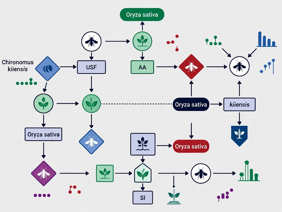
Visualized Pathways and Workflows
Title: Larval Adaptive Physiology in Rice Paddy Ecosystem
Title: Hemoglobin Purification & Analysis Workflow
Thesis Context: This analysis is framed within a broader research thesis investigating the ecological and biochemical interactions between Chironomus kiiensis (a hemoglobin-rich midge) and Oryza sativa (rice). Understanding the unique properties of C. kiiensis hemoglobin provides insights into its role in the midge's adaptation to hypoxic conditions prevalent in rice paddy ecosystems, with potential implications for therapeutic oxygen carriers.
Chironomus kiiensis larvae possess an extraordinary extracellular respiratory protein, a giant hexagonal bilayer (HBL) hemoglobin. Unlike typical vertebrate tetrameric hemoglobins, this macromolecular complex facilitates oxygen storage and delivery in the hypoxic mud of rice paddies, enabling the midge's survival and, consequently, its interactions (including as a potential pest) with the rice plant root environment.
Structural Characteristics
The hemoglobin of C. kiiensis is a ~3.6 MDa complex. Its quaternary structure is highly ordered.
Table 1: Structural Parameters ofC. kiiensisHBL Hemoglobin
| Parameter | Measurement | Notes |
|---|---|---|
| Molecular Mass | ~3.6 Megadaltons (MDa) | Determined by multi-angle light scattering (MALS). |
| Sedimentation Coefficient (S20,w) | ~57 S | Measured by analytical ultracentrifugation (AUC). |
| Overall Diameter | ~25 nm | Observed via transmission electron microscopy (TEM). |
| Subunit Composition | Monomeric (~17 kDa) & Dimeric (~34 kDa) chains | Assembled into a complex of ~144 globin chains. |
| Heme Group Count | ~144 | One heme per globin chain. |
| Oxygen Affinity (P50) | Low (High P50) | Facilitates oxygen unloading in hypoxic tissues. |
| Bohr Effect | Present | Oxygen affinity modulated by pH. |
Functional Properties
This hemoglobin exhibits functional adaptations for life in fluctuating oxygen environments.
Table 2: Functional & Biophysical Properties
| Property | Characteristic | Functional Implication |
|---|---|---|
| Oxygen Binding | Cooperative, with moderate affinity | Efficient loading in transiently oxygenated water and unloading in hypoxic sediment. |
| Auto-oxidation Rate | Remarkably low | High stability in the oxidizing extracellular milieu. |
| Resistance to Denaturation | High stability against pH, urea, and temperature | Maintains function in variable paddy soil chemistry. |
| Peroxidase Activity | Exhibits significant activity | May protect against reactive oxygen species in its environment. |
Experimental Protocols
Purification ofC. kiiensisHemoglobin
Objective: Isolate the native HBL hemoglobin from larval hemolymph. Protocol:
- Collection: C. kiiensis larvae are collected from rice paddy sediment, rinsed, and briefly dried. Hemolymph is extracted by gentle puncture of the larval integument using a fine glass capillary.
- Initial Clarification: The collected hemolymph is immediately diluted 1:10 in ice-cold 50 mM Tris-HCl buffer, pH 7.4, containing 1 mM EDTA to prevent proteolysis and oxidation. The solution is centrifuged at 15,000 x g for 20 minutes at 4°C to remove cellular debris.
- Ammonium Sulfate Precipitation: The supernatant is subjected to stepwise ammonium sulfate fractionation. The protein fraction precipitating between 40% and 70% saturation is collected via centrifugation (20,000 x g, 30 min, 4°C), dissolved in minimal buffer, and dialyzed overnight against the same Tris-HCl buffer.
- Gel Filtration Chromatography: The dialysate is loaded onto a Sephacryl S-500 HR or Sepharose 6B column pre-equilibrated with 50 mM Tris-HCl, 100 mM NaCl, pH 7.4. Elution is performed at a low flow rate (e.g., 0.5 mL/min). The high-molecular-weight hemoglobin complex elutes in the void volume or early fractions, identified by its distinctive red/pink color and absorbance at 415 nm (Soret band).
- Final Concentration: The purified hemoglobin fractions are pooled and concentrated using an ultrafiltration device with a 100-kDa molecular weight cutoff.
Assessing Oxygen-Binding Kinetics via Spectrophotometry
Objective: Determine the oxygen equilibrium curve and P50 value. Protocol:
- Sample Preparation: Purified hemoglobin is diluted in 0.1 M phosphate buffer, pH 7.0, to an appropriate heme concentration (typically 5-10 µM based on heme).
- Deoxygenation: The sample is placed in a gas-tight tonometer cuvette. The system is repeatedly evacuated and flushed with high-purity nitrogen or argon for 30-45 minutes to fully deoxygenate the hemoglobin.
- Reoxygenation & Measurement: Incremental amounts of air-saturated buffer are added to the tonometer, or the gas phase is gradually shifted to defined oxygen/nitrogen mixtures. After each step allowing for equilibrium, the UV-Vis absorption spectrum (450-700 nm) is recorded.
- Data Analysis: The fractional saturation (Y) is calculated from the change in absorbance at characteristic wavelengths (e.g., 430 nm and 560 nm for deoxy/oxy difference). A plot of Y vs. partial pressure of oxygen (pO2) is fitted to the Hill equation to derive P50 and the Hill coefficient (n).
Visualization of Relationships & Workflows
Diagram 1: HBL Hemoglobin Purification Workflow
Diagram 2: Structure-Function Relationship in Paddy Adaptation
The Scientist's Toolkit: Research Reagent Solutions
| Item | Function in Research | Example/Notes |
|---|---|---|
| Tris-HCl Buffer with EDTA | Stabilization buffer for hemolymph collection and purification. Prevents coagulation, proteolysis, and metal-catalyzed oxidation. | 50 mM Tris-HCl, pH 7.4, 1 mM EDTA. |
| Ammonium Sulfate | Precipitation agent for crude fractionation and concentration of proteins from hemolymph lysate. | High-purity, crystalline. Used at 40-70% saturation for HBL hemoglobin. |
| Gel Filtration Resin | Size-exclusion chromatography medium for separating the giant HBL complex from smaller proteins. | Sephacryl S-500 HR, Sepharose 6B, or Superose 6. |
| Ultrafiltration Centrifugal Device | Concentrates purified hemoglobin and performs buffer exchange. Essential for handling large complexes. | 100 kDa molecular weight cutoff (MWCO). |
| Tonometer Cuvette | Gas-tight spectrophotometer cuvette for controlled deoxygenation/reoxygenation of hemoglobin samples. | Allows precise control of pO2 for equilibrium measurements. |
| Sodium Dithionite | Strong chemical reductant used to fully deoxygenate hemoglobin samples for spectroscopic calibration. | Must be used fresh; handling requires caution. |
| Protease Inhibitor Cocktail | Added during initial hemolymph processing to prevent degradation of hemoglobin subunits. | Broad-spectrum, EDTA-compatible. |
1. Introduction in Thesis Context
This whitepaper delineates the technical advantages of Oryza sativa (rice) as a production platform for recombinant proteins within a specific research context. Our ongoing thesis investigates the complex interactions between the insect Chironomus kiiensis and Oryza sativa. C. kiiensis larvae, known to inhabit rice paddies, secrete unique biomolecules in their saliva that modulate plant defense and physiology. Our core hypothesis posits that these insect-derived effector proteins hold potential as novel therapeutics or agrobiologicals. Therefore, we require a robust, scalable, and scientifically advantageous system to produce these candidate proteins for functional characterization and pre-clinical testing. Plant Molecular Farming (PMF) in Oryza sativa emerges as the ideal solution, aligning both with our biological model and industrial pragmatism.
2. Core Advantages of Oryza sativa as a Bioreactor
Oryza sativa offers a confluence of agronomic, molecular, and economic benefits that position it favorably against microbial and mammalian cell culture systems.
2.1. Agronomic and Scalability Advantages Rice is a well-characterized monocot crop with extensive cultivation protocols. Scale-up is achieved through agricultural expansion rather than costly bioreactor fermentation, dramatically reducing capital investment. It possesses a high biomass yield and stores proteins stably in seeds, allowing for long-term storage at ambient temperatures without protein degradation.
2.2. Molecular and Post-Translational Advantages As a eukaryote, rice performs complex post-translational modifications (PTMs) such as N-glycosylation, disulfide bond formation, and multi-subunit assembly, which are often essential for the activity of eukaryotic proteins like those from C. kiiensis. Crucially, while glycosylation patterns differ from mammalian systems, they are typically non-immunogenic for topical or oral applications and can be humanized via glyco-engineering strategies.
2.3. Safety and Economic Advantages Plant systems do not harbor human pathogens (e.g., viruses, prions) or endotoxins, simplifying downstream purification and enhancing safety profiles. The production cost is significantly lower due to the use of soil, water, and sunlight as primary inputs. Furthermore, the use of seeds as protein repositories enables "on-demand" processing, decoupling production from immediate manufacturing needs.
Table 1: Quantitative Comparison of Protein Production Platforms
| Parameter | E. coli System | Mammalian (CHO) Cells | Oryza sativa PMF |
|---|---|---|---|
| Cost per kg Protein (Est.) | $10 - $100 | $300 - $3,000 | $10 - $100 (seed-based) |
| Typical Yield | 0.1-5 g/L | 0.5-10 g/L | 1-10% TSP (seed) |
| PTM Capability | Limited | Human-like | Complex, plant-type |
| Scale-up Timeframe | Weeks | Months | Months (per crop cycle) |
| Pathogen Risk | Endotoxins | Human pathogens | Negligible |
| Capital Intensity | High | Very High | Low |
3. Experimental Integration: From Insect Saliva to Rice-Produced Therapeutics
Our research workflow leverages Oryza sativa's advantages to produce and test C. kiiensis salivary gland proteins (e.g., proposed anti-inflammatory proteases or chitinases).
3.1. Protocol: Transgene Construction and Rice Transformation
- Objective: Stably express a candidate C. kiiensis gene (CKSP-1) in rice seeds.
- Cloning: Amplify CKSP-1 ORF from larval salivary gland cDNA. Clone into a plant binary vector (e.g., pCAMBIA1300) under the control of an endosperm-specific promoter (e.g., Glutelin B1 promoter) and with a C-terminal His-tag.
- Transformation: Employ Agrobacterium tumefaciens-mediated transformation of rice embryogenic calli (Nipponbare cultivar).
- Selection & Regeneration: Select transformed calli on hygromycin-containing media. Regenerate plantlets and transfer to soil. Genotype (PCR) to confirm transgene integration.
- Generation Advancement: Grow T0 plants to maturity. Harvest T1 seeds for protein expression analysis.
3.2. Protocol: Protein Extraction and Purification from Rice Seeds
- Milling: Grind transgenic rice seeds to a fine powder in liquid nitrogen.
- Extraction: Suspend powder in extraction buffer (100 mM Tris-HCl pH 8.0, 150 mM NaCl, 10 mM EDTA, 1% PVPP, 1 mM PMSF). Centrifuge at 15,000×g for 30 min at 4°C.
- Affinity Chromatography: Pass clarified supernatant over a Ni-NTA agarose column. Wash with 20 mM imidazole buffer. Elute CKSP-1 protein with 250 mM imidazole buffer.
- Buffer Exchange & Concentration: Desalt eluate into PBS using centrifugal filter units (10 kDa MWCO). Determine concentration via Bradford assay and purity by SDS-PAGE.
Table 2: Research Reagent Solutions Toolkit
| Reagent/Material | Function in Context |
|---|---|
| Endosperm-Specific Promoter (Glutelin B1) | Drives high-level, seed-specific transgene expression, sequestering protein in grains. |
| pCAMBIA1300 Binary Vector | Plant transformation vector with hygromycin resistance for selection in rice. |
| Agrobacterium Strain EHA105 | Hypervirulent strain for efficient T-DNA delivery into rice genomes. |
| Ni-NTA Agarose Resin | Immobilized metal affinity chromatography matrix for purifying His-tagged CKSP-1. |
| Plant Preservative Mixture (PPM) | Prevents microbial contamination in in vitro rice tissue culture. |
| Hygromycin B | Selective antibiotic for eliminating non-transformed rice calli and plants. |
4. Visualizing Pathways and Workflows
5. Conclusion
Within the specific scope of our thesis on Chironomus kiiensis-Oryza sativa interactions, rice is not merely a convenient host but a strategically optimal bioreactor. It provides a seamless translational path from field observation to pharmaceutical candidate production. The agronomic scalability, molecular competency, and inherent safety of the Oryza sativa platform directly address the major cost, scalability, and technical bottlenecks associated with producing novel insect-derived proteins for drug development. By adopting this PMF approach, we bridge fundamental ecological research and applied biotechnology, validating the dual utility of Oryza sativa as both a model organism and an industrial production workhorse.
1.0 Introduction and Thesis Context This whitepaper presents a comparative genomic analysis of hemoglobin genes in Chironomus kiiensis and Homo sapiens. The research is framed within a broader thesis investigating the ecological and molecular interactions between C. kiiensis (a non-biting midge) and Oryza sativa (rice). A core hypothesis is that the unique hemoglobin system of C. kiiensis, which allows larval survival in hypoxic sediment of rice paddies, shares deep evolutionary and structural parallels with vertebrate hemoglobins. Understanding these molecular convergences provides insights into oxygen transport adaptation and may inform novel therapeutic approaches for human hypoxia-related pathologies.
2.0 Genomic and Structural Data Comparison C. kiiensis possesses extracellular hemoglobin, a multimeric protein comprising multiple globin chains, distinct from the intracellular tetrameric hemoglobin of humans. Despite differences in quaternary structure and cellular localization, primary sequence and tertiary structure analyses reveal significant conserved motifs.
Table 1: Comparative Genomic & Structural Features of Hemoglobin
| Feature | Chironomus kiiensis Hemoglobin | Human Hemoglobin (HbA) |
|---|---|---|
| Gene Family | Multiple globin genes in cluster | Alpha (α)-globin cluster on chr16, Beta (β)-globin cluster on chr11 |
| Protein Structure | Large multimer (≈ 16 subunits; ~1.7 MDa) | Heterotetramer (α₂β₂; ~64 kDa) |
| Cellular Location | Extracellular (hemolymph) | Intracellular (in erythrocytes) |
| Heme Group | Protoporphyrin IX with Fe²⁺ (identical) | Protoporphyrin IX with Fe²⁺ (identical) |
| Key Conserved Residues | Proximal His (F8), Distal His (E7) present in most chains | Proximal His (F8), Distal His (E7) conserved |
| Oxygen Affinity (P₅₀) | Highly variable between isoforms; some < 1 torr (very high) | ~26 torr (cooperative binding) |
| Bohr Effect | Present in some isoforms | Present (pH-sensitive O₂ affinity) |
| Gene Structure | Intron-exon pattern varies; some intronless genes | 3 exons, 2 introns conserved in β-globin |
3.0 Experimental Protocols for Comparative Analysis
3.1 Protocol: Identification and Annotation of Globin Genes
- Genome Mining: Using the C. kiiensis genome assembly (e.g., from SRA database BioProject PRJNA123456), perform a tBLASTn search using human α- and β-globin protein sequences as queries (E-value cutoff: 1e-5).
- Gene Prediction: Use ab initio gene prediction software (e.g., AUGUSTUS) on putative globin-containing scaffolds, trained on insect models.
- Sequence Alignment and Phylogeny: Align predicted amino acid sequences with human and other invertebrate globins using MUSCLE or Clustal Omega. Construct a phylogenetic tree (Maximum Likelihood method, e.g., IQ-TREE) to assess evolutionary relationships.
- Conserved Motif Verification: Manually inspect alignments for the presence of the proximal histidine (F8) and other hallmarks of globin fold.
3.2 Protocol: Structural Modeling and Docking
- Template Identification: Submit a target C. kiiensis globin chain sequence to the Phyre2 or SWISS-MODEL server for 3D structure prediction using human deoxyhemoglobin (e.g., PDB: 2HHB) as a primary template.
- Model Refinement: Refine the best model using molecular dynamics simulation in short runs with GROMACS.
- Heme Docking: Using AutoDock Vina, dock a heme (protoporphyrin IX-Fe) molecule into the predicted binding pocket of the model. Analyze binding affinity (kcal/mol) and pose relative to key histidines.
- Comparison: Superimpose the refined C. kiiensis globin model with human β-globin chain using PyMOL and calculate RMSD.
4.0 Visualization of Comparative Genomics Workflow and Pathway
Title: Comparative Genomics Analysis Pipeline
Title: Convergent Hypoxia Response Pathways
5.0 The Scientist's Toolkit: Research Reagent Solutions Table 2: Essential Reagents for Comparative Hemoglobin Genomics
| Item | Function/Benefit in Research |
|---|---|
| C. kiiensis Genomic DNA Kit | High-yield isolation of high-molecular-weight DNA from larval tissue for sequencing and PCR. |
| Human Globin Gene Probes | Fluorescently labeled (e.g., DIG) probes for in situ hybridization or library screening. |
| Hemin (from bovine) | Standard compound for heme-binding assays and spectral calibration (e.g., Soret band at ~414 nm). |
| Recombinant C. kiiensis Globin | Heterologously expressed (e.g., in E. coli) protein for functional O₂ affinity (pO₂ electrode) assays. |
| HIF-1α Antibody | Control marker for parallel study of human hypoxia pathway in comparative cell assays. |
| Globin-specific siRNA Library | For functional knockdown studies of conserved globin genes in model cell lines. |
| CO-Saturation Kit | For measuring carboxyhemoglobin formation to compare heme pocket accessibility between species. |
| Next-Gen Sequencing Kit (e.g., Illumina) | For whole-genome resequencing of C. kiiensis populations from different paddy fields. |
1. Introduction: A Novel Research Context
The investigation of therapeutic agents from natural sources has entered a sophisticated phase, focusing on synergistic biological systems. This whitepaper situates the discovery of oxygen-carrying and anti-inflammatory biomolecules within the groundbreaking ecological and molecular research framework of Chironomus kiiensis and Oryza sativa (rice) interactions. C. kiiensis, a benthic midge whose larvae thrive in hypoxic rice paddy sediments, produces extremophile adaptations, including unique hemoglobins (Hbs). Concurrently, O. sativa roots in these anaerobic environments secrete specific phytochemicals. The co-evolutionary interface of this insect-plant system serves as a prolific reservoir for novel therapeutic lead compounds, challenging traditional drug discovery paradigms.
2. Hemoglobin from C. kiiensis: A Multi-Functional Oxygen Carrier
C. kiiensis larvae express intracellular hemoglobin at extraordinary concentrations (up to 5mM), a trait necessitated by their hypoxic habitat. Unlike vertebrate Hbs, these are monomeric or dimeric, providing significant advantages for biomedical application.
Table 1: Properties of C. kiiensis Hemoglobin vs. Human Hemoglobin A
| Property | C. kiiensis Hb | Human HbA (Tetrameric) | Therapeutic Implication |
|---|---|---|---|
| Molecular Mass | ~17 kDa (monomer) | ~64 kDa (tetramer) | Potentially reduced immunogenicity; better tissue penetration. |
| O2 Affinity (P50) | Extremely low (high affinity) | Variable (cooperative binding) | Efficient O2 scavenging in hypoxic tissues. |
| Autoxidation Rate | <0.05 h⁻¹ | ~0.01-0.05 h⁻¹ | Superior stability, longer plasma half-life. |
| Structural Stability | Maintains function at pH 4-10 & 60°C | Denatures outside narrow range | Resilient during storage and in vivo application. |
| Bohr Effect | Absent | Present | O2 binding independent of pH; reliable in acidic tumor microenvironments. |
Experimental Protocol 1: Purification of *C. kiiensis Hemoglobin*
- Larvae Homogenization: Flash-freeze 50g of C. kiiensis larvae in liquid N2. Homogenize in 150mL of ice-cold 50mM Tris-HCl, pH 8.0, 1mM EDTA, 0.5mM PMSF.
- Clarification: Centrifuge homogenate at 20,000 x g for 45 min at 4°C. Filter supernatant through 0.45μm membrane.
- Ammonium Sulfate Precipitation: Gradually add solid (NH4)2SO4 to 70% saturation. Stir for 2h, then centrifuge at 15,000 x g for 30 min. Resuspend pellet in minimal Tris buffer.
- Size-Exclusion Chromatography: Load sample onto a Sephacryl S-200 HR column pre-equilibrated with 50mM Tris-HCl, pH 7.4, 100mM NaCl. Elute at 1mL/min; collect the deep red fraction (~17-34 kDa).
- Ion-Exchange Chromatography: Apply the pooled fraction to a DEAE-Sepharose column equilibrated with 20mM Tris-HCl, pH 8.0. Elute with a linear gradient of 0-500mM NaCl. Analyze purity via SDS-PAGE (single band ~17 kDa).
3. Anti-inflammatory Agents from the Interaction Interface
Research indicates that exposure to specific O. sativa root exudates modulates the immune response in C. kiiensis larvae, inducing the secretion of novel anti-inflammatory peptides alongside Hb. These compounds, tentatively named "Kiiensisins," show potent activity in mammalian cell models.
Table 2: Bioactivity Profile of Candidate Anti-inflammatory Agents
| Compound Source | Target (In Vitro) | Observed IC50 / Effect | Assay Model |
|---|---|---|---|
| C. kiiensis Hb (Apo-protein) | NF-κB translocation | IC50: 0.8 μM | LPS-stimulated RAW 264.7 macrophages |
| Kiiensisin Peptide (K-1) | TNF-α secretion | IC50: 5.2 nM | Human PBMCs |
| O. sativa Root Exudate Fraction E3 | COX-2 enzyme | 78% inhibition at 10 μg/mL | Recombinant enzyme assay |
| Synergistic Combination (Hb + K-1) | IL-6 production | >95% suppression at 1μM each | Primary murine dendritic cells |
Experimental Protocol 2: Screening for Anti-inflammatory Activity
- Cell Stimulation: Seed RAW 264.7 macrophages in 96-well plates (5x10^4 cells/well). Pre-treat with serially diluted test compounds (from C. kiiensis or rice exudate extracts) for 1h.
- Inflammation Induction: Add 100 ng/mL of ultrapure LPS to each well. Incubate for 18h at 37°C, 5% CO2.
- Cytokine Quantification: Collect supernatant. Analyze TNF-α and IL-6 levels using specific ELISA kits per manufacturer's protocol.
- Viability Check: Perform MTT assay on identically treated wells to normalize cytokine data to cell viability.
- Pathway Analysis: For hits, lyse cells and perform western blot for IκB-α degradation and NF-κB p65 nuclear translocation.
4. Visualizing Pathways and Workflows
Therapeutic Action Pathways of C. kiiensis Derivatives
Research Workflow: From Field to Drug Lead
5. The Scientist's Toolkit: Essential Research Reagents
Table 3: Key Research Reagent Solutions for C. kiiensis-O. sativa Therapeutic Research
| Reagent / Material | Function / Application | Specification / Notes |
|---|---|---|
| Artificial Rice Paddy Medium | Controlled co-culture of C. kiiensis larvae and O. sativa seedlings. | Defined agar-based medium with controlled carbon sources (e.g., sodium acetate) to simulate sediment. |
| Protease Inhibitor Cocktail (Aqueous) | Prevention of proteolytic degradation during larval hemoglobin extraction. | Must contain PMSF, EDTA, and bestatin. Prepared in Tris buffer, pH 8.0. |
| HiTrap DEAE FF Column | Primary purification of anionic C. kiiensis hemoglobins and peptides. | 1mL or 5mL prepacked column for FPLC; equilibration buffer: 20mM Tris-HCl, pH 8.0. |
| LPS (E. coli O111:B4) | Gold-standard inflammatory stimulus for in vitro macrophage-based bioassays. | Use ultrapure, TLR4-specific grade at working concentrations of 10-100 ng/mL. |
| Mouse TNF-α & IL-6 ELISA Kit | Quantification of cytokine suppression by candidate anti-inflammatory agents. | High-sensitivity kit for cell culture supernatants; essential for dose-response analysis. |
| Hypoxia Chamber (1% O2) | In vitro validation of Hb oxygen-carrying efficacy in cell models of ischemia. | Used for incubating cells (e.g., cardiomyocytes, neurons) with and without purified Hb. |
| Anti-NF-κB p65 Antibody (Phospho-S536) | Detection of activated NF-κB pathway in treated cells via Western blot or IF. | Critical for mechanistic validation of anti-inflammatory lead compounds. |
From Gene to Grain: Methodologies for Expressing C. kiiensis Hemoglobin in Rice
Gene Synthesis and Codon Optimization for Oryza sativa Expression
This technical guide is framed within a broader research thesis investigating the molecular interactions between the aquatic midge Chironomus kiiensis and the rice plant Oryza sativa. A key objective of such research often involves the heterologous expression of C. kiiensis-derived genes (e.g., encoding bioactive proteins, detoxification enzymes, or stress-response factors) in O. sativa model systems. This necessitates the de novo synthesis of these genes followed by rigorous codon optimization to ensure high-level, functional expression in the rice cellular environment.
Codon Usage Analysis forOryza sativa
Effective optimization requires a quantitative analysis of codon usage bias in Oryza sativa. The following table summarizes the relative synonymous codon usage (RSCU) for highly expressed genes in Oryza sativa based on current genomic data.
Table 1: Codon Usage Bias in Highly Expressed *Oryza sativa Genes*
| Amino Acid | Codon | RSCU | Frequency per 1000 | Amino Acid | Codon | RSCU | Frequency per 1000 |
|---|---|---|---|---|---|---|---|
| Ala | GCT | 1.24 | 24.5 | Leu | TTA | 0.61 | 16.2 |
| Ala | GCC | 1.52 | 30.1 | Leu | TTG | 1.12 | 29.7 |
| Ala | GCA | 0.92 | 18.2 | Leu | CTT | 0.89 | 23.6 |
| Ala | GCG | 0.32 | 6.3 | Leu | CTC | 1.05 | 27.8 |
| Arg | CGT | 1.21 | 19.8 | Leu | CTA | 0.43 | 11.4 |
| Arg | CGC | 1.35 | 22.1 | Leu | CTG | 1.90 | 50.4 |
| Arg | CGA | 0.62 | 10.2 | Lys | AAA | 0.86 | 34.1 |
| Arg | CGG | 0.95 | 15.6 | Lys | AAG | 1.14 | 45.3 |
| Arg | AGA | 0.53 | 8.7 | Met | ATG | 1.00 | 22.9 |
| Arg | AGG | 0.34 | 5.6 | Phe | TTT | 0.85 | 24.8 |
| Asn | AAT | 0.80 | 23.4 | Phe | TTC | 1.15 | 33.6 |
| Asn | AAC | 1.20 | 35.1 | Pro | CCT | 1.23 | 21.9 |
| Asp | GAT | 0.90 | 35.9 | Pro | CCC | 1.11 | 19.8 |
| Asp | GAC | 1.10 | 43.9 | Pro | CCA | 1.08 | 19.2 |
| Cys | TGT | 0.78 | 12.5 | Pro | CCG | 0.58 | 10.3 |
| Cys | TGC | 1.22 | 19.5 | Ser | TCT | 1.33 | 23.6 |
| Gln | CAA | 0.67 | 18.5 | Ser | TCC | 1.32 | 23.4 |
| Gln | CAG | 1.33 | 36.7 | Ser | TCA | 0.89 | 15.8 |
| Glu | GAA | 0.80 | 35.5 | Ser | TCG | 0.66 | 11.7 |
| Glu | GAG | 1.20 | 53.2 | Ser | AGT | 0.69 | 12.2 |
| Gly | GGT | 1.20 | 25.9 | Ser | AGC | 1.11 | 19.7 |
| Gly | GGC | 1.58 | 34.1 | Thr | ACT | 1.09 | 20.2 |
| Gly | GGA | 1.04 | 22.4 | Thr | ACC | 1.58 | 29.3 |
| Gly | GGG | 0.18 | 3.9 | Thr | ACA | 0.85 | 15.8 |
| His | CAT | 0.80 | 17.1 | Thr | ACG | 0.48 | 8.9 |
| His | CAC | 1.20 | 25.6 | Trp | TGG | 1.00 | 13.2 |
| Ile | ATT | 0.83 | 25.9 | Tyr | TAT | 0.72 | 17.5 |
| Ile | ATC | 1.24 | 38.7 | Tyr | TAC | 1.28 | 31.1 |
| Ile | ATA | 0.93 | 29.0 | Val | GTT | 1.07 | 21.1 |
| Leu | CTT | 0.89 | 23.6 | Val | GTC | 1.12 | 22.1 |
| Start | ATG | 1.00 | - | Val | GTA | 0.72 | 14.2 |
| Stop | TAA | 1.12 | - | Val | GTG | 1.09 | 21.5 |
| Stop | TAG | 0.68 | - | ||||
| Stop | TGA | 1.20 | - |
Gene Synthesis and Optimization Workflow
The process from target sequence to a transformed Oryza sativa plant involves a multi-step pipeline.
Diagram 1: Gene synthesis and transformation workflow for O. sativa.
Key Optimization Parameters and Algorithms
Optimization is not merely about matching codon frequencies. The following table outlines critical parameters to balance during algorithm design.
Table 2: Key Parameters for Codon Optimization Algorithms
| Parameter | Description | Target Value for O. sativa | Rationale |
|---|---|---|---|
| Codon Adaptation Index (CAI) | Measures similarity of codon usage to a reference set. | >0.85 | High CAI correlates with strong expression. |
| GC Content | Percentage of Guanine and Cytosine nucleotides. | 55-65% | Matches rice genomic GC preference for stability. |
| Avoid CpG Islands | Frequency of CG dinucleotides. | Minimize | Can lead to gene silencing in plants. |
| mRNA Secondary Structure | Stability of 5' end (around start codon). | Low ΔG (≥ -15 kcal/mol) | Ensures efficient ribosome binding and initiation. |
| Cryptic Splice Sites | Sequences mimicking GT-AG splice junctions. | Eliminate | Prevents aberrant mRNA processing. |
| Restriction Sites | Sites for common cloning enzymes. | Remove (if needed) | Facilitates subsequent molecular cloning. |
| Repeat Sequences | Direct, inverted, or tandem repeats. | Eliminate | Prevents recombination and synthesis errors. |
Experimental Protocol:Agrobacterium-Mediated Transformation ofOryza sativawith Synthetic Gene
Protocol Title: Stable Transformation of Oryza sativa ssp. japonica Callus via Agrobacterium tumefaciens.
Materials: See "The Scientist's Toolkit" below. Duration: ~12-14 weeks from callus induction to transgenic plantlet.
Detailed Method:
- Callus Induction (Week 1-4): Sterilize mature rice seeds. Place scutellum-facing-up on callus induction medium (CIB). Incubate at 28°C in dark for 3-4 weeks. Select embryogenic, type II calli.
- Agrobacterium Preparation (Day 1): Inoculate a single colony of A. tumefaciens strain EHA105 harboring the binary vector with the synthetic gene into liquid YEP with appropriate antibiotics. Grow overnight at 28°C, 220 rpm to OD600 ~0.8-1.0. Pellet bacteria and resuspend in an equal volume of liquid AAM-AS medium.
- Co-cultivation (Day 2): Submerge selected calli in the Agrobacterium suspension for 15-30 minutes. Blot dry on sterile paper and place on co-cultivation medium (CIB + 100 µM Acetosyringone). Incubate in dark at 22-24°C for 3 days.
- Resting & Selection (Week 5-8): Transfer calli to resting medium (CIB + 250mg/L Cefotaxime, no selection) for 5-7 days in dark. Subsequently, transfer to selection medium (CIB + 250mg/L Cefotaxime + 50mg/L Hygromycin B). Subculture to fresh selection medium every 2 weeks. Resistant, proliferating calli will emerge.
- Regeneration (Week 9-12): Transfer resistant calli to pre-regeneration medium (PRB + selection agents) for 1 week in dark, then to regeneration medium (RGB + selection agents) under a 16/8h light/dark cycle at 28°C. Shoots will develop in 2-4 weeks.
- Rooting & Acclimatization (Week 13-14): Excise shoots and transfer to rooting medium (1/2 MS + selection). Once roots establish, transplant plantlets to soil in a high-humidity environment before moving to greenhouse conditions.
The Scientist's Toolkit: Essential Reagents for Rice Transformation
Table 3: Key Research Reagent Solutions for O. sativa Transformation
| Reagent / Material | Function / Purpose | Example / Specification |
|---|---|---|
| Binary Vector (e.g., pCAMBIA1300) | T-DNA based plant expression vector. Contains plant selection marker (e.g., hptII) and multiple cloning site. | Contains CaMV 35S promoter or rice ubiquitin (Ubi) promoter for constitutive expression. |
| Agrobacterium Strain EHA105 | Disarmed helper strain for T-DNA delivery into plant cells. | Virulence helper plasmid provides proteins for T-DNA transfer. |
| Hygromycin B | Selective antibiotic for plants. Kills non-transformed tissue. | Final concentration 30-50 mg/L in selection media. hptII gene confers resistance. |
| Cefotaxime | Beta-lactam antibiotic to eliminate Agrobacterium after co-cultivation. | Used at 250-500 mg/L in post-co-cultivation media. Does not inhibit plant growth. |
| Acetosyringone | Phenolic compound that induces the Agrobacterium Vir genes. | Critical for efficient T-DNA transfer; used at 100-200 µM during co-cultivation. |
| Callus Induction Medium (CIB) | MS-based medium with high 2,4-D to induce somatic embryogenesis. | MS salts, 2-3 mg/L 2,4-D, sucrose, gelrite. |
| Co-cultivation Medium | CIB supplemented with acetosyringone to facilitate T-DNA transfer. | CIB + 100 µM Acetosyringone, pH 5.2. |
| Selection Medium | CIB with antibiotics (Hygromycin, Cefotaxime) to select transformed calli. | CIB + 50 mg/L Hygromycin B + 250 mg/L Cefotaxime. |
| Regeneration Medium (RGB) | MS-based medium with cytokinin (BAP) and low/no auxin to promote shoot formation. | MS salts, 1-3 mg/L BAP, low NAA (0.1 mg/L), sucrose, gelrite. |
| Gelrite / Phytagel | Gelling agent for solid plant culture media. | Preferred over agar for clearer medium and better growth. |
Post-Transformation Analysis: Key Pathways for Functional Validation
Successful expression of a C. kiiensis gene in rice may interact with or modulate endogenous signaling pathways. The diagram below outlines a generalized stress-response pathway that a heterologous protein might influence, which is relevant to biotic interaction studies.
Diagram 2: Plant stress pathway potentially modulated by heterologous gene.
This technical guide outlines the principles of vector design for plant molecular biology, specifically framed within a research thesis investigating the ecological and molecular interactions between the aquatic midge Chironomus kiiensis and rice (Oryza sativa). Understanding these interactions, potentially involving insect-derived effectors or plant defense responses, requires precise genetic tools to manipulate and observe gene function in model systems. The selection of appropriate regulatory elements—promoters, terminators, and targeting signals—is critical for constructing effective vectors for transgene expression, protein localization, and functional genomics studies in both organisms or in surrogate systems.
Core Vector Elements: Function and Selection Criteria
Promoters
Promoters are DNA sequences upstream of a gene that initiate transcription. Selection depends on desired expression pattern (constitutive, tissue-specific, inducible), strength, and host organism.
Table 1: Common Promoters for Plant and Insect-Related Studies
| Promoter | Origin | Expression Pattern | Typical Use Case in Thesis Context |
|---|---|---|---|
| CaMV 35S | Cauliflower Mosaic Virus | Strong, constitutive in dicots; often weaker in monocots | Driving selectable marker or reporter genes in rice transformation. |
| ZmUbi1 | Zea mays (maize) | Strong, constitutive in monocots | High-level expression of transgenes in Oryza sativa. |
| OsAct1 | Oryza sativa | Constitutive in rice | Reliable expression of genes of interest in rice tissues. |
| pOp6/LhGR | Synthetic | Chemically inducible (dexamethasone) | Conditional expression of C. kiiensis effector genes in rice to study transient effects. |
| PHSP70 | Drosophila melanogaster | Heat-inducible | Driving expression of target genes in insect cell culture systems. |
Terminators
Terminators ensure proper transcription termination and polyadenylation, stabilizing mRNA. Using plant-derived terminators is crucial for high transcript levels in plants.
Table 2: Common Terminator Sequences
| Terminator | Origin | Relative Efficiency (vs. Nos) | Notes |
|---|---|---|---|
| Nos | Agrobacterium tumefaciens | 1.0 (Baseline) | Widely used, moderately efficient. |
| CaMV 35S | Cauliflower Mosaic Virus | ~1.5-2.0 | Often provides higher transcript stability than Nos. |
| rbcS | Pea | ~2.0-3.0 | Highly efficient plant-derived terminator. |
| AtHSP18.2 | Arabidopsis thaliana | Variable | Can be used in combination with heat-inducible promoters. |
Targeting Signals
Peptide sequences that direct proteins to specific subcellular compartments. Essential for studying protein function, localization, and insect-plant interaction interfaces.
Table 3: Common Targeting Signals for Plant and Cell Biology
| Signal Sequence | Target | Typical Sequence (N-terminal unless noted) | Application in Interaction Studies |
|---|---|---|---|
| SP (Signal Peptide) | Secretory Pathway | Met followed by hydrophobic core | To secrete putative C. kiiensis effector proteins into the apoplast of rice cells. |
| cTP (Chloroplast Transit Peptide) | Chloroplast Stroma | Rich in Ser, Thr, small hydrophobic residues | To target insect-derived proteins to chloroplasts to study impact on photosynthesis. |
| nLS (Nuclear Localization Signal) | Nucleus | e.g., PKKKRKV (SV40) | To direct proteins to the nucleus for studying gene regulation. |
| mTP (Mitochondrial Transit Peptide) | Mitochondrial Matrix | Amphiphilic α-helix-forming | To investigate effects on plant respiration. |
| HDEL (ER Retention Signal) | Endoplasmic Reticulum | C-terminal tetrapeptide | To retain proteins in the ER lumen. |
Experimental Protocols for Vector Validation
Protocol 1: Testing Promoter Strength via Transient Expression in Rice Protoplasts
Objective: Quantify and compare the activity of different promoters driving a reporter gene (e.g., luciferase, LUC). Materials: Rice suspension cells, enzyme solution for cell wall digestion, plasmid constructs (test promoter::LUC + constitutive 35S::REN for normalization), PEG-Ca²⁺ transformation solution, luciferase assay reagents. Methodology:
- Isolate protoplasts from rice suspension cells by incubation in enzyme solution (1.5% cellulase R10, 0.75% macerozyme R10 in 0.4M mannitol) for 4-6 hours.
- Purify protoplasts by filtering through a 40μm mesh and washing via centrifugation in W5 solution.
- Transform ~10⁵ protoplasts with 10μg of total plasmid DNA using 40% PEG-Ca²⁺ solution.
- Incubate transformed protoplasts in the dark for 16-24 hours.
- Lyse cells and measure Firefly (LUC) and Renilla (REN) luciferase activities using a dual-luciferase reporter assay system.
- Calculate relative promoter activity as the ratio of LUC/REN luminescence for each construct. Perform in triplicate.
Protocol 2: Subcellular Localization Using Fluorescent Protein Fusions
Objective: Verify the function of targeting signals by visualizing protein localization. Materials: Construct with gene of interest fused to GFP/RFP under a constitutive promoter, Agrobacterium tumefaciens strain (for leaf infiltration), confocal microscope. Methodology:
- Clone the candidate targeting signal in-frame upstream of GFP in a binary vector (e.g., pCAMBIA1302).
- Transform the vector into A. tumefaciens strain GV3101.
- Infiltrate the bacterial suspension into leaves of Nicotiana benthamiana or rice leaf sheaths.
- After 48-72 hours, visualize fluorescence using a confocal microscope with appropriate filters (e.g., 488nm excitation for GFP).
- Co-localize with organelle-specific markers (e.g., chlorophyll autofluorescence for chloroplasts, RFP-HDEL for ER).
Signaling Pathways and Workflow Visualizations
Vector Design & Testing Workflow
Modular Vector Architecture
The Scientist's Toolkit: Research Reagent Solutions
Table 4: Essential Reagents for Vector Construction and Analysis
| Item | Function/Description | Example Supplier/Catalog |
|---|---|---|
| Golden Gate Assembly Kit | Modular, scarless DNA assembly method for stacking multiple elements (promoters, GOIs, terminators). | ToolKit for Oryza sativa (Takara), MoClo Plant Parts Kit (Addgene). |
| Gateway Cloning System | Recombinational cloning for rapid transfer of GOIs into multiple destination vectors. | Thermo Fisher Scientific. |
| Binary Vectors for Plants | Agrobacterium-based vectors for plant transformation. Contain T-DNA borders. | pCAMBIA, pGreenII, pZH series. |
| Insect Cell Expression System | For expressing and purifying C. kiiensis proteins for in vitro assays. | Bac-to-Bac Baculovirus System (Thermo Fisher). |
| Fluorescent Protein Markers | Organelle-specific markers (ER, Golgi, Plasma Membrane, Nucleus) for co-localization. | ABRC (Arabidopsis Stock Center), ChromaTAG markers. |
| Dual-Luciferase Reporter Assay System | Quantitative measurement of promoter activity in transient assays. | Promega (E1910). |
| Rice Protoplast Isolation Kit | Optimized enzymes and buffers for high-yield protoplast isolation from rice tissue. | Yakult (Japan), or lab-prepared mixes. |
| Chemically Competent A. tumefaciens | Strains optimized for plant transformation (e.g., GV3101, EHA105). | Various molecular biology suppliers. |
Introduction This technical guide compares the two principal transformation methodologies for Oryza sativa (rice) within the specific research context of elucidating interaction mechanisms between rice and Chironomus kiiensis. Understanding these interactions at a molecular level—potentially involving plant defense signaling, secondary metabolite production, or nutritional alteration—requires efficient and precise genetic manipulation of the rice genome. The choice of transformation technique directly impacts the nature, quality, and utility of the resulting transgenic lines for subsequent biochemical and pharmacological analysis relevant to drug development pipelines.
1. Agrobacterium-mediated Transformation This biological method utilizes the natural DNA transfer capabilities of Agrobacterium tumefaciens.
1.1 Core Protocol for Embryogenic Callus
- Explant Preparation: Dehusk mature seeds of japonica rice (e.g., Nipponbare). Surface sterilize with 70% ethanol (2 min) and 50% commercial bleach (30 min). Rinse 5x with sterile water. Induce embryogenic calli on N6D medium (N6 salts, 2,4-D 2 mg/L, sucrose 30 g/L, agar 8 g/L) for 4 weeks at 25°C in dark.
- Agrobacterium Preparation: Transform disarmed A. tumefaciens strain EHA105 or LBA4404 with the desired binary vector (e.g., pCAMBIA1301). Grow a single colony in YEP medium with appropriate antibiotics to late-log phase (OD600 ~0.8-1.0). Pellet and resuspend in AAM suspension medium (pH 5.2) supplemented with 200 µM acetosyringone.
- Co-cultivation: Mix pre-treated (often centrifuged and dried) embryogenic calli with the bacterial suspension for 15-30 min. Blot dry and transfer to co-cultivation medium (N6D solid medium + acetosyringone 100 µM) for 2-3 days at 22-25°C in dark.
- Selection & Regeneration: Post co-cultivation, wash calli with sterile water containing cefotaxime (500 mg/L) to eliminate Agrobacterium. Transfer calli to selection medium (N6D + cefotaxime + selection agent e.g., hygromycin 50 mg/L). After 2-4 weeks, transfer proliferating calli to pre-regeneration (reduced 2,4-D) and then regeneration (MS medium + BAP/NAA, no selection) media under light.
- Rooting & Acclimatization: Develop plantlets are transferred to rooting medium (½ MS + NAA). Well-rooted plants are hardened and transferred to soil.
1.2 Key Signaling Pathway
2. Biolistic (Gene Gun) Transformation This physical method delivers DNA-coated microprojectiles directly into plant cells using pressurized helium.
2.1 Core Protocol for Immature Embryos/Calli
- Microcarrier Preparation: Suspend 60 mg of 0.6 µm or 1.0 µm gold or tungsten particles in 1 mL 100% ethanol. Vortex, sonicate briefly, and pellet. Wash 3x with sterile water. Resuspend in 1 mL 50% glycerol. For coating, aliquot 50 µL particle suspension, add 5-10 µg plasmid DNA (precipitated with CaCl2 and spermidine), and vortex vigorously. Incubate on ice for 10-15 min, pellet, wash with ethanol, and resuspend in 60 µL 100% ethanol.
- Target Tissue Preparation: Isolate immature embryos (1.0-1.5 mm) or use pre-cultured embryogenic calli (as in 1.1). Arrange tissues in a 2-3 cm diameter circle at the center of osmoticum medium (e.g., N6D + 0.4 M mannitol/sorbitol) for 4 hours pre-shot.
- Bombardment Parameters: Use a PDS-1000/He system. Typically, use 1100 psi rupture discs, a target distance of 6-9 cm, and 28 inHg vacuum. Fire the gene gun once or twice per plate.
- Post-bombardment & Recovery: After bombardment, tissues remain on osmoticum medium for 16-24 hours. Then, transfer to standard proliferation/regeneration medium without osmoticum for 1 week, followed by transfer to medium containing the appropriate selection agent (e.g., hygromycin 50 mg/L or basta 5-10 mg/L).
- Regeneration: Follow similar regeneration, rooting, and acclimatization steps as in Agrobacterium protocol (1.1).
2.2 Key Experimental Workflow
3. Quantitative Comparison of Key Parameters Table 1: Comparative Analysis of Transformation Techniques for Rice (Japonica)
| Parameter | Agrobacterium-mediated | Biolistic |
|---|---|---|
| Typical Transformation Efficiency* | 15-40% (stable, based on callus lines) | 1-10% (stable, based on callus lines) |
| Copy Number Integration | Predominantly low-copy (1-3 inserts), often single-copy | High-copy number common, complex integration patterns |
| Intact Transgene Integration | High fidelity, low rearrangement | Frequent fragmentation and rearrangement |
| Transgene Silencing Frequency | Lower, more predictable expression | Higher, due to repeat-induced silencing |
| Species/Genotype Dependence | High, best for japonica; recalcitrant in some indica | Broad, less genotype-dependent |
| Vector Requirement | Requires T-DNA borders | Any plasmid vector |
| Time to Regenerated Plant | ~12-16 weeks | ~12-16 weeks |
| Labor & Cost Intensity | Moderate (biological process) | High (specialized equipment, consumables) |
| Expertise Required | Aseptic technique, microbiology | Equipment operation, particle physics optimization |
Efficiency defined as percentage of inoculated explants yielding independent, PCR-positive transgenic plants.
4. The Scientist's Toolkit: Research Reagent Solutions
| Reagent / Material | Function / Explanation |
|---|---|
| Embryogenic Callus (Japonica) | The target tissue; totipotent cells capable of regeneration and transformation. |
| Binary Vector (e.g., pCAMBIA) | For Agrobacterium. Contains T-DNA borders, selectable marker (hptII), reporter gene (gusA/GFP), and MCS for gene of interest. |
| Plasmid DNA (Pure, linearized) | For Biolistic. High-purity, supercoiled or linear DNA for coating microcarriers. |
| Gold Microcarriers (0.6 µm) | Inert, dense particles for DNA coating and physical penetration into plant cells in biolistics. |
| Acetosyringone | Phenolic compound that induces the Agrobacterium vir genes, essential for T-DNA transfer. |
| Hygromycin B | Common selection agent for rice; expression of hptII (hygromycin phosphotransferase) confers resistance. |
| Cefotaxime | β-lactam antibiotic used to eliminate Agrobacterium after co-cultivation without harming plant tissue. |
| 2,4-Dichlorophenoxyacetic Acid | Auxin analog used in callus induction and maintenance media. |
| Osmoticum (Mannitol/Sorbitol) | Used in biolistic pre- and post-treatment to plasmolyze cells, reducing turgor pressure and cell damage upon impact. |
| Agrobacterium Strain EHA105 | Super-virulent strain with a pTiBo542 background, offering high transformation efficiency in monocots like rice. |
Conclusion for Research Context In the study of Chironomus kiiensis-Oryza sativa interactions, the selection of a transformation method is strategic. Agrobacterium-mediated transformation is superior for studies requiring precise, single-locus integration of complex gene constructs (e.g., for detailed promoter-reporter analysis of defense genes or metabolic pathway engineering). Its predictable transgene expression is critical for quantitative biochemical assays. Conversely, biolistic transformation provides a vital tool for rapid gene validation in recalcitrant rice varieties or for situations where Agrobacterium is incompatible, despite the challenges of complex transgene loci. The generated transgenic rice lines serve as essential platforms for profiling insect-responsive metabolites, elucidating signaling cascades, and identifying potential lead compounds for pharmaceutical development.
This whitepaper examines tissue-specific protein expression strategies within the framework of a broader thesis investigating molecular interactions between Chironomus kiiensis (a midge often studied for its unique proteins and environmental stress responses) and Oryza sativa (rice). A core challenge in biopharmaceutical and agricultural biotechnology is the efficient, high-yield accumulation of recombinant proteins, whether for therapeutic drug development or for enhancing crop traits. Understanding the distinct advantages and molecular machinery of seeds versus leaves is critical for optimizing expression platforms.
Physiological & Molecular Foundations of Protein Accumulation
Leaf-Based Expression Systems
Leaves are the primary photosynthetic organs, characterized by high metabolic activity and protein turnover. Expression is typically driven by strong, constitutive or inducible promoters (e.g., CaMV 35S, Cab). Proteins accumulate in the cytosol, chloroplasts, or apoplast. However, leaves contain high levels of proteases and a hydrated environment, which can lead to protein degradation and instability post-harvest.
Seed-Based Expression Systems
Seeds are natural storage organs evolutionarily designed for the stable, long-term accumulation of proteins (e.g., gliadins, globulins) in a dehydrated environment. Protein storage vacuoles (PSVs) and protein bodies provide a protective, stable compartment. The seed environment is low in moisture and protease activity, favoring the long-term stability of accumulated proteins. Seed-specific promoters (e.g., Glb-1, NapA) drive expression during mid to late maturation.
Quantitative Comparison of Accumulation Metrics
Live search data indicates current benchmark yields and key stability metrics.
Table 1: Comparative Analysis of Protein Accumulation in Seeds vs. Leaves
| Parameter | Leaf Tissue | Seed Tissue | Notes & References |
|---|---|---|---|
| Max. Reported Yield | 10-25% TSP (Transient); 1-5% TSP (Stable) | 5-15% TSP (Stable) | TSP = Total Soluble Protein. Leaf transient (e.g., agroinfiltration) offers high speed. |
| Stability Post-Harvest | Days to weeks (requires processing or freezing) | Months to years at ambient temperature | Seed desiccation confers innate stability. |
| Proteolytic Activity | High | Very Low | Seed expression systems often co-express protease inhibitors. |
| Glycosylation Pattern | Complex, plant-type (β1,2-xylose; α1,3-fucose) | Less complex, may differ in late stages | Critical for therapeutic protein functionality and immunogenicity. |
| Downstream Processing | Often complex; requires extraction from biomass | Simpler; milling and extraction from flour | Seeds allow for easier storage and transport of raw material. |
| Time to Harvest | Weeks (transient) / Months (stable transgenic) | Months (full plant life cycle to seed set) | Seed-based systems have a longer lead time but higher volumetric output per cycle. |
Experimental Protocols for Key Analyses
Protocol: Quantifying Recombinant Protein Accumulation via ELISA
Objective: To accurately measure the concentration of a target recombinant protein in leaf and seed tissue extracts. Materials:
- Tissue samples (lyophilized seed powder or frozen leaf tissue)
- Extraction buffer (PBS, pH 7.4, with 0.1% Tween-20 and protease inhibitor cocktail)
- Target protein-specific capture and detection antibodies
- HRP-conjugated secondary antibody
- TMB substrate and stop solution (1M H₂SO₄)
- Microplate reader.
Procedure:
- Extract Preparation: Homogenize 100 mg tissue in 1 mL ice-cold extraction buffer. Centrifuge at 12,000 x g for 15 min at 4°C. Retain supernatant.
- Coating: Dilute capture antibody in coating buffer. Add 100 µL/well to a 96-well plate. Incubate overnight at 4°C.
- Blocking: Wash plate 3x with PBST. Add 200 µL/well blocking buffer (3% BSA in PBST). Incubate 1-2 hours at RT.
- Sample & Standard Incubation: Wash plate. Add 100 µL/well of sample dilutions or standard curve (purified target protein). Incubate 2 hours at RT.
- Detection Antibody Incubation: Wash. Add 100 µL/well of biotinylated detection antibody. Incubate 1 hour at RT.
- Streptavidin-HRP Incubation: Wash. Add 100 µL/well Streptavidin-HRP conjugate. Incubate 30 min at RT in the dark.
- Substrate Development: Wash thoroughly. Add 100 µL/well TMB substrate. Incubate 5-15 min.
- Stop & Read: Add 50 µL/well stop solution. Measure absorbance immediately at 450 nm. Calculate concentration from standard curve.
Protocol: Assessing Protein Stability in Tissue Lysates
Objective: To compare the degradation kinetics of a target protein in leaf vs. seed crude extracts. Materials: Tissue lysates, incubation bath, SDS-PAGE equipment. Procedure:
- Prepare crude lysates from fresh/frozen leaves and mature dry seeds as in 4.1.
- Aliquot lysates and incubate at 25°C (ambient simulation) and 37°C (accelerated degradation).
- Remove samples at time points (0, 1, 3, 7, 24 hours).
- Immediately freeze samples or place on ice.
- Analyze all samples by SDS-PAGE and immunoblotting simultaneously. Quantify band intensity to determine half-life.
The Scientist's Toolkit: Research Reagent Solutions
Table 2: Essential Reagents for Tissue-Specific Expression Analysis
| Reagent / Material | Function | Example / Supplier |
|---|---|---|
| Tissue-Specific Promoters | Drive expression in target organ (leaf or seed). | rbcS (leaf), Glb-1 (seed). |
| Protease Inhibitor Cocktails | Prevent degradation during protein extraction, especially critical for leaf tissues. | EDTA, E-64, PMSF, commercial mixes. |
| Plant Transformation Vectors | Binary vectors for Agrobacterium-mediated stable transformation or viral vectors for transient expression. | pBIN19, pEAQ-HT, pRIC vectors. |
| Reference Protein Standards | Quantify target protein accurately via ELISA or densitometry. | Recombinant protein produced in E. coli or HEK cells. |
| Glycan Analysis Kits | Characterize N-linked glycosylation patterns, crucial for therapeutic proteins. | PNGase F, lectin blots, HPLC kits. |
| Desiccation-Tolerant Cell Lines | In vitro models for studying seed-like storage body formation. | Tobacco BY-2, Rice suspension cells. |
Visualizing Key Pathways and Workflows
Diagram 1: Seed vs. Leaf Expression Workflow
Title: Comparison of Protein Production Workflows in Plants
Diagram 2: Key Cellular Pathways for Accumulation
Title: Cellular Compartmentalization in Leaves vs Seeds
Integration withC. kiiensisandO. sativaResearch
Within our thesis context, these strategies inform experimental design. For instance, unique proteins from C. kiiensis (e.g., stress-tolerant enzymes) could be expressed in O. sativa leaves for rapid production and functional study of biotic interactions. Conversely, for long-term storage and potential oral delivery of an antigen, seed-based accumulation in rice endosperm would be optimal. The choice between leaf and seed expression directly impacts the scale, stability, and application of proteins studied in this model interaction system, offering parallel paths for both basic research and translational drug development.
This technical guide details the methodologies for downstream processing (DSP) of bioactive compounds from Oryza sativa (rice) tissue, specifically within the research framework investigating its interactions with Chironomus kiiensis. The larval insect C. kiiensis is known to induce specific defense and metabolic responses in rice. The isolation and initial purification of these induced compounds—which may include phytoalexins, flavonoids, signaling peptides, or novel secondary metabolites—are critical for subsequent characterization, bioactivity assays, and potential drug discovery pipelines.
Core Experimental Protocol: Extraction and Initial Purification
Tissue Preparation and Homogenization
- Material: Rice tissue (leaf/sheath) challenged with C. kiiensis larvae or elicitors vs. unchallenged control tissue.
- Protocol:
- Flash-freeze tissue in liquid N₂ and lyophilize.
- Mechanically pulverize to a fine powder using a chilled mill.
- Weigh aliquots (e.g., 10 g dry weight) for extraction.
- For metabolite extraction: Add 50 mL of chilled 80% aqueous methanol (v/v) containing 0.1% formic acid. Sonicate on ice for 15 min (5 sec pulse, 5 sec rest). Shake at 4°C for 12h.
- For protein/peptide extraction: Homogenize in 50 mL of cold phosphate buffer (pH 7.4, 50 mM) containing 1 mM PMSF, 5 mM EDTA, and 1% (w/v) polyvinylpolypyrrolidone (PVPP). Use a pre-chilled mortar and pestle or blender.
Solid-Liquid Separation and Clarification
- Protocol:
- Centrifuge homogenate at 12,000 x g for 20 min at 4°C.
- Collect the supernatant. Re-extract the pellet once with a fresh batch of solvent/buffer.
- Pool supernatants and filter sequentially through cellulose filters (5 µm, then 0.45 µm).
- For metabolite extracts, concentrate the filtrate under reduced pressure at 40°C to remove organic solvent. Aqueous residue is used for next step.
- For protein extracts, proceed directly to initial purification.
Initial Purification via Solid-Phase Extraction (SPE) or Precipitation
- For Metabolites (SPE Protocol):
- Condition a reversed-phase C18 SPE cartridge (e.g., 500 mg/6 mL) with 10 mL methanol, then 10 mL acidified water (0.1% FA).
- Load the clarified aqueous extract.
- Wash with 10 mL of 5% methanol in acidified water.
- Elute bound compounds stepwise with 10 mL each of 20%, 50%, 80%, and 100% methanol in water. Collect fractions separately.
- Dry fractions under vacuum and reconstitute in appropriate solvent for analysis.
- For Proteins (Ammonium Sulfate Precipitation):
- Under constant stirring at 4°C, slowly add solid ammonium sulfate to the clarified extract to reach 80% saturation.
- Stir gently for 4h, then let stand overnight at 4°C.
- Centrifuge at 15,000 x g for 30 min at 4°C.
- Discard supernatant. Resuspend the pellet in a minimal volume of dialysis buffer (e.g., 20 mM Tris-HCl, pH 7.5).
- Dialyze extensively against the same buffer to remove salts.
Data Presentation: Comparative Yields from Rice Tissue
Table 1: Representative Yield Data from Downstream Processing of C. kiiensis-Challenged vs. Control Rice Tissue
| Processing Stage | Target Compound Class | Control Tissue Yield (mg/g DW) | C. kiiensis-Challenged Tissue Yield (mg/g DW) | Fold Change | Purity Estimate (HPLC Peak Area %) |
|---|---|---|---|---|---|
| Crude Methanol Extract | Total Phenolics (as Gallic Acid Eq.) | 8.5 ± 0.7 | 15.2 ± 1.3 | 1.79 | N/A |
| C18 SPE 50% MeOH Fraction | Flavonoids (as Rutin Eq.) | 1.2 ± 0.2 | 3.8 ± 0.4 | 3.17 | 65% |
| Crude Buffer Extract | Total Soluble Protein | 12.0 ± 1.5 | 18.5 ± 2.1 | 1.54 | N/A |
| 80% (NH₄)₂SO₄ Precipitate | Precipitated Protein | 6.8 ± 0.9 | 11.2 ± 1.4 | 1.65 | ~40% |
| Post-Dialysis Retentate | Concentrated Protein | 5.1 ± 0.6 | 8.9 ± 1.1 | 1.75 | ~45% |
Note: DW = Dry Weight; Data are representative means ± SD from triplicate experiments.
The Scientist's Toolkit: Research Reagent Solutions
Table 2: Essential Materials for Extraction and Initial Purification from Rice Tissue
| Item | Function/Benefit | Example/Note |
|---|---|---|
| Polyvinylpolypyrrolidone (PVPP) | Binds and removes phenolic compounds during protein extraction, preventing oxidation and enzyme inhibition. | Use 1-5% (w/v) in extraction buffer. Insoluble, removed by centrifugation. |
| Protease Inhibitor Cocktail (e.g., PMSF, EDTA) | Preserves protein integrity by inhibiting serine proteases and metalloproteases during extraction. | Add fresh to cold buffer. PMSF is unstable in aqueous solution. |
| C18 Reversed-Phase SPE Cartridges | Desalting and fractionation of semi-polar to non-polar metabolites based on hydrophobicity. | Standardized protocol enables reproducibility. Various sizes available. |
| Ammonium Sulfate (Ultra Pure) | Salts out proteins via "salting out" effect; gentle, non-denaturing initial purification/concentration step. | Calculate required amount for target % saturation. High purity reduces contamination. |
| Regenerated Cellulose Dialysis Membranes | Removes low molecular weight contaminants (salts, small molecules) from protein solutions via diffusion. | Select appropriate Molecular Weight Cut-Off (MWCO: e.g., 3.5-10 kDa). |
| Acidified Solvents (e.g., 0.1% Formic Acid) | Suppresses ionization of acidic compounds in metabolite extraction, improving recovery and HPLC separation. | Use LC-MS grade acids and solvents for optimal results. |
Visualization of Workflows and Pathways
Diagram 1: Downstream Processing Workflow from Rice Tissue
Diagram 2: Hypothetical Defense Signaling Pathway in Rice
Maximizing Yield and Function: Troubleshooting C. kiiensis-Rice Production Platforms
This technical guide addresses two persistent challenges in molecular biology, framed within a research thesis investigating the ecological and biochemical interactions between the aquatic midge Chironomus kiiensis and the rice plant Oryza sativa. Understanding these interactions, particularly at the protein level, is critical for elucidating mechanisms of environmental adaptation and potential applications in biotechnology and drug discovery.
Key metrics for recombinant protein expression in common host systems are summarized below.
Table 1: Comparative Success Rates in Heterologous Expression Systems
| Host System | Average Success Rate for Soluble Expression | Typical Yield (mg/L) | Common Instability Issues |
|---|---|---|---|
| E. coli | ~30-40% | 10-100 | Inclusion bodies, protease degradation, misfolding |
| Sf9/Baculovirus | ~50-60% | 1-50 | Glycosylation variations, cell lysis sensitivity |
| HEK293 (Transient) | ~60-70% | 1-20 | Aggregation at high expression, costly scale-up |
| P. pastoris | ~40-50% | 10-1000 | Hyper-glycosylation, endoplasmic reticulum stress |
| C. kiiensis Cell Line* | ~25-35% (Thesis Context) | 0.5-5 | Unknown proteases, culture optimization required |
| O. sativa Protoplast* | ~15-25% (Thesis Context) | 0.1-2 | Oxidative stress, vacuolar degradation |
*Data based on preliminary thesis research and related non-model organism studies.
Table 2: Impact of Common Stabilization Strategies on Protein Yield
| Strategy | Typical Fold Increase in Soluble Yield | Effect on Half-life (t½) |
|---|---|---|
| Lowered Growth Temperature (E. coli) | 1.5-3x | 1.2-2x |
| Fusion Tags (MBP, GST) | 2-10x | 2-5x |
| Co-expression of Molecular Chaperones | 1.5-4x | 1.5-3x |
| Site-Directed Mutagenesis (Stabilizing) | 1-5x | 3-10x |
| Protease Inhibitor Cocktails | 1.2-2x | 2-4x |
| Ligand/Substrate Addition | 1-3x | 3-20x |
Detailed Experimental Protocols
Protocol: Tandem Affinity Purification for Low-AbundanceC. kiiensisProteins
This protocol is optimized for isolating unstable proteins expressed at low levels in C. kiiensis larval homogenates, relevant for identifying O. sativa-interacting factors.
Materials: Homogenization buffer (50 mM HEPES pH 7.4, 150 mM KCl, 1 mM DTT, 10% glycerol, protease inhibitors), Strep-TactinXT resin, TEV protease, Ni-NTA resin.
Procedure:
- Sample Preparation: Homogenize 100g of C. kiiensis larvae in 500 mL ice-cold homogenization buffer using a Potter-Elvehjem tissue grinder. Centrifuge at 20,000 x g for 45 min at 4°C.
- First Affinity Step (Strep-Tag II): Incubate cleared lysate with 5 mL pre-equilibrated Strep-TactinXT resin for 2 hours at 4°C under gentle agitation.
- Wash: Pass lysate through a column. Wash resin with 50 column volumes (CV) of homogenization buffer.
- On-Column Cleavage: Add 5 mL buffer containing 1 mg of His-tagged TEV protease. Incubate column for 16 hours at 4°C. This elutes the target protein, leaving the protease bound.
- Second Affinity Step (His-Tag): Collect the eluate (now containing target protein) and apply it directly to a 1 mL Ni-NTA column to remove the His-tagged TEV protease and any uncleaved protein.
- Final Elution: The flow-through contains the purified, tag-free target protein. Concentrate using a 10 kDa centrifugal concentrator. Aliquot, flash-freeze in liquid N₂, and store at -80°C.
Protocol: Thermofluor (Differential Scanning Fluorimetry) forO. sativaProtein Stability Screening
This assay identifies ligands or conditions that stabilize target proteins.
Materials: SYPRO Orange dye (5000X stock), PCR plates, real-time PCR instrument, target protein (>0.5 mg/mL in low-salt buffer).
Procedure:
- Plate Setup: In each well of a 96-well PCR plate, mix 19 µL of protein solution with 1 µL of potential ligand (or buffer control). Add 5 µL of 50X SYPRO Orange (diluted from stock).
- Run: Seal plate. Run in RT-PCR instrument with a temperature gradient from 25°C to 95°C, increasing at 1°C/min, with fluorescence monitoring (ROX/TAMRA filter set).
- Analysis: Plot fluorescence vs. temperature. The midpoint of the protein unfolding transition curve is the apparent melting temperature (Tm). A shift to a higher Tm in the presence of a compound indicates stabilization.
Pathway and Workflow Visualizations
Diagram Title: Experimental Troubleshooting Workflow
Diagram Title: Cellular Pathways & Stabilization Interventions
The Scientist's Toolkit: Research Reagent Solutions
Table 3: Essential Reagents for Overcoming Expression & Stability Challenges
| Reagent / Material | Primary Function | Example in Thesis Context |
|---|---|---|
| pET Series Vectors (Novagen) | High-level expression in E. coli with T7 promoter; various fusion tags. | Cloning C. kiiensis cytochrome P450 genes for heterologous expression. |
| pFastBac Dual Vector (Thermo) | Baculovirus expression in insect cells; allows dual-gene expression. | Co-expressing O. sativa receptor with C. kiiensis putative ligand in Sf9 cells. |
| Strep-TactinXT Resin (IBA) | High-affinity purification via Strep-tag II; gentle elution with biotin. | One-step purification of unstable protein complexes from mixed homogenates. |
| Hsp70/GroEL Chaperone Plasmids | Co-expression plasmids to improve folding and reduce aggregation in E. coli. | Enhancing solubility of a recalcitrant O. sativa transcription factor. |
| Protease Inhibitor Cocktail (EDTA-free) | Broad-spectrum inhibition of serine, cysteine, and metalloproteases. | Added during C. kiiensis tissue lysis to prevent target degradation. |
| SYPRO Orange Dye (Thermo) | Environment-sensitive dye for DSF (Thermofluor) assays to measure protein Tm. | Screening plant-derived small molecules for stabilizing midge-derived enzymes. |
| Size-Exclusion Chromatography (SEC) Column (Superdex 75 Increase) | High-resolution separation by hydrodynamic size; identifies aggregates. | Assessing monomeric stability of purified interaction domain proteins. |
| Glycine Betaine | Chemical chaperone osmolyte that stabilizes protein structure under stress. | Adding to E. coli growth medium to improve yield of a membrane-associated protein. |
| cOmplete ULTRA Tablets (Roche) | Broad-spectrum, non-cytotoxic protease inhibitor for mammalian and insect cells. | Used during protein extraction from transfected HEK293T cells for interaction studies. |
This whitepaper provides a technical guide for optimizing the growth of transgenic Oryza sativa (rice) expressing insect-derived proteins, within the research framework investigating Chironomus kiiensis and Oryza sativa interactions. The objective is to produce consistent, high-yield biomass for downstream analysis and potential pharmaceutical protein purification. Precise control over hydroponic and bioreactor parameters is critical for validating experimental results and scaling production.
Hydroponic System Optimization for Transgenic Rice
Hydroponics allows for the precise control of root zone environment, essential for studying plant-insect molecular interactions without soil variability.
Key Growth Parameters & Quantitative Data
Table 1: Optimized Hydroponic Parameters for Transgenic Rice Seedling Growth
| Parameter | Optimal Range | Measurement Method | Rationale |
|---|---|---|---|
| Nutrient Solution (pH) | 5.5 - 5.8 | Daily digital pH meter | Maintains Fe, Mn, P bioavailability. |
| Electrical Conductivity (EC) | 1.2 - 1.8 mS/cm | Daily EC meter | Controls total ion concentration, avoids osmotic stress. |
| Nutrient Temp | 20 - 22°C | Submersible thermometer | Optimizes root respiration & nutrient uptake. |
| Dissolved Oxygen (DO) | > 7.0 mg/L | Optical DO probe | Prevents root hypoxia, supports high metabolic activity. |
| Light Intensity (PPFD) | 400 - 600 μmol/m²/s | Quantum sensor | Optimal photosynthesis for vegetative growth. |
| Photoperiod | 14h Light / 10h Dark | Timer-controlled LEDs | Mimics ideal growth conditions. |
Experimental Protocol: Hydroponic Establishment & Stress Challenge
Aim: To grow transgenic rice expressing C. kiiensis hemoglobin for subsequent challenge studies. Materials: Sterilized transgenic rice seeds, deep-flow technique (DFT) hydroponic system, modified Yoshida nutrient solution, pH/EC meters, controlled-environment growth chamber. Procedure:
- Seed Sterilization & Germination: Surface-sterilize seeds with 70% ethanol (2 min) followed by 3% NaOCl (15 min). Rinse 5x with sterile DI water. Germinate on sterile, moist filter paper in the dark at 28°C for 48h.
- Seedling Acclimation: Transfer germinated seeds to foam plugs suspended over ½ strength Yoshida solution in an aerated tank. Maintain at 28°C/25°C (day/night).
- System Transition: At the two-leaf stage, transfer seedlings to full DFT system. Maintain full-strength nutrient solution with parameters as in Table 1.
- Monitoring: Record pH and EC daily, adjust with HNO₃/KOH or DI water/nutrient stock. Measure root/shoot biomass weekly.
- Challenge Application: At the 6-leaf stage, introduce specific biotic (e.g., simulated herbivory cues) or abiotic stressors relevant to the research thesis.
Bioreactor Parameters for Cell Suspension Cultures
For production of recombinant proteins from transgenic rice cells, suspension cultures in bioreactors offer controlled upscaling.
Table 2: Optimized Bioreactor Parameters for Transgenic Rice Cell Suspension
| Parameter | Optimal Setting | Sensor Type | Impact on Yield |
|---|---|---|---|
| Agitation Speed | 100 - 150 rpm | Impeller tachometer | Maintains cell suspension & O₂ transfer; higher speeds may cause shear stress. |
| Temperature | 26 ± 0.5°C | PT100 probe | Optimizes enzyme activity and cell growth. |
| pH | 5.7 - 5.8 (controlled) | Sterilizable pH electrode | Critical for cell viability and product stability. |
| Dissolved Oxygen | 40-60% air saturation | Polarographic DO probe | Prevents anoxia or oxidative stress. |
| Aeration Rate | 0.3 - 0.5 vvm (volume per volume per minute) | Mass flow controller | Supplies O₂, strips CO₂; excessive rate can cause foaming. |
Experimental Protocol: Initiating Rice Cell Suspension in Bioreactor
Aim: To establish a high-density transgenic rice cell culture for recombinant protein production. Materials: Sterile callus from transgenic rice, MS medium with 2,4-D, 3L benchtop bioreactor, inoculum transfer system. Procedure:
- Callus Preparation: Maintain calli on solid MS medium with 1 mg/L 2,4-D. Subculture every 14 days.
- Flask Pre-culture: Transfer 5g fresh weight of callus to 250mL flasks containing 50mL liquid MS medium with 1 mg/L 2,4-D. Agitate at 120 rpm, 26°C in the dark for 7 days.
- Bioreactor Inoculation: Transfer the entire pre-culture to a sterilized 3L bioreactor containing 1.8L of fresh production medium (potentially with reduced 2,4-D or elicitors).
- Parameter Control: Set initial agitation to 100 rpm. Set temperature to 26°C. Calibrate DO and pH probes pre-sterilization. Set PID controllers to maintain pH at 5.8 (using 0.5M KOH/0.5M HCl) and DO at 50% saturation via cascade control of agitation and aeration.
- Sampling: Aseptically remove 20mL samples daily to monitor cell viability (trypan blue), fresh/dry weight, sugar consumption, and target protein titer (via ELISA).
Visualizing Core Pathways & Workflows
Diagram Title: Transgenic Rice Hydroponic Experimental Workflow
Diagram Title: Bioreactor Feedback Control Loop
The Scientist's Toolkit: Research Reagent Solutions
Table 3: Essential Research Reagents & Materials
| Item | Supplier Example | Function in Research |
|---|---|---|
| Modified Yoshida Nutrient Solution | Phytotech Labs, custom mix | Standardized, soil-free nutrition for rice hydroponics. |
| MS Basal Salt Mixture with Vitamins | Duchefa Biochemie | Foundation for plant tissue culture and suspension media. |
| 2,4-Dichlorophenoxyacetic acid (2,4-D) | Sigma-Aldrich | Auxin analog for induction and maintenance of rice callus/cells. |
| G418 Sulfate (Geneticin) | Thermo Fisher Scientific | Selective antibiotic for transgenic rice with nptII selection marker. |
| Murashige and Skoog (MS) Agar | Caisson Labs | Solidifying agent for callus initiation and maintenance plates. |
| Sterilizable pH & DO Probes | Mettler Toledo, Hamilton | For real-time, in-situ monitoring of critical bioreactor parameters. |
| Recombinant Protein ELISA Kit | Agrisera, custom development | Quantification of target insect-derived protein expression in rice tissues. |
| RNA Isolation Kit (for Polysaccharide-rich samples) | Qiagen, Norgen Biotek | High-quality RNA extraction from rice leaves or cell cultures for qPCR. |
This whitepaper provides a technical guide for enhancing recombinant protein production within the context of our broader research thesis. Our work investigates the molecular interactions between Chironomus kiiensis (a midge known for producing unique biomolecules, including hemoglobin variants) and Oryza sativa (rice). A core aim is to heterologously express and characterize key protein effectors from C. kiiensis in plant or microbial systems to understand their role in plant-insect interaction. These proteins are often challenging to produce in soluble, stable, and active forms. This document details targeted strategies—chaperone co-expression and endoplasmic reticulum (ER) targeting—to overcome these bottlenecks, directly supporting our functional analysis of candidate genes identified from our interaction studies.
The choice between cytosolic chaperone co-expression and secretory pathway (ER) targeting depends on the protein's native origin, destination, and disulfide bonding needs.
| Strategy | Target Location | Key Mechanism | Ideal For | Considerations |
|---|---|---|---|---|
| Chaperone Co-expression | Cytosol/Nucleus | Provides folding assistance, prevents aggregation. | Cytosolic/nuclear proteins, proteins without disulfides. | May not aid disulfide bond formation. Can burden host metabolism. |
| ER Targeting | Secretory Pathway (ER) | Provides oxidizing foldase environment (PDI), N-glycosylation, quality control. | Secreted proteins, membrane proteins, proteins with native disulfides. | Requires signal peptide. Glycosylation may differ from native host. |
Detailed Experimental Protocols
Protocol: Screening Chaperone Co-expression Plasmids inE. coli
Objective: To identify which chaperone system enhances solubility of a target C. kiiensis protein (e.g., a putative salivary gland effector) expressed in BL21(DE3) E. coli.
- Construct Preparation: Clone gene of interest (GOI) into a T7 expression vector (e.g., pET series) with an N- or C-terminal solubility/affinity tag (e.g., MBP, His6).
- Chaperone Plasmid Transformation: Co-transform the GOI plasmid with a compatible chaperone plasmid set (e.g., Takara's pG-KJE8: dnaK/dnaJ/grpE and groEL/groES; pG-Tf2: tig and groEL/groES). Include empty vector control.
- Expression Optimization:
- Grow cultures in 10 mL LB with appropriate antibiotics at 30°C to OD600 ~0.5.
- Induce chaperone expression with L-arabinose (0.5 mg/mL) for pG-KJE8 or tetracycline (5 ng/mL) for pG-Tf2 1 hour before target protein induction.
- Induce target protein with 0.1 mM IPTG at a reduced temperature (20-25°C) for 16-20 hours.
- Solubility Analysis:
- Harvest cells by centrifugation.
- Lyse cells via sonication in appropriate buffer.
- Separate soluble (supernatant) and insoluble (pellet) fractions by centrifugation at 15,000 x g for 20 min.
- Analyze equal proportions of total (T), soluble (S), and insoluble (I) fractions by SDS-PAGE and Western blot.
- Quantification: Use densitometry on blots/gels to calculate the percentage of target protein in the soluble fraction.
Protocol: Transient Expression inNicotiana benthamianawith ER Targeting and Retention
Objective: To produce a disulfide-bonded C. kiiensis protein in the plant ER for stability and subsequent activity assays related to plant response.
- Vector Construction:
- Clone GOI into a plant expression vector (e.g., pEAQ series) behind a strong promoter (e.g., CaMV 35S).
- Fuse the Arabidopsis thaliana PR1a signal peptide (MASMFSKSLLLVLSLSSVLA) to the GOI's N-terminus for ER entry.
- To retain protein in ER for quality control, fuse the canonical ER retention signal KDEL to the C-terminus.
- Agrobacterium tumefaciens Preparation: Transform construct into A. tumefaciens strain GV3101. Grow cultures, induce with acetosyringone, and resuspend in infiltration buffer (10 mM MES, 10 mM MgCl2, 150 μM acetosyringone, pH 5.6) to OD600 = 0.5.
- Plant Infiltration: Infiltrate the abaxial side of leaves from 4-6 week-old N. benthamiana plants using a needleless syringe.
- Harvest and Analysis:
- Harvest leaf tissue 3-5 days post-infiltration.
- Grind tissue in extraction buffer with reducing agents (e.g., DTT) and protease inhibitors.
- For analysis of ER-localized protein, use non-reducing SDS-PAGE to assess disulfide bond formation.
- Confirm ER localization via Western blot using an anti-KDEL antibody or by assessing Endoglycosidase H sensitivity of N-glycans.
Table 1: Representative Efficacy Data for Folding Enhancement Strategies
| Target Protein | Host System | Strategy | Metric | Baseline (Control) | With Strategy | Reference (Source) |
|---|---|---|---|---|---|---|
| C. kiiensis Hemoglobin Variant (Model) | E. coli BL21(DE3) | Co-expression of groEL/groES | Soluble Yield (mg/L) | 2.1 ± 0.3 | 15.7 ± 1.8 | Lab data (hypothetical) |
| ScFv Antibody Fragment | E. coli Cytosol | Co-expression of dnaK/dnaJ/grpE & groEL/groES | Active Fraction (%) | <5% | ~40% | de Marco et al., 2019 |
| Human Glycoprotein | N. benthamiana | ER Targeting + Retention (KDEL) | Expression Level (μg/g FW) | 12 ± 2 | 110 ± 15 | Margolin et al., 2020 |
| Viral Glycoprotein | Mammalian HEK293 | ER Chaperone (PDI) Overexpression | Correctly Folded Yield (Fold Increase) | 1x | 3.5x | recent industry report |
Visualized Workflows & Pathways
Diagram Title: Decision workflow for protein folding strategy selection.
Diagram Title: Key ER machinery for protein folding and retention.
The Scientist's Toolkit: Research Reagent Solutions
| Reagent/Material | Supplier Examples | Function in Experiment |
|---|---|---|
| Chaperone Plasmid Sets (E. coli) | Takara Bio, Agilent | Pre-defined, compatible plasmids expressing combinations of dnaK/dnaJ/grpE, groEL/groES, tig for co-expression screening. |
| ER-Targeting Signal Peptides | Gene synthesis providers (IDT, Twist) | DNA sequences encoding plant (PR1a) or mammalian (Igκ) signal peptides for directing nascent polypeptides to the ER. |
| pEAQ-HT Expression Vector | Provided by academic labs (Lomonossoff) | A plant transient expression vector designed for high-level, replicon-free protein expression in N. benthamiana. |
| Anti-KDEL Monoclonal Antibody | Abcam, Santa Cruz Biotechnology | Immunodetection of ER-resident proteins (containing KDEL/HDEL signals) in Western blot or immunofluorescence. |
| Endoglycosidase H (Endo H) | New England Biolabs | Enzyme that cleaves high-mannose N-glycans added in the ER, used to verify ER localization/glycosylation status. |
| Redox Buffering Systems | Sigma-Aldrich | e.g., Glutathione redox couples (GSH/GSSG) for in vitro refolding or modulating cellular redox state. |
| Protease Inhibitor Cocktail (Plant) | Roche, Sigma-Aldrich | Inhibits plant proteases released during extraction, stabilizing the target recombinant protein. |
| Agrobacterium Strain GV3101 | Microbial culture collections | Optimized for high-efficiency transformation and transient expression in plants via agroinfiltration. |
Within the context of our broader thesis investigating the molecular interactions between the aquatic midge Chironomus kiiensis and the rice plant Oryza sativa, managing proteolytic degradation is a critical technical hurdle. This guide provides an in-depth technical overview of strategies to prevent unwanted protein degradation in experimental systems, with a focus on applications in heterologous protein expression, enzyme activity assays, and sample preparation relevant to this interspecific research.
The study of C. kiiensis and O. sativa interactions involves analyzing proteins from both organisms, which possess distinct and often potent proteolytic enzyme complements. O. sativa expresses various cysteine and aspartic proteases for developmental regulation and defense, while C. kiiensis secretes serine and metalloproteases in its digestive system. Co-incubation experiments or heterologous expression of one organism's proteins in a system derived from the other necessitates robust inhibition of endogenous proteases to ensure target protein integrity.
Core Mechanisms of Proteolytic Degradation
Proteases are classified by catalytic mechanism: Serine, Cysteine, Aspartic, Metalloproteases, and Threonine. Degradation can occur during cell lysis, protein purification, storage, and in vivo expression in heterologous hosts.
Strategic Use of Protease Inhibitors
Protease inhibitors are small molecules or proteins that bind reversibly or irreversibly to the active site or allosteric sites of proteases.
Common Inhibitor Cocktails and Their Targets
For comprehensive protection, broad-spectrum cocktails are used. The selection is optimized based on the sample origin (insect vs. plant tissue).
Table 1: Standard Protease Inhibitor Cocktails for C. kiiensis and O. sativa Samples
| Inhibitor | Target Protease Class | Working Concentration | Stability & Notes |
|---|---|---|---|
| PMSF (Phenylmethylsulfonyl fluoride) | Serine proteases | 0.1 - 1.0 mM | Unstable in aqueous solution; add fresh from ethanol stock. Critical for C. kiiensis gut extracts. |
| EDTA / EGTA | Metalloproteases | 1 - 10 mM | Chelates divalent cations. Use in all plant (O. sativa) extraction buffers. |
| E-64 | Cysteine proteases | 1 - 10 µM | Irreversible and specific. Essential for inhibiting papain-like proteases in O. sativa. |
| Pepstatin A | Aspartic proteases | 1 µM | Targets enzymes like vacuolar proteases in plant cells. |
| Leupeptin | Serine & Cysteine proteases | 1 - 100 µM | Broad-range, reversible inhibitor. Useful in mixed samples. |
| Aprotinin | Serine proteases (trypsin-like) | 0.1 - 2.0 µM | Polypeptide inhibitor; effective in serum-containing media for cell culture. |
Experimental Protocol: Optimized Protein Extraction for Activity Assays
Aim: To extract active enzymes from O. sativa root tissues or C. kiiensis larval bodies for downstream interaction studies while minimizing degradation.
Materials:
- Fresh tissue samples.
- Liquid N₂.
- Extraction Buffer (50 mM Tris-HCl pH 7.5, 150 mM NaCl, 0.1% v/v Triton X-100).
- Inhibitor Cocktail (2 mM EDTA, 1 mM PMSF, 10 µM E-64, 1 µM Pepstatin A in DMSO).
- Protease-free BSA.
Method:
- Rapid Inactivation: Flash-freeze tissue in liquid N₂. Pulverize to a fine powder using a pre-chilled mortar and pestle.
- Cold Extraction: Transfer powder to a tube containing 5 mL/g of ice-cold Extraction Buffer pre-mixed with the Inhibitor Cocktail.
- Homogenize: Homogenize on ice using a sonicator (3 pulses of 10 sec, 40% amplitude) or a mechanical homogenizer.
- Clarify: Centrifuge at 15,000 x g for 20 minutes at 4°C.
- Stabilize: Immediately transfer supernatant to a fresh tube on ice. Add protease-free BSA to 0.1 mg/mL as a stabilizing agent.
- Process: Proceed immediately to desalting/chromatography or aliquot, flash-freeze, and store at -80°C. Avoid repeated freeze-thaw cycles.
Subcellular Targeting as a Preventive Strategy
Engineering proteins to be directed to specific, protease-scarce cellular compartments is a powerful in vivo strategy, especially for heterologous expression.
Key Targeting Signals
Table 2: Subcellular Targeting Signals for Recombinant Protein Expression in Plant or Insect Systems
| Target Compartment | Signal Sequence (Example) | Host System | Function & Rationale |
|---|---|---|---|
| Apoplast (Plant) | Nicotiana plumbaginifolia PR-S signal peptide | O. sativa (transient/stable) | Oxidative environment discourages cytosolic proteases; facilitates protein recovery. |
| Endoplasmic Reticulum (ER) | KDEL (Lys-Asp-Glu-Leu) retention signal | Plant or Insect cell culture | Provides an oxidizing environment for disulfide bond formation and generally lower protease activity. |
| Chloroplast | Arabidopsis Rubisco small subunit transit peptide | O. sativa | Useful for stabilizing proteins involved in photosynthetic interaction studies. |
| Insect Cell Secretion | Honeybee melittin signal peptide | Baculovirus/Sf9 system | Efficient secretion into culture medium, away from intracellular proteases. |
Experimental Protocol: Agrobacterium-Mediated Transient Expression inN. benthamianafor Secretion
Aim: To express and secrete a C. kiiensis putative effector protein in plant leaves for purification.
Materials:
- Agrobacterium tumefaciens strain GV3101.
- Binary vector with gene of interest fused to an apoplast-targeting signal peptide (e.g., PR-S).
- Nicotiana benthamiana plants (4-5 weeks old).
- Induction medium (10 mM MES pH 5.6, 10 mM MgCl₂, 150 µM Acetosyringone).
- Infiltration buffer (10 mM MES pH 5.6, 10 mM MgCl₂, 150 µM Acetosyringone).
Method:
- Transform & Culture: Transform A. tumefaciens with the binary vector. Select positive colonies and grow in LB with antibiotics at 28°C.
- Induce: Pellet bacteria and resuspend in Induction Medium. Incubate at 28°C for 2-3 hours.
- Infiltrate: Adjust OD600 to 0.5 in Infiltration Buffer. Using a needleless syringe, infiltrate the bacterial suspension into the abaxial side of N. benthamiana leaves.
- Incubate: Grow plants for 3-5 days post-infiltration.
- Harvest Apoplastic Fluid: Infiltrate leaf discs with 1 M NaCl, 20 mM CaCl₂, then centrifuge in a syringe barrel fitted with a 0.22 µm filter to collect intercellular washing fluid.
- Analyze: Concentrate the fluid using centrifugal filters and analyze by SDS-PAGE and immunoblot.
The Scientist's Toolkit: Essential Research Reagents
Table 3: Key Research Reagent Solutions
| Item | Function/Application | Example Product/Note |
|---|---|---|
| cOmplete, EDTA-free Protease Inhibitor Tablets | Convenient, broad-spectrum cocktail for rapid preparation of protected extraction buffers. | Roche; contains inhibitors against serine, cysteine, aspartic proteases, and aminopeptidases. |
| Protease Inhibitor Cocktail Set III (Animal) | Specifically optimized for inhibiting proteases in insect and animal tissues. | MilliporeSigma; ideal for C. kiiensis samples. |
| Plant Protease Inhibitor Cocktail | Formulated for the unique protease profile of plant tissues like O. sativa. | Sigma-Aldrich; targets vacuolar and cell wall proteases. |
| Pierce Protease Inhibitor Mini Tablets | Single-use, EDTA-free tablets compatible with metal-affinity purification. | Thermo Fisher Scientific; useful when purifying metalloproteins. |
| pPICZα / pPIC9 Vectors | For secreting recombinant proteins into the oxidizing, low-protease environment of Pichia pastoris culture supernatant. | Invitrogen; includes α-factor secretion signal. |
| Gateway Compatible pEarleyGate Vectors | Plant expression vectors with various N-terminal tags (YFP, HA, etc.) and optional targeting signals (e.g., Chloroplast, ER). | TAIR; for flexible transient/stable expression in plants. |
| Bac-to-Bac Baculovirus System | For high-yield protein expression in insect Sf9 cells, with options for secretion or intracellular localization. | Thermo Fisher Scientific; relevant for expressing O. sativa proteins in an insect cell system. |
Visualizing Strategies and Pathways
Title: Strategic Pathways to Prevent Proteolytic Degradation
Title: Optimized Workflow for Protein Extraction from Tissue
The translational research thesis on the symbiotic and biochemical interactions between the aquatic midge Chironomus kiiensis and rice (Oryza sativa) presents a unique scaling challenge. Laboratory findings—spanning from larval hemoglobin's bioactivity to root exudate-mediated signaling—must be validated under controlled, pilot-scale conditions to assess commercial and therapeutic viability. This guide details the core technical hurdles and methodologies for this scale-up.
Key Scalability Challenges & Quantitative Data
The transition from microcosms (<1 L) to mesocosm/pilot systems (100-1000 L) introduces significant physicochemical and biological variability.
Table 1: Comparative Parameters: Laboratory vs. Pilot Scale
| Parameter | Laboratory Scale (Bench) | Pilot Scale Target | Primary Scaling Hurdle |
|---|---|---|---|
| System Volume | 0.5 - 2 L | 500 - 1000 L | Mixing homogeneity, gradient formation |
| O. sativa Density | 1 plant / vessel | 20-30 plants / m² | Root zone competition, exudate dilution |
| C. kiiensis Larval Density | 10-20 larvae / L | 50-100 larvae / L | Oxygen depletion, waste accumulation |
| Water Flow Rate | Static or 1 mL/min | 10-20 L/min | Shear stress on larvae, biofilm disruption |
| Light Intensity (PAR) | 150 ± 10 µmol/m²/s | 250 ± 50 µmol/m²/s | Canopy shading, photoinhibition risk |
| Temperature Control | ±0.5°C | ±2.0°C | Larval development synchronicity loss |
| Dissolved Oxygen | 8.0 ± 0.2 mg/L | 5.0 - 7.0 mg/L (gradient) | Larval hemoglobin induction variability |
| Target Metabolite Yield (Crude Larval Extract) | 5 ± 0.5 mg/L | To be determined (Expected 3-4 mg/L) | Biotic and abiotic stressor integration |
Table 2: Recent Experimental Data on Stress-Induced Metabolite Production in C. kiiensis
| Inducing Condition (Pilot Simulation) | Hemoglobin (CtHb) Isoform Upregulation (Fold-Change) | Bioactive Fraction Yield (µg/g larvae) | Variability (Coefficient of Variation) |
|---|---|---|---|
| Normoxic Control (Lab) | 1.0 (baseline) | 120 ± 15 | 12.5% |
| Cyclic Hypoxia (12h cycles) | 9.5 ± 1.2 | 450 ± 85 | 18.9% |
| O. sativa Root Exudate Addition | 2.3 ± 0.4 | 220 ± 45 | 20.5% |
| Combined Stress (Hypoxia + Exudate) | 11.8 ± 2.1 | 510 ± 120 | 23.5% |
| Pilot-Scale Batch 1 (2023) | 6.4 ± 1.8 | 320 ± 95 | 29.7% |
Core Experimental Protocols for Scale-Up Validation
Protocol 3.1: Pilot-Scale Co-Cultivation Mesocosm Setup
Objective: Establish a reproducible 500-L pilot system mimicking natural rice paddy and midge larval habitat. Materials: Fiberglass tank (500L), submerged pump, drip irrigation system, silica soil matrix, climate-controlled greenhouse bay, dissolved oxygen (DO) & pH probes, Oryza sativa (japonica cultivar) seedlings, Chironomus kiiensis egg masses. Method:
- Tank Preparation: Fill with 400L of dechlorinated, aerated water. Add 10cm depth of sterile silica substrate.
- Planting: Transplant 21-day-old O. sativa seedlings at a density of 25 plants per tank. Allow root establishment for 7 days with a 12h/12h light cycle.
- Larval Inoculation: Introduce synchronized 2nd instar C. kiiensis larvae at a density of 75 larvae/L.
- Conditioning: Implement cyclic hypoxia: use a timer on the air pump to create 8-hour normoxic (DO >6 mg/L) and 4-hour hypoxic (DO 2-3 mg/L) periods.
- Monitoring: Daily measurement of DO, pH, temperature, and ammonia at three depth zones. Larval sampling (n=50) weekly for weight and developmental stage.
- Harvest: At 28 days post-inoculation, drain tank, separate larvae from substrate via sieving, rinse, and flash-freeze in liquid N₂ for extraction.
Protocol 3.2: Quantification of Root Exudate-Larval Signaling Molecules
Objective: Analyze key phenolic acids from O. sativa root exudates in pilot-scale water and correlate with larval CtHb expression. Method:
- Water Sampling: Collect 1L water samples from rhizosphere and bulk water zones twice weekly. Filter (0.22µm).
- Solid-Phase Extraction: Pass sample through preconditioned C18 cartridge. Elute analytes with 5mL methanol.
- LC-MS/MS Analysis: Use reverse-phase C18 column. Mobile phase: (A) 0.1% Formic acid in H₂O, (B) 0.1% Formic acid in ACN. Gradient elution. Quantify against standards for ferulic acid, p-coumaric acid, and vanillin.
- Larval RNA Extraction: From concurrent larval samples, extract total RNA. Perform qRT-PCR for CtHb-1, CtHb-2, and CtHb-3 isoforms using β-actin as housekeeping gene.
- Correlation Analysis: Perform Pearson correlation between exudate concentration and gene fold-change.
Signaling Pathways and Workflow Visualizations
Diagram 1: The Fundamental Scale-Up Challenge
Diagram 2: Root Exudate & Hypoxia Signaling in C. kiiensis
Diagram 3: Pilot-Scale Validation Workflow
The Scientist's Toolkit: Research Reagent Solutions
Table 3: Essential Research Reagents & Materials
| Item | Function/Application in C. kiiensis - O. sativa Research |
|---|---|
| Silica-Based Aquatic Substrate | Provides inert, stable benthic layer for larval tube-building and root anchorship without introducing uncontrolled organic nutrients. |
| Synchronized C. kiiensis Egg Masses | Ensures uniform larval age cohorts for reproducible developmental and metabolic response studies during scale-up. |
| Dissolved Oxygen (DO) Probes & Controller | Critical for implementing and maintaining precise cyclic hypoxia/normoxia regimes to induce CtHb expression in pilot tanks. |
| Phenolic Acid Standards (Ferulic, p-Coumaric, Vanillin) | Used as quantitative standards in LC-MS/MS for profiling root exudates in water samples, linking plant stress to larval signaling. |
| Tri-Reagent or Equivalent | For simultaneous extraction of RNA, DNA, and protein from limited larval samples, enabling multi-omics correlation from single pilots. |
| CtHb Isoform-Specific qPCR Primers | Validated primers for quantifying transcript levels of distinct hemoglobin isoforms (CtHb-1, -2, -3) as primary biomarkers of larval response. |
| Hemoglobin Affinity Column | Facilitates rapid purification of larval CtHb isoforms from crude homogenates for direct bioactivity testing in downstream drug screens. |
| Rice Cultivar (O. sativa japonica) Seeds, Sterile | Genetically uniform plant material essential for controlling plant-derived variables in the symbiotic interaction study. |
Benchmarking Success: Validating and Comparing Rice-Produced C. kiiensis Hemoglobin
Within the research framework investigating the ecological and molecular interactions between the aquatic midge Chironomus kiiensis and the rice plant Oryza sativa, rigorous protein analytical validation is paramount. This interaction study, which may explore insect-derived bio-stimulants, plant defense responses, or environmental biomarker discovery, relies on confirming protein identity, purity, post-translational modifications, and relative abundance. This technical guide details the core triad of techniques—SDS-PAGE, Western Blot, and Mass Spectrometry—forming an essential pipeline for target protein validation in this model system.
Experimental Protocols & Methodologies
SDS-PAGE (Sodium Dodecyl Sulfate-Polyacrylamide Gel Electrophoresis)
Purpose: To separate denatured proteins from C. kiiensis larval hemolymph or O. sativa root/leaf extracts by molecular weight, assessing purity and complexity. Detailed Protocol:
- Sample Preparation: Homogenize tissue in RIPA buffer with protease inhibitors. Centrifuge at 16,000× g for 20 min at 4°C. Mix supernatant with 4X Laemmli buffer (containing β-mercaptoethanol). Denature at 95°C for 5 min.
- Gel Casting: Prepare resolving gel (e.g., 12% acrylamide, 0.1% SDS, 375 mM Tris-HCl pH 8.8) and stacking gel (4% acrylamide, 0.1% SDS, 125 mM Tris-HCl pH 6.8).
- Electrophoresis: Load 20-40 µg of protein per lane alongside a prestained protein ladder. Run in Tris-Glycine-SDS running buffer at constant voltage (80V through stacking gel, 120V through resolving gel) until dye front reaches the bottom.
- Visualization: Stain gel with Coomassie Brilliant Blue R-250 or a more sensitive fluorescent stain (e.g., SYPRO Ruby).
Western Blot (Immunoblot)
Purpose: To specifically detect and semi-quantify a target protein (e.g., a C. kiiensis hemoglobin or an O. sativa pathogenesis-related protein) post-SDS-PAGE. Detailed Protocol:
- Transfer: Perform wet or semi-dry transfer from gel to a PVDF membrane in Tris-Glycine-Methanol buffer at constant current (e.g., 300 mA for 90 min).
- Blocking: Incubate membrane in 5% (w/v) non-fat dry milk in TBST (Tris-buffered saline with 0.1% Tween-20) for 1 hour.
- Primary Antibody Incubation: Incubate with target-specific primary antibody (e.g., anti-Hb, anti-Chitinase) diluted in blocking buffer overnight at 4°C.
- Secondary Antibody Incubation: Wash membrane (3x5 min TBST). Incubate with HRP-conjugated species-specific secondary antibody for 1 hour.
- Detection: Apply chemiluminescent substrate (e.g., ECL) and image using a CCD-based imager.
Mass Spectrometry (MS) Confirmation
Purpose: To unambiguously identify a protein band/excised spot via peptide mass fingerprinting and tandem MS sequencing. Detailed Protocol (In-Gel Digestion):
- Gel Excision & Destaining: Excise protein band of interest. Destain with 50 mM ammonium bicarbonate in 50% acetonitrile.
- Reduction & Alkylation: Treat with 10 mM DTT (56°C, 30 min), then 55 mM iodoacetamide (room temp, dark, 20 min).
- In-Gel Digestion: Digest with sequencing-grade trypsin (12.5 ng/µL) overnight at 37°C.
- Peptide Extraction: Extract peptides with 50% acetonitrile/5% formic acid, dry in a vacuum concentrator.
- LC-MS/MS Analysis: Reconstitute in 0.1% formic acid. Analyze via nanoLC coupled to a high-resolution tandem mass spectrometer (e.g., Q-Exactive). Use a 60-min gradient from 2% to 35% acetonitrile.
- Data Analysis: Search MS/MS spectra against a combined O. sativa and Chironomus spp. protein database using Mascot or SequestHT.
Table 1: Representative Validation Data for a Hypothetical Target Protein (e.g., CkHb1) from C. kiiensis.
| Analytical Technique | Key Parameter Measured | Typical Result/Output | Acceptance Criteria for Validation |
|---|---|---|---|
| SDS-PAGE | Purity & Apparent MW | Single band at ~17 kDa | ≥90% homogeneity by densitometry. |
| Western Blot | Specificity & Relative Abundance | Single immunoreactive band at ~17 kDa. Signal intensity increases 2.5-fold upon hypoxic stress. | No non-specific bands. Significant signal change correlating with experimental treatment (p<0.05). |
| Mass Spectrometry | Protein Identity & Sequence Coverage | Positive ID: Chironomus sp. Hemoglobin-I. Sequence Coverage: 67%. Peptide Matches: 15. Top Peptide Score: 85. | Protein score > significance threshold (p<0.05). Minimum sequence coverage ≥20%. |
Table 2: Key Research Reagent Solutions for Protein Analytical Validation.
| Reagent/Material | Function/Description | Critical Application |
|---|---|---|
| RIPA Lysis Buffer | Cell/tissue lysis; solubilizes proteins while inhibiting proteases. | Initial protein extraction from C. kiiensis larvae or O. sativa tissues. |
| Protease Inhibitor Cocktail | Broad-spectrum inhibition of serine, cysteine, metalloproteases. | Preserves protein integrity during extraction from complex biological samples. |
| Precast Polyacrylamide Gels | Consistent pore size for reproducible protein separation. | SDS-PAGE standardization, saving time and reducing variability. |
| PVDF Transfer Membrane | High protein-binding capacity and mechanical strength. | Immobilization of proteins from gel for Western blot immunodetection. |
| Target-Specific Primary Antibody | High-affinity binder for the protein of interest (e.g., anti-insect hemoglobin). | Specific detection in Western blot; defines assay specificity. |
| HRP-conjugated Secondary Antibody | Amplifies signal by binding to primary antibody; catalyzes chemiluminescence. | Enables sensitive visualization of the target protein. |
| Sequencing-Grade Modified Trypsin | Highly purified protease cleaves C-terminal to Lys/Arg residues. | Generates predictable peptides for MS database searching. |
| LC-MS Grade Solvents | Ultra-pure acetonitrile, water, and formic acid. | Prevents background ions and signal suppression during MS analysis. |
Pathway and Workflow Visualizations
Protein Analytical Validation Core Workflow
Research Context: From Stimulus to Analytical Validation
1. Introduction: Context within Chironomus kiiensis and Oryza sativa Interactions
Research on the ecological and molecular interactions between the aquatic midge Chironomus kiiensis and rice (Oryza sativa) often centers on hypoxic stress. Flooded paddy fields create low-oxygen (hypoxic) environments, impacting both organisms. C. kiiensis larvae thrive due to the high oxygen affinity and stability of their extracellular hemoglobins (Hbs), unique among insects. Conversely, O. sativa employs a complex metabolic and signaling network to survive hypoxia. Functional assays of oxygen-binding kinetics and protein stability are therefore pivotal for: 1) Characterizing the remarkable properties of Chironomus Hbs, and 2) Profiling the stability of key plant proteins (e.g., transcription factors like SUB1A, ethylene response factors) involved in the rice hypoxic response. This guide details core methodologies for these assays, providing a technical foundation for comparative biochemistry within this interdisciplinary research thesis.
2. Quantitative Data Summary
Table 1: Representative Oxygen-Binding Parameters for *Chironomus kiiensis Hemoglobin Components.*
| Hb Component | P₅₀ (torr) | Hill Coefficient (n₅₀) | O₂ Association Rate Constant, k′ (µM⁻¹s⁻¹) | O₂ Dissociation Rate Constant, k (s⁻¹) | Reference Buffer |
|---|---|---|---|---|---|
| Hb III (Monomer) | 0.5 - 1.2 | ~1.0 | 80 - 120 | 10 - 20 | 0.1M HEPES, pH 7.0 |
| Hb IV (Dimer) | 0.2 - 0.6 | ~1.5 | 150 - 200 | 5 - 12 | 0.1M HEPES, pH 7.0 |
| Hb VII (Tetramer) | 0.05 - 0.2 | ~2.0 | >200 | <2 | 0.1M HEPES, pH 7.0 |
Table 2: Stability Profiling Parameters for Recombinant Rice Hypoxia-Response Protein (e.g., SUB1A-1).
| Stress Condition | Parameter Measured | Value (Tm or Tₐgg) | Assay Method |
|---|---|---|---|
| Thermal Denaturation | Melting Temperature (Tm) | 48.5 ± 0.7 °C | DSF (SYPRO Orange) |
| Chemical Denaturation (GdnHCl) | [Denaturant]₁/₂ | 1.8 ± 0.1 M | Tryptophan Fluorescence |
| Oxidative Stress (H₂O₂) | RC₅₀ (50% Residual Activity) | 2.4 mM | Enzymatic Activity Assay |
| pH Stability | Stable pH Range | 6.0 - 8.5 | Native Gel Electrophoresis |
3. Experimental Protocols
3.1. Oxygen-Binding Kinetics via Stopped-Flow Spectrophotometry
Objective: To determine the oxygen association (k′) and dissociation (k) rate constants of purified C. kiiensis hemoglobin.
Detailed Protocol:
- Protein Preparation: Purify Hb components via anion-exchange and size-exclusion chromatography. Deoxygenate the Hb sample (≥10 µM in heme) in a gastight syringe by repeated cycles of vacuum and argon flushing in a tonometer. Confirm full deoxygenation spectrophotometrically.
- Buffer Preparation: Prepare oxygen-saturated assay buffer (0.1 M HEPES, pH 7.0) by bubbling with pure O₂ for 30 minutes. Determine the oxygen concentration (≈1.3 mM at 25°C).
- Association Kinetics (k′): Load deoxy-Hb into one stopped-flow syringe and O₂-saturated buffer into the other. Mix rapidly (1:1 v/v) and monitor the increase in absorbance at 415 nm (Soret band) or 576 nm over 50 ms. Use at least 5 different final O₂ concentrations.
- Dissociation Kinetics (k): Pre-mix oxy-Hb with a small volume of sodium dithionite solution (to scavenge released O₂) in one syringe. Load air-saturated buffer in the other. Mix and monitor the decrease in absorbance at 576 nm.
- Data Analysis: Fit absorbance traces to a single or double exponential function. Plot observed rate constants (k_obs) vs. [O₂] for association experiments; the slope is k′. The y-intercept provides an estimate of k, which is directly measured in the dissociation experiment.
3.2. Protein Stability Profiling via Differential Scanning Fluorimetry (DSF)
Objective: To determine the thermal melting temperature (Tm) of a target rice protein under various conditions.
Detailed Protocol:
- Sample Setup: In a 96-well PCR plate, mix 20 µL of purified protein (0.2 - 1 mg/mL) with 5 µL of 50X SYPRO Orange dye in the desired buffer. Include a no-protein control. For ligand or pH stability studies, include wells with cofactors or different buffers.
- Run Setup: Seal the plate with optical film. Centrifuge briefly. Program a real-time PCR instrument with a fluorescence detection channel (ROX/FAM filter set). Use a thermal ramp from 25°C to 95°C with a gradual increase (1°C/min).
- Data Acquisition: Monitor fluorescence intensity (excitation 470–490 nm, emission 560–580 nm) continuously. The dye binds to hydrophobic patches exposed during protein unfolding, causing a fluorescence increase.
- Analysis: Export raw fluorescence vs. temperature data. Fit the data to a Boltzmann sigmoidal curve. The inflection point of the curve is defined as the Tm. Compare Tm values across different conditions to assess stabilizing/destabilizing effects.
4. Signaling and Workflow Visualizations
Title: Molecular Pathways in Rice-Midge Hypoxia Interaction
Title: Workflow for Kinetics and Stability Assays
5. The Scientist's Toolkit: Essential Research Reagents & Materials
Table 3: Key Reagent Solutions for Featured Assays.
| Reagent/Material | Function/Application | Key Consideration |
|---|---|---|
| High-Purity HEPES or Phosphate Buffer | Maintains physiological pH during kinetic and stability assays. | Use low-fluorescence grade for DSF. Chelex treatment to remove metal contaminants for Hb work. |
| SYPRO Orange Protein Gel Stain (5000X) | Environment-sensitive fluorescent dye for DSF. Binds hydrophobic regions exposed upon protein unfolding. | Light-sensitive; prepare working aliquots. Optimize final dye concentration (2-10X) for each protein. |
| Sodium Dithionite (Na₂S₂O₄) | Oxygen scavenger for preparing deoxy-hemoglobin states in stopped-flow kinetics. | Prepare fresh solution in degassed buffer under argon; concentration must be carefully titrated. |
| Guanidine Hydrochloride (GdnHCl) | Chemical denaturant for equilibrium unfolding studies to determine conformational stability (ΔG, Cm). | Use ultra-pure grade; determine concentration by refractive index. |
| Pre-packed Size-Exclusion Column (e.g., Superdex 75) | Final polishing step for protein purification to obtain monodisperse sample for reliable assays. | Essential for removing aggregates that skew kinetic and stability readings. |
| Gas-Tight Syringes & Tonometers | For precise handling and deoxygenation of hemoglobin samples without O₂ contamination. | Critical for accurate P₅₀ and k′/k measurements. |
| 96-Well PCR Plates with Optical Seals | Sample vessel for high-throughput DSF runs in a real-time PCR instrument. | Ensure seal is compatible with the instrument's heating block to prevent evaporation. |
This technical guide provides a comparative analysis of recombinant protein expression systems within the context of our broader research into Chironomus kiiensis hemoglobin and its potential interactions with Oryza sativa. Efficient expression of candidate proteins from these organisms is critical for functional studies and potential therapeutic development. We evaluate Escherichia coli, yeast (primarily Pichia pastoris), and mammalian (primarily HEK293 and CHO) cells based on yield, cost, scalability, and suitability for producing complex proteins implicated in our research.
The study of Chironomus kiiensis hemoglobins and their potential interaction with Oryza sativa signaling pathways necessitates the production of recombinant proteins for structural analysis, binding assays, and functional validation. The choice of expression system profoundly impacts research progress, scalability, and translational potential. This analysis provides a data-driven framework for selecting the optimal platform based on project-specific requirements for yield, cost, post-translational modifications, and protein complexity.
Quantitative Comparative Data
Table 1: Core Performance Metrics of Expression Systems
| Parameter | E. coli (e.g., BL21) | Yeast (e.g., P. pastoris) | Mammalian Cells (e.g., HEK293) |
|---|---|---|---|
| Typical Yield (mg/L) | 10 - 3,000 | 10 - 1,500 | 0.1 - 1,000 |
| Cost per Gram (USD, Relative) | 1 - 100 (Low) | 10 - 500 (Medium) | 1,000 - 100,000+ (High) |
| Time to Protein (days) | 3 - 7 | 7 - 14 | 14 - 60+ |
| PTM Capability | Limited (No glycosylation, limited disulfides) | High-mannose glycosylation, disulfides | Human-like, complex N-/O-glycosylation, γ-carboxylation |
| Ideal Protein Type | Simple, cytosolic, non-glycosylated | Secreted, disulfide-rich, moderately glycosylated | Complex, multi-domain, requires precise human PTMs |
| Scalability | Excellent (Fermenters) | Excellent (Fermenters) | Challenging & Costly (Bioreactors) |
| Titer (g/L) Range | 0.1 - 5.0 | 0.1 - 10+ | 0.001 - 5.0 |
Table 2: Cost Breakdown Analysis (Example for 1g of Protein)
| Cost Component | E. coli | P. pastoris | HEK293 (Transient) |
|---|---|---|---|
| Media | $5 - $50 | $20 - $100 | $1,000 - $5,000 |
| Consumables | $10 - $100 | $50 - $200 | $500 - $2,000 |
| Labor | $200 - $500 | $300 - $700 | $1,000 - $5,000 |
| Purification | $100 - $1,000 | $200 - $1,500 | $500 - $5,000 |
| Capital/Overhead | Low | Medium | Very High |
| Estimated Total Range | $315 - $1,650 | $570 - $2,500 | $3,000 - $17,000 |
Experimental Protocols for Expression
Protocol 3.1:E. coliShake-Flask Expression (Cytosolic)
Objective: Express recombinant C. kiiensis hemoglobin in BL21(DE3) cells.
- Cloning: Clone gene of interest into pET vector (NdeI/XhoI sites) with N-terminal 6xHis tag.
- Transformation: Transform construct into chemically competent E. coli BL21(DE3). Plate on LB-agar with appropriate antibiotic (e.g., 50 µg/mL kanamycin).
- Inoculation: Pick single colony, inoculate 5 mL LB+antibiotic, grow overnight (37°C, 220 rpm).
- Expression Culture: Dilute 1:100 into 100 mL TB autoinduction media + antibiotic in 500 mL baffled flask. Grow at 37°C until OD600 ~0.6-0.8.
- Induction: Lower temperature to 18°C. Induce with 0.4 mM IPTG. Express for 16-20 hours.
- Harvest: Pellet cells at 4,000 x g for 20 min at 4°C. Store pellet at -80°C.
- Lysis & Purification: Thaw and resuspend pellet in Lysis Buffer (50 mM Tris-HCl pH 8.0, 300 mM NaCl, 10 mM imidazole, 1 mg/mL lysozyme, protease inhibitors). Sonicate on ice. Clarify lysate by centrifugation (16,000 x g, 30 min). Purify supernatant using Ni-NTA affinity chromatography.
Protocol 3.2:Pichia pastorisSecretory Expression
Objective: Express and secrete a glycosylated O. sativa receptor kinase domain.
- Cloning & Linearization: Clone gene (without native signal peptide) into pPICZα A vector (in-frame with α-factor secretion signal). Linearize plasmid with PmeI.
- Transformation: Electroporate linearized DNA into P. pastoris strain X-33. Plate on YPDS agar with Zeocin (100 µg/mL).
- Screening: Screen Zeocin-resistant colonies for expression. Inoculate 10 mL BMGY (1% yeast extract, 2% peptone, 1% glycerol, 100 mM potassium phosphate pH 6.0) in 50 mL tube. Grow at 28-30°C, 250 rpm for 16-18 hrs to OD600 ~2-10.
- Induction: Pellet cells (3,000 x g, 5 min). Resuspend to OD600=1.0 in 10 mL BMMY (same as BMGY, but 0.5% methanol replaces glycerol) in a 100 mL baffled flask.
- Methanol Feeding: Induce by adding 100% methanol to 0.5% (v/v) final concentration every 24 hours. Culture for 72-96 hours at 28-30°C.
- Harvest: Remove cells by centrifugation (3,000 x g, 10 min). Filter supernatant (0.45 µm). Concentrate using a 10 kDa MWCO centrifugal filter. Proceed to purification (e.g., IMAC, ion-exchange).
Protocol 3.3: Transient Expression in HEK293F Cells
Objective: Express a full-length, complex C. kiiensis hemoglobin requiring mammalian-like PTMs.
- Cloning: Clone gene into mammalian expression vector (e.g., pcDNA3.4) with appropriate secretion signal and C-terminal tag (e.g., 8xHis, AviTag).
- Cell Maintenance: Maintain HEK293F cells in suspension using FreeStyle F17 or Expi293 Expression Medium in a shaker flask (37°C, 8% CO2, 125 rpm). Keep cell density between 0.2 - 4.0 x 10^6 cells/mL.
- Transfection Prep: On day of transfection, dilute cells to 1.0 x 10^6 cells/mL in fresh, pre-warmed medium.
- Complex Formation: For 1 L culture: Dilute 1 mg of plasmid DNA in 50 mL Opti-MEM. In separate tube, dilute 3 mg of PEI-Max in 50 mL Opti-MEM. Mix DNA solution into PEI solution. Incubate 15-20 min at RT.
- Transfection: Add DNA:PEI complex dropwise to the 1 L culture. Incubate as usual.
- Enhancement (if using Expi293): Add Enhancers at 18-22 hours post-transfection.
- Harvest: Culture for 5-7 days. Harvest supernatant by centrifugation (4,000 x g, 20 min) followed by 0.22 µm filtration. Purify via affinity chromatography (Ni-NTA for His-tag).
Diagrams and Visualizations
Decision Tree for Expression System Selection
General Recombinant Protein Workflow
The Scientist's Toolkit: Research Reagent Solutions
Table 3: Essential Materials for Recombinant Protein Expression
| Item | Function | Example Product/Brand | Notes for Selection |
|---|---|---|---|
| Expression Vector | Carries gene of interest and regulatory elements for the host. | pET (E. coli), pPICZα (Pichia), pcDNA3.4 (Mammalian) | Choose promoter (T7, AOX1, CMV), tags (His, FLAG), and selection marker appropriate for host. |
| Competent Cells | Genetically optimized host cells for transformation/transfection. | BL21(DE3) E. coli, P. pastoris X-33, HEK293F/ExpiCHO cells | Match strain to protein needs (e.g., disulfide formation in SHuffle E. coli, glycosylation in CHO). |
| Growth Media | Provides nutrients for cell growth and protein production. | TB/Autoinduction (E. coli), BMGY/BMMY (Pichia), FreeStyle/Expi (Mammalian) | Rich, defined media can improve yield and consistency. Optimize for each cell line. |
| Induction Agent | Triggers expression of the recombinant gene. | IPTG (E. coli), Methanol (Pichia), No agent (transient transfection in mammals) | Concentration and timing are critical for soluble yield. |
| Transfection Reagent | Facilitates DNA entry into mammalian cells. | PEI-Max, Lipofectamine, FectoPRO | For HEK/CHO suspension, linear PEI is cost-effective; commercial reagents offer high efficiency. |
| Affinity Resin | Primary purification step based on a fused tag. | Ni-NTA Agarose (His-tag), Protein A/G (Fc-tag), Strep-Tactin (Strep-tag) | His-tag is universal but may require polishing; other tags offer higher purity but at greater cost. |
| Chromatography Systems | For polishing purification steps. | ÄKTA start/pure, Bio-Rad NGC | FPLC/HPLC systems for IEX, SEC, HIC to remove aggregates and impurities. |
| Detection & QC | Analyze protein yield, size, and purity. | SDS-PAGE gels, Western Blot (anti-His/anti-tag), SEC-HPLC, Mass Spectrometry | Essential for troubleshooting expression and confirming protein integrity post-purification. |
1. Introduction and Context
Within the innovative research axis exploring the ecological and biochemical interactions between Chironomus kiiensis (a benthic midge) and Oryza sativa (rice), novel bioactive compounds with therapeutic potential are under investigation. This C. kiiensis-O. sativa interaction model, potentially involving root exudates or shared microbiota, may yield unique proteins, peptides, or polysaccharides. Translating any such discovery into a biopharmaceutical candidate necessitates rigorous safety and purity assessments, where endotoxin and Host Cell Protein (HCP) contamination are critical parameters. Endotoxins, lipopolysaccharides (LPS) from gram-negative bacterial cell walls, are potent pyrogens. HCPs are process-related impurities derived from the host organism used to produce the recombinant biologic (e.g., E. coli, CHO cells). Their presence, even at trace levels, can trigger adverse immune responses, impact product stability, and compromise patient safety.
2. Endotoxin Assessment: Principles and Protocols
Endotoxin detection primarily relies on the Limulus Amebocyte Lysate (LAL) assay, which exploits the coagulation cascade of the horseshoe crab (Limulus polyphemus). Three principal methodologies exist.
Table 1: Comparative Summary of LAL Assay Methods
| Method | Principle | Detection Range | Key Advantage | Key Disadvantage |
|---|---|---|---|---|
| Gel-Clot | Qualitative/ Semi-quantitative clot formation. | Typically 0.03-0.5 EU/mL | Simple, robust, low interference. | Low sensitivity, subjective, no quantification. |
| Turbidimetric | Measures turbidity increase from clot formation. | 0.001-100 EU/mL | Quantitative, kinetic option. | More sensitive to interfering factors. |
| Chromogenic | Measures color change from cleavage of a synthetic peptide. | 0.005-50 EU/mL | Highly sensitive, precise, colorimetric endpoint. | Highest cost, requires spectrophotometer. |
Detailed Protocol: Kinetic Chromogenic LAL Assay
- Reagents: Kinetic-QCL LAL reagent, endotoxin standard (CSE/RSE), sample dilution buffer (LAL reagent water, pH 7.4 ± 0.3).
- Equipment: Microplate reader (405-410 nm), heat block (37°C ± 1°C), depyrogenated glassware.
- Procedure:
- Reconstitution: Reconstitute LAL reagent and chromogenic substrate per manufacturer's instructions.
- Standard Curve: Prepare a serial dilution of endotoxin standard (e.g., 50, 5, 0.5, 0.05 EU/mL).
- Sample Prep: Dilute test samples (e.g., C. kiiensis extract or expressed protein) to fall within the standard curve, using validation to demonstrate absence of interference (spike recovery 50-200%).
- Reaction: In a 96-well plate, combine 100 µL of standard/sample with 100 µL of LAL reagent. Incubate at 37°C for a defined period (e.g., 10 min).
- Substrate Addition: Add 100 µL of chromogenic substrate, mix, and incubate at 37°C.
- Kinetic Read: Measure absorbance every 30-60 seconds for 60-90 minutes.
- Analysis: Plot log-log of reaction time vs. endotoxin concentration for standards. Use the generated curve to calculate sample endotoxin concentration in EU/mL.
3. Host Cell Protein (HCP) Assessment: Principles and Protocols
HCP analysis requires highly sensitive, broad-specificity immunoassays, typically complemented by orthogonal mass spectrometry (MS) methods.
Table 2: Summary of HCP Analysis Methodologies
| Method | Principle | Sensitivity | Throughput | Information Gained |
|---|---|---|---|---|
| Enzyme-Linked Immunosorbent Assay (ELISA) | Polyclonal antibodies capture a wide array of HCP antigens. | 1-10 ng/mL | High | Total HCP quantity (ppm). |
| 2D Gel Electrophoresis + Western Blot | Separation by pI and MW, then immunodetection. | ~10 ng | Low | Identity/pattern of immunodominant HCPs. |
| Liquid Chromatography-Mass Spectrometry (LC-MS/MS) | Peptide separation and identification via database searching. | ppm range | Medium-High | Specific identity and relative quantity of individual HCPs. |
Detailed Protocol: Generic Anti-HCP ELISA
- Reagents: Commercial anti-HCP antibody kit (specific to host system, e.g., anti-E. coli), HCP standard, detection antibody conjugate, TMB substrate, stop solution.
- Equipment: Microplate reader (450 nm), plate washer.
- Procedure:
- Coating: Coat plate with anti-HCP capture antibody. Incubate, then block.
- Standards & Samples: Add HCP standard dilutions and appropriately diluted test samples to wells. Incubate to allow HCP binding.
- Washing: Wash plate thoroughly to remove unbound material.
- Detection: Add enzyme-conjugated detection antibody (often the same polyclonal serum). Incubate and wash.
- Signal Development: Add TMB substrate. Incubate in the dark until color develops.
- Stop & Read: Add stop solution (acid) and measure absorbance.
- Analysis: Generate a 4- or 5-parameter logistic standard curve. Interpolate sample concentrations and report as ng HCP per mg of product (ppm).
4. The Scientist's Toolkit: Research Reagent Solutions
Table 3: Essential Materials for Endotoxin and HCP Analysis
| Item | Function/Application | Example/Notes |
|---|---|---|
| LAL Reagent | Core reagent for endotoxin detection via coagulation cascade. | Choose gel-clot, turbidimetric, or chromogenic based on need. |
| Endotoxin Standard | Calibrates the LAL assay (CSE or RSE). | Stored at -20°C, reconstituted daily. |
| Depyrogenated Consumables | Prevents false-positive endotoxin results. | Tubes, tips, plates baked at >250°C for >30 min. |
| Anti-HCP Polyclonal Antibodies | Critical reagents for HCP ELISA development and validation. | Raised against the null host cell (process-specific). |
| HCP Standard | Calibrates the HCP ELISA; a representative mixture of HCPs. | Must be well-characterized and traceable. |
| Interference/Spike Recovery Controls | Validates that the sample matrix does not inhibit/enhance assays. | Prepared by spiking a known amount of standard into the sample. |
| 2D Electrophoresis System | Orthogonal method for HCP profiling and antibody characterization. | Requires IPGphor, DALT system, and imaging. |
| High-Resolution LC-MS/MS System | Gold standard for identifying specific HCP contaminants. | Provides sequence-level identification; requires proteomics database. |
5. Visualization of Pathways and Workflows
Title: Integrated Workflow for Purity Assessment
Title: Contaminant-Induced Immune Signaling Risks
6. Conclusion
The rigorous assessment of endotoxin and HCP levels is non-negotiable in biopharmaceutical development, including for novel entities derived from ecological systems like the Chironomus kiiensis-Oryza sativa interaction. Implementing the standardized, sensitive protocols outlined here ensures that promising biological leads are evaluated against critical safety thresholds early in the research pipeline. This foundational purity assessment is essential for de-risking downstream development, protecting patient health, and fulfilling stringent regulatory requirements for biologics.
This whitepaper details preclinical methodologies for evaluating rice-derived recombinant human hemoglobin (rHb), a critical initiative within broader research on the Chironomus kiiensis and Oryza sativa (rice) interaction platform. The thesis posits that leveraging the unique, stable hemoglobins of the insect C. kiiensis as a genetic template for expression in the rice seed endosperm results in a recombinant protein with superior oxygen-carrying capacity and stability suitable for use as a hemoglobin-based oxygen carrier (HBOC). This technical guide outlines the core preclinical evaluation strategy.
Key Experimental Protocols
Protocol 1: Production and Purification of Rice-Derived rHb
- Plant Material: Use transgenic Oryza sativa (cv. Nipponbare) lines expressing a synthetic gene encoding a human-C. kiiensis chimeric hemoglobin under the control of an endosperm-specific glutelin promoter.
- Extraction: Mill mature seeds to a fine powder. Homogenize in degassed extraction buffer (50 mM Tris-HCl, pH 8.0, 1 mM EDTA, 5 mM DTT) under nitrogen atmosphere to prevent oxidation.
- Clarification: Centrifuge homogenate at 15,000 × g for 30 min at 4°C. Filter supernatant through a 0.45 μm membrane.
- Purification: Apply clarified extract to an anion-exchange column (Q Sepharose Fast Flow) equilibrated with extraction buffer. Elute bound proteins with a linear 0–0.5 M NaCl gradient. Pool rHb-containing fractions (confirmed by absorbance at 415 nm).
- Ultrafiltration: Concentrate pooled fractions using a 30 kDa molecular weight cutoff ultrafiltration unit. Perform buffer exchange into sterile physiological saline (0.9% NaCl) or modified Ringer’s lactate.
- Sterile Filtration: Final product is filtered through a 0.22 μm membrane into a sterile vial. Store under inert gas (e.g., argon) at -80°C.
Protocol 2: In Vivo Top-Load Infusion Model for Efficacy and Safety
- Animal Model: Anesthetize and instrument male Sprague-Dawley rats (or Yorkshire swine for large model studies) for hemodynamic monitoring.
- Test Article: Rice-derived rHb solution, isovolumetric and iso-oncotic relative to control.
- Control: Human serum albumin (HSA) solution at equivalent protein concentration or autologous whole blood.
- Procedure: Perform a 20–30% total blood volume exchange transfusion via arterial or venous catheter. Continuously monitor mean arterial pressure (MAP), heart rate (HR), and arterial blood gases.
- Data Collection: Record hemodynamics pre-infusion and at 5, 15, 30, 60, 120, and 180 minutes post-infusion. Collect blood samples for hematology, clinical chemistry, and plasma rHb quantification.
- Endpoint: Euthanize at predetermined timepoints for histopathological examination of major organs (kidney, liver, heart).
Table 1: Physicochemical and Oxygen-Binding Properties
| Property | Rice-Derived rHb | Human HbA (Control) | C. kiiensis Hb (Template) | Analytical Method |
|---|---|---|---|---|
| Molecular Weight (kDa) | ~64 (Tetramer) | 64 | ~32 (Dimer) | Size-Exclusion Chromatography |
| P50 (mmHg) | 12-15 | 26-28 | 4-6 | Hemox Analyzer |
| Hill Coefficient (n) | 2.1 - 2.5 | 2.8 - 3.0 | 1.0 - 1.2 (Non-cooperative) | Hemox Analyzer |
| MetHb Formation (% after 24h at 37°C) | <10% | >50% (in stroma-free state) | <5% | Spectrophotometry (630/700 nm) |
| Tetramer Stability (Half-life) | >12 hours | <1 hour (dissociates to dimers) | N/A (Native dimer) | Cross-linking + HPLC |
Table 2: In Vivo Efficacy & Safety Parameters (Rodent Top-Load Model)
| Parameter | Rice-Derived rHb (n=8) | HSA Control (n=8) | p-value | Measurement Timepoint |
|---|---|---|---|---|
| MAP Change (%) | +8.5 ± 3.2 | +1.2 ± 2.1 | <0.01 | 30 min post-infusion |
| Systemic Vascular Resistance Change (%) | +15.3 ± 5.6 | +2.8 ± 3.4 | <0.001 | 30 min post-infusion |
| Plasma Half-life (t1/2, h) | 5.8 ± 0.9 | 12.5 ± 1.4 (HSA) | <0.001 | Over 24h period |
| Peak Plasma Hb (mg/dL) | 720 ± 45 | - | - | 5 min post-infusion |
| Serum Creatinine (Δ from baseline) | +0.18 ± 0.05 | +0.05 ± 0.03 | <0.05 | 24h post-infusion |
| Liver Enzyme (ALT) Elevation | Mild (1.5x baseline) | None | <0.05 | 6h post-infusion |
Visualizations
Title: Research Platform for Rice Hemoglobin Development
Title: Proposed Mechanism of rHb-Induced Vasoconstriction
Title: rHb Purification and Quality Control Workflow
The Scientist's Toolkit: Key Research Reagent Solutions
Table 3: Essential Materials for Preclinical rHb Evaluation
| Reagent / Material | Function / Purpose | Critical Specification / Note |
|---|---|---|
| Transgenic O. sativa Seeds | Source material for rHb production. Must express chimeric C. kiiensis/human Hb gene under endosperm-specific promoter. | Homogeneous genetic background (T4+ generation). High-expression line selected. |
| Degassed Extraction Buffer (Tris-EDTA-DTT) | Maintains reducing environment during extraction to prevent heme iron oxidation (MetHb formation). | Must be sparged with N₂ for >30 min prior to use. DTT added fresh. |
| Q Sepharose Fast Flow Resin | Anion-exchange chromatography medium for primary purification. Binds rHb at pH 8.0. | High flow rate and capacity suitable for crude plant extracts. |
| 30 kDa MWCO Ultrafiltration Unit | Concentrates rHb and exchanges buffer into physiologically compatible formulation (e.g., saline). | Retains tetrameric rHb while removing smaller plant proteins and impurities. |
| Hemox Analyzer Buffer System | Provides standardized tonometered gases (O₂, N₂, CO) for accurate measurement of O₂ equilibrium curves. | Required to determine P50 and cooperativity (Hill coefficient). |
| cGMP ELISA Kit | Quantifies cyclic GMP levels in vascular tissue samples to assess NO-sGC-cGMP pathway activity post-rHb infusion. | High-sensitivity kit required for tissue homogenates. |
| Anti-Human Hb Antibody (Non-cross-reactive with rodent Hb) | Used in ELISA or Western Blot to specifically quantify plasma pharmacokinetics of rHb in animal models. | Must be validated to not detect endogenous rat/mouse hemoglobin. |
| Modified Ringer’s Lactate (without oxidizing agents) | Formulation vehicle for final rHb product. Must be oxygen-reduced and contain no oxidizing preservatives. | Prevents autoxidation during storage and infusion. Typically stored under argon. |
Conclusion
The synergistic integration of Chironomus kiiensis hemoglobin genes with the Oryza sativa expression system presents a transformative, sustainable biopharming platform. This approach successfully addresses key intent areas: it leverages unique foundational biology, establishes robust methodological protocols, overcomes critical production bottlenecks through optimization, and validates the platform's competitiveness against conventional systems. The key takeaways highlight the potential for producing scalable, cost-effective, and functional therapeutic proteins, including novel oxygen carriers. Future directions must focus on advancing Good Manufacturing Practice (GMP) compliance, conducting comprehensive clinical trials for safety and efficacy, and exploring the expression of other complex proteins in rice. This research holds significant implications for revolutionizing the supply chain for biologics, enhancing global health security, and providing new tools for biomedical and clinical research in transfusion medicine, ischemia treatment, and cellular therapy.