Chironomus kiiensis in Rice Paddies: A Unique Biochemical Habitat for Biomedical Research
This article provides a comprehensive scientific analysis of the habitat characteristics of the non-biting midge *Chironomus kiiensis* in rice paddy ecosystems.
Chironomus kiiensis in Rice Paddies: A Unique Biochemical Habitat for Biomedical Research
Abstract
This article provides a comprehensive scientific analysis of the habitat characteristics of the non-biting midge *Chironomus kiiensis* in rice paddy ecosystems. Targeted at researchers and drug development professionals, we explore the unique biochemical and environmental parameters defining its niche, establish optimal methods for field sampling and laboratory culture, address key challenges in population maintenance and hemoglobin extraction, and validate its significance through comparative analysis with related species. The synthesis underscores the paddy habitat's role in shaping the species' remarkable hemoglobin physiology, presenting implications for novel oxygen therapeutics, biosensors, and hypoxia research.
Defining the Niche: Essential Biotic and Abiotic Characteristics of C. kiiensis Paddy Habitats
Taxonomic Identity and Unique Hemoglobin Profile ofChironomus kiiensis
This whitepaper, framed within a broader thesis on Chironomus kiiensis rice paddy habitat characteristics, provides an in-depth technical examination of the species' taxonomic classification and its distinctive extracellular hemoglobin system. The unique oxygen-binding properties of C. kiiensis hemoglobin, an adaptation to hypoxic paddy environments, present significant implications for comparative physiology and potential therapeutic applications in oxygen transport and drug delivery systems.
Taxonomic Identity ofChironomus kiiensis
Chironomus kiiensis Tokunaga, 1936 is a species of non-biting midge (Diptera: Chironomidae) endemic to East Asia. Its taxonomy is delineated by specific morphological and genetic markers, crucial for distinguishing it from sympatric species in rice paddy ecosystems.
Diagnostic Morphological Characteristics
Key identifying features reside in the adult male genitalia and larval morphology (cephalic capsule, mentum, and antennae). Recent molecular barcoding using the mitochondrial COI gene has solidified its placement within the Chironomus genus.
Quantitative Taxonomic Data
Table 1: Morphometric and Genetic Diagnostic Characters for C. kiiensis
| Character | State in C. kiiensis | Comparison to Congener C. plumosus |
|---|---|---|
| Male Inferior Appendage | Broad, with distinct medial lobe | Narrower, lobe less pronounced |
| Larval Mentum Teeth | Central tooth pair slightly recessed | Central teeth prominent |
| Larval Antennal Ratio | 2.1 - 2.4 | Typically > 2.5 |
| COI Sequence Divergence | 8-12% from closest congener | N/A |
| Polytene Chromosome | Arm combination AB, CD, EF, G | Arm combination AE, BF, CG, D |
Unique Hemoglobin Profile: Structure and Function
C. kiiensis larvae possess an extraordinary adaptation: high concentrations of extracellular hemoglobin (Hb) dissolved in their hemolymph, enabling survival in the anoxic mud of rice paddies.
Hemoglobin Multiplicity and Properties
The hemoglobin is a multi-component system of high-molecular-weight polymers. The primary components are differing aggregates of subunits, yielding distinct oxygen-affinity properties.
Table 2: Characteristics of Major C. kiiensis Hemoglobin Components
| Component | Approx. Molecular Weight (kDa) | Quaternary Structure | O₂ Affinity (P₅₀) | Autoxidation Rate (Relative) |
|---|---|---|---|---|
| Hb I (Cathodal) | ~ 500 | Dodecamer (12-mer) | Low (~5 mmHg) | High |
| Hb II | ~ 400 | Hetero-tetramer (4-mer) | Moderate (~2 mmHg) | Moderate |
| Hb III (Anodal) | ~ 310 | Truncated dimer | High (~0.5 mmHg) | Low |
| Human Hb A | 64 | Tetramer (α₂β₂) | ~26 mmHg | Reference |
Functional Significance in Paddy Habitat
The heterogeneity allows for efficient oxygen loading across a range of microhabitat oxygen tensions—from the nearly anoxic sediment to the oxygenated water surface. The high oxygen affinity ensures oxygen scavenging under hypoxia, while the large size prevents renal loss and provides oncotic pressure.
Experimental Protocols for Hemoglobin Analysis
Protocol: Hemolymph Collection and Hemoglobin Purification
- Collection: Fourth-instar larvae are blotted dry. A proleg is gently snipped, and hemolymph is collected via microcapillary into ice-cold 50 mM Tris-HCl buffer, pH 7.4, containing protease inhibitors (e.g., 1 mM PMSF).
- Clarification: Centrifuge at 15,000 × g for 20 min at 4°C to remove hemocytes and debris.
- Gel Filtration Chromatography: Load supernatant onto a Sephacryl S-300 HR column pre-equilibrated with 50 mM Tris-HCl, pH 7.4, 100 mM NaCl. Elute at 0.5 mL/min. Monitor absorbance at 280 nm (protein) and 415 nm (heme Soret band).
- Ion-Exchange Chromatography: Pooled Hb fractions are dialyzed against 20 mM Tris-HCl, pH 8.0, and applied to a DEAE-Sepharose column. Elute with a linear NaCl gradient (0 to 300 mM). Anodal (Hb III) and cathodal (Hb I, II) components separate based on charge.
Protocol: Oxygen Equilibrium Measurement
- Sample Preparation: Purified Hb component is dialyzed into 100 mM HEPES buffer, pH 7.0, at 20°C.
- Tonometry: The Hb solution is deoxygenated in a tonometer by repeated cycles of evacuation and flushing with humidified nitrogen (< 5 ppm O₂).
- Spectrophotometry: Using a dual-wavelength spectrophotometer, record the absorbance difference between 430 nm (deoxy-Hb sensitive) and 421 nm (isosbestic point) as small aliquots of oxygenated buffer are introduced stepwise.
- Data Analysis: Plot oxygen saturation (Y) vs. partial pressure of oxygen (pO₂). Fit data to the Hill equation: Y = (pO₂ⁿ) / (P₅₀ⁿ + pO₂ⁿ), where P₅₀ is the half-saturation pressure and n is the Hill coefficient (cooperativity index).
Visualizations
Diagram 1: Hemoglobin Adaptation to Paddy Hypoxia
Diagram 2: Hemoglobin Isolation & Analysis Workflow
The Scientist's Toolkit: Research Reagent Solutions
Table 3: Essential Reagents and Materials for C. kiiensis Hemoglobin Research
| Item | Function/Application | Key Consideration |
|---|---|---|
| Protease Inhibitor Cocktail | Prevents hemoglobin degradation during hemolymph collection and purification. | Must be broad-spectrum; include serine/cysteine protease inhibitors (e.g., PMSF, leupeptin). |
| Sephacryl S-300 HR Resin | Size-exclusion chromatography for initial separation of high-MW hemoglobin polymers. | Superior for large globular proteins (5-1500 kDa). Pre-calibrate with standard proteins. |
| DEAE-Sepharose (Weak Anion Exchanger) | Separation of hemoglobin components by charge (isoelectric point). | Hb III is anodal (binds weakly), Hb I/II are cathodal (bind strongly). Use pH 8.0 buffer. |
| HEPES Buffer | For oxygen equilibrium measurements. | Preferred over phosphate buffers due to minimal interaction with heme iron and stable pH. |
| Gas-Tight Tonometry System | Precisely controls pO₂ for measuring oxygen-binding curves. | Requires connection to humidified N₂/O₂ gas mixing system. Must be scrupulously clean. |
| Dual-Wavelength Spectrophotometer | Measures minute absorbance changes during Hb oxygenation/deoxygenation. | Eliminates light scattering artifacts. Key wavelengths: ~430 nm and an isosbestic point (~421 nm). |
| RNAlater Stabilization Solution | Preserves RNA for gene expression studies of hemoglobin subunits. | Critical for field collection in paddy habitats before freezing. |
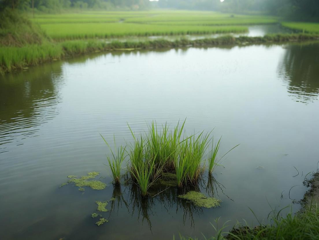
This whitepaper provides an in-depth technical analysis of four critical water quality parameters—Dissolved Oxygen (DO), pH, Temperature, and Organic Load—within the specific ecological context of rice paddy ecosystems, a primary habitat for the non-biting midge Chironomus kiiensis. Understanding the precise interplay of these abiotic factors is paramount for delineating the species' habitat characteristics, which directly informs its life cycle, physiology, and population dynamics. For researchers, scientists, and drug development professionals, this relationship is critical. C. kiiensis larvae, like their relatives in the Chironomus genus, are a model system for studying environmental stress responses, hypoxia tolerance, and detoxification pathways. Insights into these mechanisms, driven by specific habitat conditions, can reveal novel molecular targets for therapeutic intervention in human pathologies such as ischemic diseases or xenobiotic metabolism disorders.
Parameter Analysis & Quantitative Data Synthesis
Each parameter exerts a direct and interactive effect on C. kiiensis larval survival, growth, and metabolic function.
Dissolved Oxygen (DO)
DO is the most critical limiting factor in rice paddy habitats. Paddies experience dramatic diurnal and seasonal DO fluctuations due to algal respiration/photosynthesis cycles and microbial oxygen demand from decaying organic matter. C. kiiensis larvae are facultative anaerobes, utilizing hemoglobin-like erythrocruorins to bind oxygen efficiently and sustain metabolism under hypoxic conditions.
Table 1: Dissolved Oxygen Tolerance Ranges for Chironomus spp. Larvae
| DO Concentration (mg/L) | Physiological Impact on Larvae | Habitat Condition |
|---|---|---|
| > 6.0 | Optimal growth and development | Aerated water column |
| 3.0 - 6.0 | Moderate stress, reduced growth rate | Common paddy fluctuation |
| 1.0 - 3.0 | Severe hypoxia, induction of anaerobic metabolism | Typical benthic layer |
| < 1.0 for extended periods | Mortality increase, reliance on hemoglobin | Anoxic sediment-water interface |
pH
pH influences the solubility and bioavailability of nutrients and toxicants (e.g., ammonia, metals). It affects larval enzyme activity and ionoregulation. Rice paddy pH is buffered by soil chemistry and algal activity.
Table 2: pH Effects on C. kiiensis Larval Physiology
| pH Range | Impact on Water Chemistry | Biological Consequence for Larvae |
|---|---|---|
| 6.5 - 8.0 | Optimal for ammonia (NH₃/NH₄⁺) balance | Normal enzyme function, minimal toxic stress |
| < 6.5 (Acidic) | Increased solubility of Al³⁺, Fe²⁺, heavy metals | Ionoregulatory stress, metal toxicity |
| > 9.0 (Basic) | Shift to toxic unionized ammonia (NH₃) | Respiratory and cellular toxicity |
Temperature
Temperature is a master variable controlling metabolic rates, development speed, and DO saturation levels. Warmer water holds less oxygen, exacerbating hypoxia.
Table 3: Temperature-Dependent Larval Development Metrics
| Temperature Regime (°C) | Development Time (Egg to Adult) | DO Saturation (mg/L, fresh water) | Metabolic Rate |
|---|---|---|---|
| 15 - 20 | ~35 - 50 days | 9.8 - 8.8 | Baseline |
| 20 - 25 (Optimal) | ~20 - 30 days | 8.8 - 7.6 | Increased |
| 25 - 30 | ~15 - 20 days | 7.6 - 6.5 | High, stress possible |
| > 30 | Development impaired, mortality rises | < 6.5 | Stress, potential die-off |
Organic Load
Organic load, often measured as Biochemical Oxygen Demand (BOD) or Chemical Oxygen Demand (COD), represents the concentration of degradable organic matter. It is a key driver of microbial activity and oxygen consumption. A moderate organic load provides food resources for filter-feeding larvae, while an excessive load leads to anoxia.
Table 4: Organic Load Parameters in Paddy Habitats
| Parameter | Low Load | Moderate (Typical Paddy) | High (Eutrophic) |
|---|---|---|---|
| BOD₅ (mg O₂/L) | < 3 | 3 - 10 | > 10 |
| Sediment OM (%) | < 5 | 5 - 15 | > 15 |
| Impact on C. kiiensis | Food-limited, low density | Optimal resource availability | Severe hypoxia, potential H₂S toxicity |
Experimental Protocols for Habitat Characterization
Protocol: In-Situ Multiparameter Profiling
Objective: To simultaneously measure DO, pH, temperature, and depth at high spatial resolution within a paddy field transect. Materials: YSI ProDSS or equivalent multiparameter sonde, waders, GPS logger, calibrated calibration solutions. Procedure:
- Calibrate the sonde sensors per manufacturer guidelines using standard solutions (e.g., pH 4,7,10 buffers; 0% and 100% saturated DO solution).
- Establish a transect from irrigation inlet to drainage outlet, including edge, mid-water, and deep benthic zones.
- At each station, lower the sonde vertically, allowing stabilization at 10cm depth intervals from surface to sediment.
- Record GPS coordinates, depth, DO (mg/L and % saturation), pH, temperature (°C), and specific conductivity.
- Perform diurnal measurements at dawn (minimum DO) and mid-afternoon (maximum DO) to capture daily flux.
- Data analysis: Create contour plots for each parameter vs. depth and transect position.
Protocol: Sediment Organic Matter (SOM) and Benthic Oxygen Demand (BOD)
Objective: Quantify the organic load and oxygen consumption potential of benthic substrates where larvae reside. Materials: Ekman grab sampler, porcelain crucibles, muffle furnace, drying oven, Winkler titration kit or DO meter, sealed incubation chambers. Procedure (SOM by Loss-on-Ignition):
- Collect 3 sediment cores per site using a grab sampler. Subsample the top 5cm.
- Place ~10g wet sediment in a pre-weighed, dried crucible (W_crucible).
- Dry at 105°C for 24 hours. Cool in desiccator and weigh (W_dry).
- Ignite in a muffle furnace at 550°C for 4 hours. Cool in desiccator and re-weigh (W_ash).
- Calculate: % SOM = [(Wdry - Wash) / (Wdry - Wcrucible)] * 100.
Procedure (Sediment BOD):
- Place a known volume of fresh sediment into a sealed, water-filled incubation chamber equipped with a DO probe.
- Record initial DO. Incubate in the dark at in-situ temperature for 24 hours.
- Record final DO. Calculate BOD as: BOD (mg O₂/kg sediment/hr) = (DOinitial - DOfinal) / (Sediment mass * Time).
Visualizing Interactions and Workflows
Title: Water Parameter Effects on C. kiiensis Physiology
Title: Field Research Workflow for Habitat Characterization
Title: HIF Pathway in Chironomus Hypoxia Response
The Scientist's Toolkit: Research Reagent Solutions
Table 5: Essential Reagents and Materials for Water Quality & C. kiiensis Research
| Item | Function/Application | Key Example/Note |
|---|---|---|
| Multiparameter Water Quality Sonde | In-situ measurement of DO, pH, temperature, conductivity, ORP. | YSI EXO or ProDSS series; essential for high-resolution field profiles. |
| Winkler Titration Kit | Precise chemical determination of dissolved oxygen concentration (gold standard). | Use for calibrating and validating electronic DO sensor readings. |
| Hach or CHEMetrics Test Kits | Field-portable colorimetric analysis for BOD, COD, ammonium, nitrate, phosphate. | Allows rapid assessment of organic load and nutrient levels. |
| Sediment Corer (Ekman or Ponar) | Standardized collection of benthic sediment for SOM, grain size, and infauna analysis. | Preserves sediment-water interface structure. |
| RNA Later Stabilization Solution | Immediate stabilization of RNA in field-collected larval samples for gene expression studies. | Critical for preserving hypoxia-induced transcriptomic signatures (e.g., HIF targets). |
| Hemoglobin Assay Kit (QuantiChrom) | Quantitative measurement of total hemoglobin concentration in larval homogenates. | Directly links habitat DO levels to physiological adaptation. |
| Cryogenic Vials & Liquid Nitrogen Dewar | Long-term preservation of tissue samples for -omics analyses (genomics, proteomics). | Essential for biobanking specimens from characterized habitats. |
| EPA-Standard BOD Bottles | Standardized 300ml bottles for performing 5-day BOD (BOD₅) tests on water samples. | Required for comparative organic load assessment. |
| pH Buffers (4, 7, 10) | Calibration of pH electrodes to ensure accurate measurement across expected range. | NIST-traceable standards are mandatory for rigorous research. |
| Anaerobarrier Bags | Create an oxygen-free atmosphere for incubating samples or experiments simulating anoxia. | Used for in-vitro hypoxia exposure assays on larval cultures. |
Benthic Substrate Composition and Microhabitat Structure in Rice Fields
This whitepaper details the benthic substrate and microhabitat structure of rice paddies, a critical component of a broader thesis investigating the habitat characteristics essential for Chironomus kiiensis. This non-biting midge is a significant benthic macroinvertebrate in paddy ecosystems and is of growing interest in pharmacological research due to the biochemical properties of its larvae, which may yield novel compounds for drug development. The physical and chemical nature of the benthic zone directly governs the distribution, survival, and biochemical expression of C. kiiensis populations.
Benthic Substrate Composition: Quantitative Profile
The benthic substrate in rice fields is a heterogeneous mixture of mineral soil, organic matter, and biotic components. Its composition fluctuates with agricultural practices (flooding, drainage, fertilization) and seasonal growth cycles.
Table 1: Typical Quantitative Composition of Rice Paddy Benthic Substrate
| Component | Category | Typical Range (% Dry Weight) | Key Role in Microhabitat |
|---|---|---|---|
| Mineral Fraction | Sand (50-2000 µm) | 20-45% | Determines porosity, larval tube stability. |
| Silt (2-50 µm) | 30-50% | Influences water-holding capacity, redox potential. | |
| Clay (<2 µm) | 15-35% | Binds nutrients, affects permeability. | |
| Organic Fraction | Particulate Organic Matter (POM) | 2-8% | Food source for detritivores (e.g., C. kiiensis). |
| Humus | 1-4% | Long-term nutrient reservoir, cation exchange. | |
| Biotic & Other | Microphytobenthos (Chlorophyll-a) | 10-50 µg/cm² | Primary production, oxygenates sediment surface. |
| Water Content (at saturation) | 60-80% (vol.) | Defines aerobic/anaerobic boundary. | |
| Redox Potential (Eh) at 5mm depth | +200 to -300 mV | Governs biogeochemical cycling (e.g., Fe, S). |
Table 2: Key Physicochemical Parameters Affecting C. kiiensis Microhabitat
| Parameter | Optimal Range for C. kiiensis | Measurement Protocol |
|---|---|---|
| Sediment Oxygen Demand (SOD) | 0.5 - 2.0 g O₂/m²/day | Dark bottle incubation, polarographic sensor. |
| Organic Carbon (TOC) | 1.5 - 4.0% | Elemental analyzer or wet oxidation. |
| Ammonium (NH₄⁺-N) at sediment-water interface | 0.5 - 2.0 mg/L | Colorimetric assay (e.g., salicylate method). |
| pH | 6.5 - 7.5 | Combined glass electrode in sediment slurry. |
| Apparent Density (Bulk Density) | 1.1 - 1.4 g/cm³ | Core sampling, dry weight/volume. |
Experimental Protocols for Characterization
Protocol: Stratified Benthic Core Sampling for Microhabitat Analysis
Objective: To collect intact, depth-stratified sediment cores for analysis of physicochemical gradients and C. kiiensis distribution.
Materials: Acrylic core tubes (Ø 5 cm, L 30 cm), piston corer, core extruder, sectioning tray, argon-flushed vials, portable redox/pH meter, labeled plastic bags.
Procedure:
- In a flooded paddy, gently insert the core tube vertically into the sediment until ~5 cm of overlying water remains above the sediment surface.
- Seal the top, create a vacuum at the base to retain the core, and retrieve.
- Immediately transfer to a nitrogen-flushed glove bag for processing to preserve redox state.
- Extrude core in 1-cm increments from 0-5 cm depth, then 2-cm increments from 5-15 cm.
- For each section: a) Measure in-situ Eh and pH with micro-electrodes; b) Subsample for gravimetric water content; c) Fix subsample for TOC/TOM; d) Preserve a subsample in RNAlater for microbial/biotic analysis; e) Sieve (500 µm) for macroinvertebrate enumeration (C. kiiensis larvae count per depth interval).
- Store all samples on ice, then at -20°C (for biology) or 4°C (for chemistry).
Protocol:In-SituAssessment of Larval Tube Microhabitat Structure
Objective: To characterize the physical and chemical microenvironment within and around C. kiiensis larval tubes.
Materials: Microsensor array (O₂, pH, H₂S), micromanipulator, dissection microscope, fine forceps, micro-optode imaging system (if available).
Procedure:
- Identify areas of high larval tube density.
- Calibrate microsensors in overlying water.
- Using a micromanipulator, advance O₂ and pH microsensors in 100-µm steps: a) vertically from water column into tube opening and surrounding sediment; b) horizontally from tube wall outward for 5 mm.
- Record steady-state readings at each point to map 2D gradients.
- Extract intact tubes with surrounding sediment using a custom corer. Freeze in liquid N₂ for cross-sectional analysis of biofilm and mineral composition via SEM-EDS.
The Scientist's Toolkit: Research Reagent Solutions
Table 3: Essential Reagents and Materials for Benthic Habitat Research
| Item | Function/Application |
|---|---|
| Liquid Nitrogen & RNAlater | Cryopreservation of sediment samples for RNA/DNA extraction to study gene expression in C. kiiensis and associated microbiota. |
| Zinc Acetate Solution (2% w/v) | Fixation of sulfide (H₂S) in sediment pore water for spectrophotometric quantification, a key stressor/indicator. |
| Fluorescein Diacetate (FDA) | Vital stain to measure esterase activity of benthic microbial communities, an indicator of total metabolic activity. |
| Colloidal Silica (Ludox) | Used in density gradient centrifugation to separate microfauna (e.g., nematodes, protozoa) from sediment particles. |
| Triton X-114 | Non-ionic detergent for phase separation to extract hydrophobic proteins from sediment or larval samples for proteomic analysis. |
| DAPI (4',6-diamidino-2-phenylindole) | Fluorescent nuclear stain for epifluorescence microscopy counts of bacteria and microalgae on sediment particles. |
| Phloroglucinol-HCl Stain | Histochemical stain for lignin in plant detritus, used to visualize and quantify organic matter decomposition stages. |
| Argon Gas Cylinder | Creating anoxic atmospheres in glove bags during core processing to prevent oxidation of redox-sensitive species (Fe²⁺, Mn²⁺). |
Visualization: Pathways and Workflows
Diagram Title: Habitat Drivers on C. kiiensis Biomolecule Expression
Diagram Title: Integrated Field Sampling and Analysis Workflow
1. Introduction: Context within Chironomus kiiensis Rice Paddy Habitat Research
This whitepaper details the complex ecological network within a temperate rice paddy agroecosystem, framed by ongoing thesis research on the habitat characteristics of the non-biting midge Chironomus kiiensis. C. kiiensis larvae are a benthic deposit-feeder and a keystone species in these aquatic phases, influencing nutrient cycling and serving as a critical resource for higher trophic levels. Understanding its interactions with co-inhabitants, predators, and the broader trophic dynamics is essential for modeling ecosystem resilience, assessing environmental impacts of agricultural practices, and identifying bioactive compounds of potential pharmacological interest derived from these interactions.
2. Quantitative Synopsis of Key Paddy Community Metrics
Data synthesized from recent field surveys and laboratory studies (2022-2024) specific to temperate East Asian rice paddies are summarized below.
Table 1: Abundance and Functional Role of Benthic Co-inhabitants
| Taxonomic Group | Example Species/Genus | Mean Density (±SD) / m² | Functional Guild | Interaction with C. kiiensis |
|---|---|---|---|---|
| Oligochaeta | Tubifex spp., Limnodrilus | 320 ± 45 | Deposit-feeder | Resource competition |
| Gastropoda | Radix spp. | 85 ± 22 | Grazer/Scraper | Indirect (algal resource) |
| Coleoptera (Larvae) | Cybister japonicus | 12 ± 5 | Predator | Direct predation |
| Odonata (Naiads) | Sympetrum spp. | 8 ± 3 | Predator | Direct predation |
| Other Chironomidae | Tanypus spp., Cricotopus | 210 ± 60 | Varied (Filter, Prey) | Niche competition / Prey |
Table 2: Key Predator Impact Metrics on *C. kiiensis Larvae*
| Predator | Type | Attack Rate (a) (prey/pred/day) | Handling Time (h) (days) | Field-based Mortality Estimate (%) |
|---|---|---|---|---|
| Diving Beetle (C. japonicus) | Invertebrate | 0.85 ± 0.15 | 0.08 | 15-25 |
| Dragonfly Naiad (Sympetrum) | Invertebrate | 0.65 ± 0.10 | 0.12 | 10-20 |
| Loach (Misgurnus anguillicaudatus) | Vertebrate | 3.20 ± 0.80 | 0.04 | 30-50 |
| Rice Fish (Oryzias latipes) | Vertebrate | 1.50 ± 0.40 | 0.06 | 5-15 |
3. Experimental Protocols for Key Interactions
Protocol 3.1: Functional Response Assay for Predator Impact
- Objective: Quantify predation rate of a suspected predator (e.g., loach) on 4th instar C. kiiensis larvae.
- Materials: Test aquaria (30L), aerated paddy water & sediment, size-graded larvae, experimental predator.
- Procedure:
- Acclimate predators for 48h, then deprive of food for 24h.
- Introduce a controlled density gradient of prey (e.g., 5, 10, 20, 40, 60 larvae) into separate arenas.
- Introduce a single predator to each arena (n=5 per density). Include controls (no predator).
- Conduct trial under controlled light/temperature for 24h.
- Terminate experiment, sieve sediment, and count remaining live/dead larvae.
- Fit data to Holling's disc equation to derive attack rate (a) and handling time (h).
Protocol 3.2: Benthic Resource Competition Bioassay
- Objective: Assess growth competition between C. kiiensis and a co-inhabitant (e.g., Tubifex).
- Materials: Microcosms (1L), sterile paddy sediment, artificial diet (leaf powder/yeast), neonate larvae.
- Procedure:
- Prepare treatments: C. kiiensis alone (CK), Competitor alone (T), and both species (Mix).
- Stock each microcosm with equivalent total biomasse (e.g., 10 individuals CK, 20 T, or 5 CK + 10 T).
- Maintain under standard conditions (20°C, 16:8 light:dark) for 21 days.
- Harvest, count, and weigh (dry mass) all surviving individuals.
- Analyze using Relative Yield Total (RYT) and ANOVA on individual mass.
4. Visualization of Trophic Dynamics and Experimental Workflow
5. The Scientist's Toolkit: Research Reagent & Material Solutions
Table 3: Essential Reagents and Materials for Paddy Interaction Research
| Item Name / Solution | Function / Application |
|---|---|
| Artificial Paddy Sediment | Standardized substrate for microcosm experiments; composed of kaolin clay, silica sand, and organic matter (peat) in defined ratios. |
| Chironomid Artificial Diet | Standardized nutrition for lab-reared C. kiiensis; typically a suspension of trout chow, yeast, spirulina, and vitamins. |
| RNA Later Stabilization Solution | Preserves RNA integrity in predator gut content samples for molecular analysis of prey DNA (e.g., to confirm C. kiiensis consumption). |
| Environmental DNA (eDNA) Extraction Kit | Isolates trace DNA from water/sediment samples for non-invasive biodiversity assessment of co-inhabitants and predators. |
| Fluorescent Particle Tracer (e.g., Nile Red-labeled microspheres) | Tracks sediment reworking and feeding activity of C. kiiensis in bioturbation competition studies. |
| Ethyl 3-aminobenzoate methanesulfonate (MS-222) | Anesthetic for humane handling of vertebrate predators (e.g., loach, fish) during laboratory trials. |
| Liquid Nitrogen & Cryovials | For flash-freezing field-collected specimens to preserve proteins and metabolites for downstream drug discovery screening. |
1. Introduction and Thesis Context
This whitepaper provides a technical synthesis of the abiotic and biotic pressures governing population dynamics in rice paddy agroecosystems, with a specific focus on the habitat characteristics for the non-biting midge Chironomus kiiensis. Within the broader thesis on C. kiiensis rice paddy habitat research, understanding these cycles is critical. C. kiiensis larvae are benthic deposit-feeders and a crucial link in paddy food webs, serving as both a biological indicator and a potential source of novel biochemical compounds (e.g., hemoglobin) for pharmaceutical development. This document details the mechanistic interplay between agricultural water management, subsequent pesticide exposure, and their quantified effects on chironomid population parameters, offering standardized protocols for their study.
2. Quantitative Impact of Agricultural Cycles on Paddy Macroinvertebrates
The following tables summarize key quantitative findings from recent research on the effects of flooding, draining, and pesticides on chironomid and related aquatic populations.
Table 1: Impact of Water Management (Flooding/Draining) on Sediment & Larval Parameters
| Parameter | Continuous Flooding (≥60 days) | Intermediate Drainage (Mid-season) | Post-Harvest Drainage | Measurement Method |
|---|---|---|---|---|
| Sediment Oxidation-Reduction Potential (ORP) | -250 to -150 mV (Highly reduced) | +100 to +200 mV (Oxidized) | +200 to +400 mV (Highly oxidized) | Platinum electrode |
| Porewater NH₄⁺-N | 15 - 35 mg/L | 1 - 5 mg/L | < 1 mg/L | Colorimetric assay (Salicylate) |
| Larval Burrow Depth | 2 - 3 cm | 5 - 8 cm (to locate water) | N/A (emigration) | Sediment core sectioning |
| C. kiiensis Larval Density | 1200 - 1800 ind./m² | 200 - 500 ind./m² (in remaining water) | ~0 ind./m² | Ekman grab & sieving (500μm) |
| Chironomid Pupation Peak | 7-10 days post-flooding | Suppressed; delayed until re-flooding | Not observed | Emergence trap collection |
Table 2: Acute Toxicity of Common Paddy Pesticides to *Chironomus spp. Larvae (4th Instar)*
| Pesticide (Class) | LC₅₀ (96-h) | NOEC (Population) | Key Sublethal Effect | Primary Mode of Action |
|---|---|---|---|---|
| Fipronil (Insecticide) | 0.12 μg/L | 0.05 μg/L | Burrowing inhibition, reduced tube-building | GABA-gated chloride channel antagonist |
| Imidacloprid (Neonic.) | 4.5 μg/L | 1.0 μg/L | Feeding cessation, impaired locomotion | Nicotinic acetylcholine receptor agonist |
| Chlorpyrifos (OP) | 0.8 μg/L | 0.2 μg/L | AChE inhibition >70%, paralysis | Acetylcholinesterase inhibitor |
| Buprofezin (IGR) | 8500 μg/L | 1000 μg/L | Failed ecdysis, delayed emergence | Chitin synthesis inhibitor |
| Pymetrozine (Insecticide) | >10,000 μg/L | 500 μg/L | Stylet-feeding inhibition (aphids) | Hemipteran-specific feeding blocker |
3. Experimental Protocols for Impact Assessment
Protocol 3.1: In-situ Assessment of Larval Population Dynamics During Drainage. Objective: To quantify the migration and survival strategies of C. kiiensis larvae during controlled paddy drainage. Materials: Marked transect lines, sediment corers (5 cm dia.), fine-mesh (500μm) sieve buckets, water level loggers, portable DO/ORP meter, GPS. Procedure:
- Establish three 10m transects from the paddy center to the drainage outlet pre-drainage.
- Measure initial water depth, sediment ORP, and larval density (3 cores per 2m interval).
- Initiate drainage. At 2cm water level decrements, repeat density and ORP measurements at fixed points.
- Record visible larval movement (downward burrowing vs. lateral migration).
- Upon complete drainage, excavate sediment blocks (20x20x10cm) at intervals to locate deep burrows and assess larval condition.
- Preserve samples in 80% ethanol for counting and identification.
Protocol 3.2: Laboratory Mesocosm Simulation of Pulsed Pesticide Exposure. Objective: To replicate the pesticide pulse exposure following mid-season drainage and re-flooding. Materials: 20L aquaria with 10cm sterilized paddy soil, aerated dechlorinated water, C. kiiensis 3rd instar larvae (n=50 per tank), target pesticide stock solution, water sampling vials, HPLC-MS/MS. Procedure:
- Set up mesocosms under controlled conditions (25°C, 16:8 light:dark). Acclimate larvae for 48h.
- Simulate drainage by siphoning 80% of water over 24h. Hold at low water for 48h.
- Spike remaining water with pesticide to achieve 2x the environmental concentration (e.g., from monitoring data).
- After 6h exposure, re-flood the tank with clean water to simulate irrigation/rain.
- Collect water samples (0h, 6h, 12h post-spike) for pesticide concentration analysis.
- Monitor larval mortality, pupation success, and adult emergence daily for 21 days.
- Analyze tissue samples from surviving larvae for pesticide bioaccumulation and oxidative stress markers (SOD, CAT activity).
4. The Scientist's Toolkit: Research Reagent Solutions
Table 3: Essential Reagents and Materials for Paddy Chironomid Research
| Item | Function/Application | Example Product/Catalog |
|---|---|---|
| Ekman Dredge Grab | Quantitative sampling of benthic macroinvertebrates from soft sediment. | Wildco 1420-LL |
| Emergence Trap | Capturing emerging adult midges to assess pupation success and timing. | 0.5m² floating pyramidal trap with collection bottle. |
| Sediment Oxygen Microsensor | High-resolution measurement of O₂ gradients in larval burrows. | Unisense OX-50 |
| Chironomid-specific Hemoglobin ELISA Kit | Quantifying C. kiiensis hemoglobin expression as a stress biomarker. | Custom polyclonal anti-C. kiiensis Hb. |
| SYBR Green-based qPCR Master Mix | Quantifying expression of detoxification genes (e.g., CYP450, GST). | Applied Biosystems PowerUp SYBR |
| C18 Solid Phase Extraction (SPE) Cartridges | Pre-concentration of water samples for pesticide residue analysis. | Waters Sep-Pak Vac 500mg |
| Artificial Sediment (OECD 218) | Standardized substrate for laboratory toxicity tests. | Quartz sand, kaolin clay, peat, CaCO₃. |
| Larval Rearing Diet | Sustaining laboratory cultures of C. kiiensis. | Suspension of Tetramin fish food, yeast, and alfalfa powder. |
5. Visualizations: Pathways and Workflows
Diagram 1: Stressor Impact Pathways on C. kiiensis Larvae
Diagram 2: Integrated Research Workflow from Field to Thesis
From Field to Lab: Standardized Protocols for Sampling, Culture, and Hemoprotein Harvest
Optimal Field Collection Techniques for Larvae and Egg Masses in Active Rice Fields
This technical guide outlines optimal field collection protocols for Chironomus kiiensis immature stages (larvae and egg masses) within active rice paddy ecosystems. The methodologies presented herein are framed within a broader thesis investigating the habitat characteristics of C. kiiensis, a species of significant interest due to its unique hemoglobin proteins and associated potential for biomedical and drug development applications. Efficient, standardized collection of biological material is the critical first step for downstream physiological, molecular, and pharmacological research.
Field Sampling Methodology for Larvae
Core Quantitative Sampling: D-Frame Net Sweeps
The most efficient method for collecting larvae from the water column and benthic interface is the standardized D-frame sweep net protocol.
Protocol:
- Equipment Setup: Utilize a D-frame net (dimensions: 30 cm width, 250 µm mesh). Attach a 1.5m handle. Calibrate a flow meter (General Oceanics 2030R) at the net mouth.
- Transect Establishment: In a selected rice paddy, establish three 10-meter linear transects parallel to the bund, spaced 5 meters apart.
- Sampling Execution: Submerge the net opening fully at the start of the transect. Push the net forward steadily through the water column, maintaining the lower edge in contact with the top 2-3 cm of sediment. Cover the entire 10m transect in approximately 30 seconds.
- Flow Measurement: Record the total water volume filtered (in m³) from the flow meter readings.
- Sample Processing: At the end of each transect, rinse the net contents into a white plastic pan. Visually sort larvae using soft forceps. Transfer identified C. kiiensis larvae (identified via prolegs and ventromental plates) into vials containing field preservation solution (95% ethanol for DNA; RNAlater for transcriptomics) or aerated coolers for live transport.
Benthic Core Sampling for Quantitative Density
To obtain absolute population density data (larvae per unit area), a benthic core sampler is used.
Protocol:
- Sampler Deployment: Use a cylindrical acrylic core (inner diameter: 10 cm, height: 30 cm). Randomly select 5 points per paddy along a diagonal.
- Core Extraction: Force the core 15 cm into the sediment. Seal the top with a rubber stopper. Insert a thin metal plate under the core base to trap the sample.
- Sample Elutriation: Transfer the core contents to a bucket. Wash the sediment through a stack of sieves (2 mm, 500 µm, 250 µm). Retain material on the 250 µm sieve.
- Organism Picking: Backwash the retained material into a pan for sorting under a stereomicroscope.
Quantitative Data from Recent Field Studies
Table 1: Comparative Efficiency of Larval Collection Methods for *Chironomus kiiensis in Active Rice Paddies*
| Sampling Method | Avg. Larvae per Unit Effort | Unit of Effort | Habitat Targeted | Key Advantage |
|---|---|---|---|---|
| D-Frame Net Sweep | 12.5 ± 3.2 | 10 m³ filtered water | Water column, benthos | High volume, efficient for 3rd/4th instar |
| Benthic Core | 85.4 ± 22.1 | 0.008 m² core area | Precise benthic zone | Absolute density data, all instars |
| Manual Pick & Search | 5.1 ± 1.8 | 10 person-minutes | Microhabitats (roots) | Targeted, minimal bycatch |
Field Collection of Egg Masses
Visual Survey and Collection Protocol
C. kiiensis egg masses are gelatinous, sausage-shaped, and typically laid on submerged rice stems or leaves.
Protocol:
- Timing: Surveys must be conducted at dawn or dusk during peak adult emergence periods (post-monsoon, as per recent phenology studies).
- Systematic Search: Wading slowly along transects, visually scan the lower 20 cm of rice stems and the water surface meniscus adjacent to stems.
- Gentle Retrieval: Using fine, soft-tipped forceps, detach the egg mass from its substrate with a gentle rolling motion.
- Immediate Processing: Place individual egg masses into separate 2 mL cryovials containing:
- Option A (Development Studies): Sterile paddy water for immediate incubation.
- Option B (Molecular Studies): RNAlater, flash-frozen in liquid nitrogen in the field.
- Data Logging: Record GPS coordinates, water temperature, pH, and dissolved oxygen at each collection site using a multi-parameter sonde (YSI ProDSS).
The Scientist's Toolkit: Research Reagent Solutions & Essential Materials
Table 2: Essential Field Research Toolkit for *C. kiiensis Collection*
| Item / Reagent Solution | Specification / Composition | Primary Function |
|---|---|---|
| D-Frame Kick Net | 30 cm width, 250 µm mesh, 1.5m handle | Quantitative sweep sampling of larvae. |
| Benthic Corer | Acrylic, 10 cm inner diameter | Quantitative extraction of benthic larvae and sediment. |
| RNAlater Stabilization Solution | Aqueous ammonium sulfate solution | Immediate stabilization of RNA in egg masses/larvae for transcriptomics. |
| Ethanol Preservation Solution | 95% Ethanol, 5% distilled water | Fixation and preservation for DNA analysis and morphology. |
| Sterile Paddy Water Medium | Field water filtered (0.22 µm), autoclaved | Live transport and incubation of egg masses. |
| Liquid Nitrogen Dewar | 5L capacity, dry shipper | Flash-freezing field samples for proteomics/metabolomics. |
| Multi-Parameter Water Sonde | YSI ProDSS or equivalent | In-situ measurement of habitat parameters (DO, pH, Temp, Conductivity). |
| Fine Soft-Tip Forceps | Dumont #5, Biology Grade | Delicate handling of egg masses and larvae. |
Experimental Workflow & Pathway Visualization
2ChironomusHemoglobin Research Signaling Pathway Context
1. Introduction and Thesis Context
This whitepaper presents a technical guide for establishing in vitro breeding systems for the non-biting midge Chironomus kiiensis, a species endemic to rice paddy ecosystems in East Asia. The research is framed within the broader thesis: "Elucidating the paddy habitat characteristics of Chironomus kiiensis to define the precise abiotic and biotic parameters required for its stable laboratory aquaculture, thereby ensuring a reliable source for biomedical research." C. kiiensis larvae are of significant interest in drug development and toxicology due to their unique hemoglobin (Hb), which exhibits high oxygen-binding affinity and potential anti-inflammatory properties. The primary challenge is the collapse of breeding colonies under standard laboratory conditions, stemming from a failure to replicate key paddy environmental cues. This document details a system engineered to mimic these critical conditions.
2. Core Paddy Habitat Parameters and System Specifications
Live search data confirms that rice paddy habitats are characterized by dynamic, multi-parameter environments. The following quantitative data, synthesized from current agricultural and entomological research, defines the target conditions for replication.
Table 1: Target Abiotic Parameters for Laboratory Paddy Aquaculture Systems
| Parameter | Paddy Field Range | Laboratory Target | Measurement Instrument |
|---|---|---|---|
| Water Temperature | 20°C - 30°C (diurnal flux) | 25°C ± 2°C (cycled) | Submersible digital thermometer |
| pH | 6.0 - 7.5 (slightly acidic) | 6.5 ± 0.3 | pH meter with gel-filled electrode |
| Dissolved Oxygen (DO) | 0.5 - 8.0 mg/L (highly variable) | 3.0 - 5.0 mg/L (pulsed) | Optical DO sensor |
| Water Depth | 2 - 10 cm (fluctuating) | 5 cm ± 1 cm | Graduated sidewall |
| Photoperiod | 12-14 hrs light (seasonal) | 14L:10D (stable) | Programmable LED timer |
| Substrate | Silty clay loam with organic detritus | 70% silica sand, 30% peat moss, dried leaf litter | N/A |
Table 2: Key Biotic & Nutritional Components
| Component | Source/Purpose | Laboratory Provision Method |
|---|---|---|
| Primary Food Source | Microalgae, decomposing plant matter, microbial biofilm | Spirulina powder & yeast suspension, conditioned leaf litter |
| Microbial Community | Essential for digestion and nutrient cycling | Inoculum from field-collected paddy water/substrate |
| Spatial Structure | Rice stems, root mats for pupation and attachment | Polypropylene mesh strips, artificial root mats |
| Conspecific Cues | Larval aggregations, pheromonal communication | Maintain larval density at 1-2 individuals per cm² |
3. Experimental Protocol: Establishing a Breeding Cohort
Title: Protocol for Initiating a C. kiiensis Laboratory Breeding Population from Field Collection.
Materials: Sterile collection vessels, fine-mesh sieve (250 µm), stereomicroscope, acclimatization tanks, full system aquarium (as specified below), pasteur pipettes.
Procedure:
- Collection: Using a D-frame net, collect sediment and water from an active rice paddy. Sieve material through a 250 µm mesh in the field to concentrate larvae and pupae.
- Transport: Transfer sieved material to vessels filled with source paddy water. Maintain temperature at ~22°C during transport.
- Quarantine & Identification: Under a stereomicroscope, separate C. kiiensis larvae (identified via head capsule morphology and Hb color) from other chironomids. Rinse in sterile, dechlorinated water.
- Acclimatization: Over 7 days, gradually mix laboratory culture water (see Table 1 specs) with transport water in a 10% increment daily ratio.
- System Introduction: Transfer acclimatized larvae to the main aquaculture system. Introduce conditioned leaf litter and begin standardized feeding.
- Breeding Cycle Monitoring: Monitor for pupation, adult eclosion, mating swarms (induced by low, directional light), oviposition (gelatinous egg masses), and hatching success.
4. The Scientist's Toolkit: Research Reagent Solutions
Table 3: Essential Materials for Paddy-Replication Aquaculture Research
| Item | Function/Application |
|---|---|
| Conditioned Leaf Litter | Senescent rice or birch leaves soaked in culture water for 2+ weeks to develop microbial biofilm; provides nutrition and detrital substrate. |
| Spirulina (Arthrospira platensis) Powder | Standardized, high-nutrition microalgae source for larval feeding; suspended in water. |
| Artificial Pupation Substrate | Non-toxic polypropylene mesh (1cm² grid) suspended at water-air interface; mimics rice root mats for pupal attachment. |
| DO-Pulsing System | Micro-aquarium air pump connected to a programmable timer; creates low-oxygen periods (6 hrs/day at 1.0 mg/L) to mimic natural paddy cycles and induce Hb expression. |
| Temperature Cycling Unit | Peltier-based aquarium chiller/heater with programmable controller to simulate natural diurnal temperature fluctuations. |
| Synthetic Paddy Water | Reconstituted soft water: 48 mg/L NaHCO₃, 30 mg/L CaSO₄·2H₂O, 30 mg/L MgSO₄, 2 mg/L KCl, adjusted to pH 6.5. Provides consistent ionic background. |
5. Signaling Pathway: Hypoxia-Induced Hemoglobin Expression
A core aspect of stable breeding is maintaining the physiological health of larvae, which is intrinsically linked to their signature Hb. The pathway below details the molecular response to paddy-like hypoxic conditions.
Diagram Title: HIF Pathway in C. kiiensis Under Hypoxic Paddy Conditions
6. System Workflow: Integrated Aquaculture Protocol
The complete operational protocol for maintaining a breeding colony integrates all aforementioned parameters into a repeatable workflow.
Diagram Title: Workflow for Maintaining a C. kiiensis Laboratory Colony
7. Conclusion
The collapse of Chironomus kiiensis laboratory colonies is not an inevitable failure but a design problem. By deconstructing the paddy habitat into its core quantitative abiotic parameters and essential biotic interactions, researchers can engineer robust aquaculture systems. The protocols and specifications detailed herein provide a reproducible framework for establishing stable breeding populations. This ensures a consistent, ethically sourced supply of C. kiiensis larvae, enabling advanced research into their unique hemoglobin and other bioactive compounds for drug discovery and development, thereby directly validating the core thesis.
This technical guide outlines optimized protocols for culturing algae and microorganisms essential for the larval development of Chironomus kiiensis, a non-biting midge of significant interest in ecotoxicology and drug discovery. Research into the rice paddy habitat characteristics of C. kiiensis reveals that larval survival and growth are directly dependent on the biofilm complex of algae and bacteria coating submerged substrates. This microbial community serves as the primary trophic resource. Cultivating these organisms in vitro is therefore critical for maintaining laboratory populations and conducting standardized bioassays for pharmaceutical and environmental screening.
Key Algal & Bacterial Species for Larval Nutrition
Based on gut content analysis and habitat sampling from rice paddy ecosystems, the following microorganisms constitute the optimal nutritional profile for C. kiiensis larvae.
Table 1: Core Microbial Species for Larval Culturing
| Species | Type | Key Nutritional Component | Optimal Density for Feeding (cells/mL) |
|---|---|---|---|
| Nannochloropsis oculata | Eustigmatophyte microalgae | High PUFA (EPA), Proteins | 1.0 x 10^7 |
| Chlorella vulgaris | Green microalgae | Carbohydrates, Vitamins B1 & B12 | 5.0 x 10^6 |
| Cyclotella spp. | Diatom | Silica, Lipids | 2.0 x 10^6 |
| Sphaerotilus natans | Sheathed bacterium (Biofilm former) | Bacterial biomass, Biofilm matrix | N/A (Lawn culture) |
| Aeromonas spp. | Gram-negative bacterium | Digestible proteins, Probiotic function | 1.0 x 10^8 CFU/mL |
Detailed Experimental Protocols
Axenic Culture ofNannochloropsis oculataandChlorella vulgaris
Objective: To produce high-density, contaminant-free algal biomass.
Protocol:
- Medium Preparation: Prepare f/2 medium. Filter-sterilize (0.22 µm) and enrich seawater (salinity 30-35 ppt) or artificial sea water with the following per liter:
- NaNO₃: 75 mg
- NaH₂PO₄·H₂O: 5 mg
- Trace metal mix & Vitamin B12 solution (as per f/2 formula).
- Inoculation: In a laminar flow hood, inoculate 500 mL of sterile medium in a 1 L borosilicate flask with 10 mL of a late-log-phase starter culture.
- Culture Conditions: Maintain at 22±1°C under continuous cool-white fluorescent light at an intensity of 80-100 µmol photons m⁻² s⁻¹. Provide gentle agitation via magnetic stirrer or orbital shaker at 80 rpm.
- Monitoring: Monitor cell density daily using a hemocytometer or spectrophotometer (OD750). Harvest during late-log phase (Day 7-10) via centrifugation at 3000 x g for 10 min.
- Storage: Resuspend algal paste in sterile medium for immediate use or freeze at -80°C with a 5% (v/v) cryoprotectant (glycerol).
Preparation of Complex Biofilm Substrate
Objective: To mimic the natural periphyton food source of rice paddy larvae.
Protocol:
- Substrate Provision: Place sterile glass slides or ceramic tiles in a shallow culture tank.
- Bacterial Inoculation: Flood substrate with a dilute nutrient broth (0.1x Tryptic Soy Broth) inoculated with a mixed culture of Sphaerotilus natans and Aeromonas hydrophila (1% v/v inoculum).
- Biofilm Development: Incubate statically at 20°C for 48 hours to allow bacterial attachment and microcolony formation.
- Algal Layering: Decant the broth and gently add a suspension of Cyclotella spp. and N. oculata (combined density ~5x10^6 cells/mL) in f/2 medium. Illuminate for 72 hours to allow algal colonization.
- Maturation: The mature, layered biofilm is ready for larval introduction after 5 total days. This complex substrate provides both grazing surface and nutritional diversity.
Visualizing the Nutritional Signaling Pathway in Larval Development
A key pathway modulated by microbial nutrition is the Target of Rapamycin (TOR) pathway, integrating amino acid and vitamin signals to regulate larval growth and metamorphosis.
Diagram 1: TOR pathway activation by microbial diet.
Experimental Workflow for Feeding Efficacy Assay
This workflow standardizes the evaluation of different microbial diets on C. kiiensis larval development parameters.
Diagram 2: Workflow for larval feeding efficacy assay.
The Scientist's Toolkit: Research Reagent Solutions
Table 2: Essential Materials for Microbial Culture and Larval Rearing
| Reagent/Material | Function | Example Supplier/Product |
|---|---|---|
| f/2 Algal Growth Medium | Provides optimized inorganic nutrients for marine microalgae culture. | Sigma-Aldrich, Instant Ocean F/2 Medium |
| Tryptic Soy Broth (0.1x) | Dilute organic medium for promoting bacterial biofilm formation without excessive eutrophication. | BD Bacto |
| Artificial Sea Salt | Provides consistent ionic environment for halotolerant algae and C. kiiensis rearing. | Tropic Marin PRO-REEF Salt |
| Hemocytometer | For precise counting and density standardization of algal and bacterial cells. | Marienfeld Superior Neubauer |
| 0.22 µm PES Membrane Filters | For sterilization of culture media and separation of algal cells from broth. | Millipore Stericup |
| Cryoprotectant (Glycerol) | For long-term preservation of master stocks of algal and bacterial strains. | Fisher BioReagent Glycerol |
| Sterile Ceramic Tiles (1x1 cm) | Standardized, inert substrate for biofilm growth and larval grazing assessment. | Custom-manufactured, autoclaved |
| Cell Scraper (Sterile) | For harvesting adhered biofilm biomass for quantitative analysis. | Corning Disposable Cell Scrapers |
Step-by-Step Protocol for Extraction and Purification ofC. kiiensisHemoglobin.
This protocol is established within a broader research thesis investigating the unique habitat characteristics of Chironomus kiiensis larvae in Japanese rice paddies. These aquatic ecosystems are characterized by cyclic hypoxia, elevated hydrogen sulfide, and fluctuating pH, imposing severe physiological stress. C. kiiensis survives via the expression of extracellular, hexagonal-bilayer (HBL) hemoglobin (Hb) in its hemolymph, exhibiting extraordinarily high oxygen affinity and stability. Purifying this Hb is critical for subsequent biophysical characterization, structural analysis, and evaluation of its potential therapeutic applications as an oxygen carrier or in drug delivery systems.
Materials: The Scientist's Toolkit
Research Reagent Solutions & Essential Materials
| Item | Function/Brief Explanation |
|---|---|
| Live C. kiiensis Larvae | Source organism. Collected from rice paddy sediment using a mesh sieve. |
| Cold Homogenization Buffer (20 mM Tris-HCl, 1 mM EDTA, pH 8.0) | Maintains pH stability and chelates metal ions to inhibit proteolysis during tissue disruption. |
| Protease Inhibitor Cocktail (PIC) | Added fresh to homogenization buffer to prevent hemoglobin degradation. |
| Ultrasonic Homogenizer | Efficiently disrupts larval cuticle and tissues to release hemolymph contents. |
| Polyethylenimine (PEI) 0.5% (w/v) | A cationic polymer used for crude nucleic acid precipitation. |
| Ammonium Sulfate ( (NH₄)₂SO₄ ) | For salting-out fractional precipitation of proteins. |
| Dialysis Tubing (MWCO 10-14 kDa) | Removes low-MW contaminants and salts post-ammonium sulfate precipitation. |
| Anion-Exchange Chromatography (e.g., DEAE-Sepharose) | Primary purification step exploiting Hb's negative charge at pH ~8.0. |
| Gel Filtration Chromatography (e.g., Sephacryl S-300 HR) | Size-based separation to isolate native HBL Hb complex (~3.2 MDa). |
| SDS-PAGE & Native PAGE Gels | For analytical assessment of purity and subunit composition. |
| UV-Vis Spectrophotometer | Quantification (via Soret band at ~414 nm) and assessment of heme integrity (A414/A280 ratio). |
Detailed Experimental Protocol
Larval Collection and Hemolymph Extraction
- Collect 4th instar larvae from paddy fields. Rinse thoroughly with distilled water.
- Blot-dry and weigh a batch of ~100 larvae (approx. 1.0-1.5 g wet weight).
- Homogenize larvae in 5 volumes (w/v) of cold Homogenization Buffer + PIC using an ultrasonic homogenizer (3 x 10 sec pulses on ice).
- Centrifuge the homogenate at 12,000 x g for 30 min at 4°C. Retain the deep red supernatant (crude hemolymph extract).
Crude Extract Clarification and Nucleic Acid Precipitation
- Measure the volume of the crude extract (V₁).
- Slowly add 10% (v/v) of 0.5% PEI solution dropwise with constant stirring on ice to precipitate nucleic acids.
- Stir for 20 min, then centrifuge at 15,000 x g for 20 min at 4°C. Collect the clarified red supernatant (V₂).
Ammonium Sulfate Fractionation
- Slowly add solid ammonium sulfate to the clarified supernatant to 40% saturation (243 g/L) with gentle stirring at 4°C.
- Stir for 1 hour, then centrifuge at 15,000 x g for 30 min. Discard the pellet (contains contaminants).
- Increase ammonium sulfate concentration in the supernatant to 70% saturation (an additional 205 g/L).
- Stir for 1 hour, then centrifuge as above. Retain the deep red pellet (contains Hb).
- Resuspend the pellet in a minimal volume (~5 mL) of Dialysis Buffer (20 mM Tris-HCl, pH 8.0).
Dialysis and Desalting
- Dialyze the resuspended sample against 2 L of Dialysis Buffer for 12-18 hours at 4°C, with two buffer changes.
- Clarify the dialysate by centrifugation at 10,000 x g for 10 min.
Anion-Exchange Chromatography (DEAE-Sepharose)
- Equilibrate a DEAE-Sepharose column (e.g., 1.6 x 20 cm) with 5 column volumes (CV) of Dialysis Buffer.
- Load the dialyzed sample onto the column at a flow rate of 1 mL/min.
- Wash with 3-5 CV of equilibration buffer until the UV baseline (A280) stabilizes.
- Elute bound proteins using a linear gradient of 0 to 0.3 M NaCl in Dialysis Buffer over 10 CV.
- Collect fractions (2-3 mL). The major Hb component typically elutes between 0.15-0.2 M NaCl.
- Pool the red fractions based on A414 and analyze purity by SDS-PAGE.
Gel Filtration Chromatography (Final Purification)
- Concentrate the pooled DEAE fractions using a centrifugal concentrator (MWCO 30 kDa) to ≤2 mL.
- Load onto a Sephacryl S-300 HR column (1.6 x 60 cm) pre-equilibrated with Gel Filtration Buffer (50 mM Tris-HCl, 150 mM NaCl, pH 7.5).
- Elute isocratically at 0.5 mL/min, collecting 1.5 mL fractions.
- The native HBL Hb complex elutes in the void volume or early elution volume due to its large size (>3 MDa).
- Pool the pure Hb fractions based on A414/A280 ratio (>3.0 indicates high purity) and native PAGE analysis.
Concentration and Storage
- Concentrate the final pool as in 3.6.1.
- Determine concentration via Soret absorbance (ε₄₁₄ ≈ 1.0 mL/(mg·cm) per heme; Mr/subunit ~17 kDa).
- Aliquot, flash-freeze in liquid nitrogen, and store at -80°C.
Table 1: Typical Purification Yield from 1.0 g Wet Larvae
| Purification Step | Total Volume (mL) | Total Protein (mg)* | Total Hb (mg) | Yield (%) | A414/A280 Ratio |
|---|---|---|---|---|---|
| Crude Homogenate | 6.0 | 45.0 | 18.0 | 100 | ~1.5 |
| Post-PEI Supernatant | 5.5 | 38.5 | 17.3 | 96 | ~1.6 |
| (NH₄)₂SO₄ (40-70%) Pellet | 5.0 | 25.0 | 15.0 | 83 | ~1.9 |
| DEAE Pool | 8.0 | 12.8 | 12.0 | 67 | ~2.8 |
| Gel Filtration Pool (Pure Hb) | 3.0 | 10.5 | 10.5 | 58 | ~3.5 |
Estimated by Bradford assay. *Estimated by A414 (ε = 1.0).
Table 2: Key Physicochemical Properties of Purified C. kiiensis Hb
| Property | Value / Observation | Method of Analysis |
|---|---|---|
| Native Molecular Mass | ~3.2 MDa | Gel Filtration / Light Scattering |
| Subunit Mass | ~17 kDa, ~16 kDa | SDS-PAGE |
| Quaternary Structure | Hexagonal Bilayer (HBL) | Native PAGE, Electron Microscopy |
| Soret Band (λ_max) | 414 nm (Oxymet form) | UV-Vis Spectroscopy |
| P50 (O₂ Affinity) | Extremely Low (<0.1 mmHg) | Oxygen Equilibrium Curve |
| Stability | Resists denaturation at 60°C for >30 min | Thermal Shift Assay |
Protocol Visualization
Title: C. kiiensis Hemoglobin Purification Workflow
Title: From Paddy Stress to HBL Hemoglobin Therapeutics
This technical guide delineates a biomolecular application pathway, framed within the broader ecological and biochemical thesis on Chironomus kiiensis in rice paddy habitats. Research into C. kiiensis, a midge species possessing extracellular hemoglobin (Hb) with unique oxygen-binding properties adapted to hypoxic paddy conditions, provides a foundational biological template. The characterization of this specialized hemoglobin initiates a translational pipeline, moving from ecological adaptation to in vitro and in silico platforms for modern drug discovery, particularly in oxygen-sensing and oxidative stress pathways.
Pathway Workflow: From Ecological Specimen to Drug Screen
The integrated pathway follows a logical progression from field sample to high-throughput screening (HTS).
Diagram 1: Translational research pathway from ecology to drug screening.
Hemoglobin Characterization: Core Protocols & Data
3.1 Specimen Collection & Homogenization Protocol: Larvae collected from defined rice paddy transects are flash-frozen in liquid N₂. Tissues are homogenized in ice-cold phosphate buffer (20 mM, pH 7.4) with protease inhibitors. The homogenate is centrifuged at 12,000 x g for 30 min at 4°C. The red, Hb-rich supernatant is collected.
3.2 Purification via Fast Protein Liquid Chromatography (FPLC) Protocol: The supernatant is applied to an anion-exchange column (e.g., HiTrap Q HP) equilibrated with 20 mM Tris-HCl, pH 8.0. Elution uses a linear 0-1 M NaCl gradient over 20 column volumes. Hb fractions (detected at 415 nm) are pooled and further purified via size-exclusion chromatography (Superdex 200 Increase) in phosphate-buffered saline.
3.3 Spectroscopic & Functional Characterization Protocol:
- Oxygen Equilibrium Curve (OEC): Use a tonometer with a spectrophotometric cell. Record absorbance at 415 nm (Soret band) vs. stepwise increased pO₂. Fit data to the Hill equation.
- Autoxidation Rate: Record decay of oxy-Hb (A415) in air-saturated buffer at 25°C over 24 hours.
- CO-binding Kinetics: Use stopped-flow apparatus to mix deoxy-Hb with CO-saturated buffer, monitor A430.
Table 1: Characterization Data for C. kiiensis Hemoglobin
| Parameter | C. kiiensis Hb | Human Hb A | Method/Notes |
|---|---|---|---|
| Molecular Mass (kDa) | ~31 (monomer) | ~64 (tetramer) | SEC-MALS |
| P₅₀ (mmHg) | 0.15 ± 0.03 | 26.0 | 25°C, pH 7.4 |
| Hill Coefficient (n₅₀) | 1.0 - 1.2 | 2.8 - 3.0 | Indicates cooperativity |
| Autoxidation Rate (h⁻¹) | 0.008 ± 0.002 | 0.06 ± 0.01 | 25°C, pH 7.4 |
| Soret λₘₐₓ (Oxy, nm) | 414 | 415 | UV-Vis Spectroscopy |
Target Pathway Identification & Assay Development
The extraordinarily high oxygen affinity and stability of C. kiiensis Hb suggest utility in modulating human hypoxia-inducible factor (HIF-1α) pathway, which is central to cancer, anemia, and ischemia.
4.1 HIF-1α Stabilization Assay Protocol Principle: Under normoxia, HIF-1α is rapidly degraded. Compounds mimicking or influencing high-affinity O₂ binding may stabilize HIF-1α in cultured cells. Method:
- Seed HEK293 or HeLa cells in 24-well plates.
- At ~80% confluency, treat with test compounds or vehicle control. Use desferrioxamine (DFO, 100 µM) as positive control.
- Incubate for 4-6 hours under normoxia (21% O₂).
- Lyse cells in RIPA buffer. Resolve 30 µg protein by SDS-PAGE.
- Perform Western blot with anti-HIF-1α and anti-β-actin antibodies.
- Quantify band intensity; HIF-1α/β-actin ratio indicates stabilization.
Diagram 2: HIF-1α stabilization pathway under normoxia and inhibition.
High-Throughput Screening (HTS) Implementation
A cell-based HTS assay is developed to identify compounds that stabilize HIF-1α.
5.1 HTS Protocol using HIF-1α Reporter Cell Line Protocol:
- Cell Line: HEK293 cells stably transfected with a plasmid containing HIF-responsive elements (HRE) driving firefly luciferase.
- Plating: Dispense 5,000 cells/well in 384-well white, clear-bottom plates using automated liquid handler.
- Compound Addition: At 24h, pin-transfer compounds from a library (e.g., 10,000-drug diversity set) to test wells. Final DMSO concentration <0.5%. Include DFO (100 µM) and DMSO-only controls on each plate.
- Incubation: 18 hours, 37°C, 5% CO₂, normoxia.
- Luciferase Assay: Add One-Glo Luciferase Reagent, incubate 10 min, read luminescence on plate reader.
- Viability Counter-Screen: In parallel plate, use CellTiter-Glo after 18h to measure ATP as viability proxy.
Table 2: HTS Assay Performance Metrics
| Parameter | Value | Acceptance Criterion |
|---|---|---|
| Z'-Factor | 0.72 ± 0.08 | >0.5 |
| Signal-to-Noise Ratio | 18:1 | >10:1 |
| Coefficient of Variation (CV) | 8.5% | <20% |
| Assay Window | 25-fold (DFO/DMSO) | >5-fold |
| Library Screened | 10,240 compounds | N/A |
The Scientist's Toolkit: Essential Research Reagents & Materials
Table 3: Key Research Reagent Solutions for the Pathway
| Item/Category | Specific Example | Function in Workflow |
|---|---|---|
| Protein Purification | HiTrap Q HP column, Superdex 200 Increase column | Anion-exchange and size-exclusion chromatography for Hb purification. |
| Spectroscopy Standards | Sodium dithionite, Carbon monoxide (CO) gas | Generate deoxy-Hb and carbonmonoxy-Hb for spectroscopic calibration. |
| Cell Culture | DMEM, Fetal Bovine Serum (FBS), Penicillin-Streptomycin | Maintenance of mammalian cell lines for target validation and HTS. |
| HIF Pathway Modulators | Desferrioxamine (DFO), Dimethyloxalylglycine (DMOG) | Positive control compounds for HIF-1α stabilization assays. |
| HTS Reporter System | HRE-luciferase stable cell line, One-Glo Luciferase Assay | Core components for the high-throughput, luminescence-based screening assay. |
| Viability Assay | CellTiter-Glo Luminescent Cell Viability Assay | Counterscreen to identify cytotoxic false positives in HTS. |
| Western Blot | Anti-HIF-1α antibody, Anti-β-actin antibody, HRP-conjugated secondary Ab | Validation of HIF-1α protein stabilization in hit confirmation. |
Utilizing Habitat Data to Predict and Enhance Hemoglobin Yield and Quality
This technical guide is framed within a broader thesis investigating the unique habitat characteristics of Chironomus kiiensis in rice paddy ecosystems. The larvae of this species produce unique, extracellular hemoglobins (Hbs) with potential therapeutic applications (e.g., oxygen carriers, cell culture supplements). This document posits that abiotic and biotic habitat parameters are predictive variables for larval hemoglobin yield and quality, enabling targeted bioprospecting and optimized culturing protocols.
Core Habitat Parameters and Hemoglobin Correlation
Primary field research on C. kiiensis in Japanese rice paddies identifies critical habitat variables. Quantitative data linking these parameters to hemoglobin metrics (yield [mg/g larval biomass] and purity [A414/A280 ratio]) are synthesized below.
Table 1: Key Habitat Parameters and Their Correlation with Hemoglobin Output in C. kiiensis
| Parameter | Optimal Range for High Hb Yield/Quality | Measured Impact on Hemoglobin | Data Source (Field Study) |
|---|---|---|---|
| Water Dissolved Oxygen (DO) | 0.5 - 2.0 mg/L | Strong Negative Correlation (r = -0.89) with Hb yield. Chronic hypoxia induces Hb overexpression. | Continuous logger data from 15 paddy sites, July-August 2023. |
| Sediment Redox Potential (Eh) | -150 to -300 mV | Positive Correlation (r = 0.78) with Hb yield. Highly reducing conditions correlate with higher production. | Platinum electrode measurements at larval tube depth (5-10cm). |
| Organic Matter Content | 15-25% (loss on ignition) | Non-linear relationship. Peak yield at ~20%. Below 10%, low biomass; above 30%, poor larval health. | Sediment core analysis from 50 sampling points. |
| Water Temperature | 20-25°C | Optimal window. Yield declines sharply outside 18-28°C range. Quality (A414/A280) peaks at 22°C. | Seasonal monitoring over three cultivation cycles. |
| Larval Density | 800-1200 individuals/m² | Density-dependent yield. Positive correlation up to ~1000/m², then declines due to resource competition. | Quadrat sampling and subsequent larval Hb extraction. |
Experimental Protocols for Validation and Application
Protocol A: Field-Based Habitat Profiling and Larval Sampling
Objective: To systematically correlate in-situ habitat data with hemoglobin metrics from wild C. kiiensis populations.
- Site Selection: Grid-based selection of rice paddy plots (e.g., 10m x 10m) representing a gradient of DO and sediment types.
- Parameter Measurement:
- DO/Temp: Use a calibrated multiparameter sonde at the sediment-water interface.
- Sediment Eh: Insert platinum electrodes at 5, 10, and 15cm depths; allow 1-hour stabilization.
- Core Sampling: Extract sediment cores (10cm diameter) for larval collection and organic matter analysis.
- Larval Processing: Sort C. kiiensis larvae from cores, rinse, bloat-dry, and weigh for biomass. Preserve in liquid N₂ for Hb extraction.
Protocol B: Controlled Mesocosm Cultivation for Yield Enhancement
Objective: To test and refine habitat parameters for maximizing Hb yield in controlled environments.
- Mesocosm Setup: Establish 50L tanks with layered paddy soil and dechlorinated water. Maintain temperature gradient (15-30°C) across tanks.
- Hypoxia Induction: Use N₂ bubbling or controlled bacterial respiration (via added rice straw) to titrate DO levels within the 0.5-2.0 mg/L range.
- Larval Inoculation & Harvest: Introduce equal biomass of 3rd instar larvae. Harvest cohorts weekly for 4 weeks.
- Hemoglobin Extraction & Analysis: Follow Protocol C.
Protocol C: Hemoglobin Extraction and Quality Assessment
Objective: To quantify yield and purity of hemoglobin from larval samples.
- Homogenization: Homogenize larval biomass (1g) in 10mL of ice-cold 0.1M sodium phosphate buffer (pH 7.0) with protease inhibitors.
- Clarification: Centrifuge at 15,000 x g for 30 min at 4°C. Filter supernatant through 0.45μm.
- Concentration: Use tangential flow filtration (10 kDa MWCO) to concentrate the hemoglobin solution.
- Quality Analysis:
- Yield: Determine Hb concentration via pyridine hemochromogen assay (using ε557 = 34.7 mM⁻¹cm⁻¹).
- Purity: Measure absorbance at 414 nm (Soret band) and 280 nm (protein). Calculate A414/A280 ratio.
- Structural Integrity: Perform native PAGE and size-exclusion chromatography.
Visualization of Workflows and Relationships
Diagram 1: Habitat-Driven Hb R&D Workflow (67 chars)
Diagram 2: Proposed Hb Induction Signaling Pathway (60 chars)
The Scientist's Toolkit: Research Reagent Solutions
Table 2: Essential Materials for Habitat-Based Hemoglobin Research
| Item | Function/Application | Key Consideration |
|---|---|---|
| Multiparameter Water Quality Sonde | In-situ measurement of DO, temperature, pH, and conductivity in paddy water. | Must have low-range (0-5 mg/L) DO sensor for hypoxic conditions. |
| Redox (Eh) Electrode (Pt/Ag/AgCl) | Direct measurement of sediment reducing potential at larval depth. | Requires frequent calibration with ZoBell's solution; stable reading is critical. |
| Pyridine Hemochromogen Assay Kit | Specific quantification of heme concentration, used to calculate Hb yield. | More specific for hemoglobin than Bradford or BCA assays. |
| Tangential Flow Filtration (TFF) System | Gentle concentration and buffer exchange of crude larval hemoglobin extract. | 10 kDa MWCO membrane retains Hb monomers/tetramers while removing contaminants. |
| Protease Inhibitor Cocktail (Broad-Spectrum) | Added during larval homogenization to prevent hemoglobin degradation. | Essential for maintaining protein integrity and accurate yield measurement. |
| Native PAGE Kit | Assessment of hemoglobin oligomeric state and purity without denaturation. | Allows visualization of functional Hb complexes (e.g., tetramers, dimers). |
| Size-Exclusion Chromatography (SEC) Columns | High-resolution separation of Hb isoforms and aggregates for quality control. | Coupled with UV-Vis detector to monitor A414 for specific Hb detection. |
Solving Common Challenges in C. kiiensis Research: Culture Stability and Biomolecule Integrity
This in-depth guide is framed within a broader thesis investigating the habitat characteristics of Chironomus kiiensis in rice paddy ecosystems. The larvae of C. kiiensis are of significant interest for their potential in drug discovery (e.g., hemoglobin-derived pharmaceuticals) and as environmental bioindicators. High larval mortality poses a major bottleneck for both ecological stability and bioprospecting efforts. This whitepaper details technical strategies to mitigate mortality through pathogen control and stabilization of aquatic parameters.
Pathogen Profiles and Quantitative Impact onC. kiiensisLarvae
Pathogens are a primary driver of larval die-offs. The following table summarizes key pathogens, their observed mortality rates, and critical life stages.
Table 1: Major Pathogens Affecting Chironomus kiiensis Larvae
| Pathogen Type | Specific Agent/Group | Reported Mortality (%) in Susceptible Stages | Target Larval Stage | Primary Transmission Route |
|---|---|---|---|---|
| Bacterial | Vibrio spp. | 60-85% (in lab challenge) | 2nd - 4th instar | Oral ingestion, Cuticular abrasion |
| Fungal | Coelomomyces spp. | 40-70% (field epizootics) | 1st - 3rd instar | Motile zoospores in water |
| Microsporidian | Parathelohania spp. | 30-50% (chronic infection) | All instars | Spore ingestion, Vertical |
| Viral | Chironomid Iridovirus | 70-100% (acute outbreaks) | Late 3rd - 4th instar | Unknown, likely oral/cuticular |
| Protistan | Cryptobia spp. | 20-40% | All instars | Direct waterborne transmission |
Core Experimental Protocols for Pathogen Challenge & Control
Protocol 2.1: Standardized Larval Pathogen Challenge Assay
Purpose: To quantify pathogen-specific virulence and evaluate biocontrol agents.
- Larval Source: Obtain 4th instar C. kiiensis larvae from a certified axenic culture.
- Pathogen Preparation: Grow bacterial (Vibrio cholerae non-O1 strain) cultures in TCBS broth to OD600 = 1.0. Centrifuge, wash, and resuspend in sterile pond water to a concentration of 1 x 10^7 CFU/mL.
- Exposure: Distribute 10 larvae into each well of a 12-well plate containing 5 mL of test suspension. Control wells receive sterile pond water only.
- Environmental Conditions: Maintain at 25±1°C, 12:12 light:dark cycle.
- Monitoring: Record larval mortality, evidenced by lack of movement upon gentle prodding, every 12 hours for 96 hours. Remove dead larvae promptly.
- Analysis: Calculate cumulative mortality percentage and determine LC50 values using probit analysis.
Protocol 2.2: Evaluation of Probiotic Application
Purpose: To assess the efficacy of probiotic strains in suppressing Vibrio-induced mortality.
- Probiotic Strains: Select candidate bacteria (e.g., Bacillus subtilis AB1, Pseudomonas fluorescens PF2).
- Pre-colonization: Expose larvae to probiotic suspension (1 x 10^5 CFU/mL) for 24 hours prior to pathogen challenge.
- Co-challenge: Transfer pre-colonized larvae to wells containing both the probiotic (maintained concentration) and the pathogen (Vibrio at 1 x 10^6 CFU/mL).
- Control Groups: Include pathogen-only, probiotic-only, and water-only controls.
- Endpoint Assay: After 72 hours, homogenize surviving larvae and plate on selective media to quantify pathogen and probiotic load.
Critical Water Parameters and Mortality Thresholds
Fluctuations in rice paddy water chemistry, driven by agricultural practices, directly stress larvae and exacerbate pathogen susceptibility.
Table 2: Lethal and Optimal Ranges for Key Water Parameters for C. kiiensis Larvae
| Parameter | Optimal Range (Stable Growth) | Sub-Lethal Stress Range | Lethal Threshold (≥50% Mortality) | Primary Fluctuation Source in Paddies |
|---|---|---|---|---|
| Dissolved Oxygen (DO) | 4.0 - 6.0 mg/L | <3.0 mg/L | <1.0 mg/L for >4 hrs | Microbial respiration, algal die-offs |
| pH | 6.5 - 7.8 | <6.0 or >8.5 | <5.0 or >9.5 | Fertilizer (urea) hydrolysis, algal blooms |
| Ammonia (NH3-N) | <0.1 mg/L | 0.1 - 2.0 mg/L | >2.5 mg/L | Fertilizer runoff, organic decay |
| Temperature | 20 - 25°C | 15-18°C or 28-30°C | <12°C or >32°C | Diurnal cycle, water depth changes |
| Oxidation-Reduction Potential (ORP) | +50 to +150 mV | -100 to +50 mV | <-200 mV | Organic matter loading, anoxic conditions |
Diagram: Integrated Stress Pathway Leading to Larval Mortality
Title: Stress-Pathogen Interaction Pathway in C. kiiensis Larvae
The Scientist's Toolkit: Key Research Reagent Solutions
Table 3: Essential Reagents and Materials for Larval Health Research
| Item Name/Type | Function & Application | Example Product/Specification |
|---|---|---|
| Axenic Larval Culture Media | Provides standardized, pathogen-free nutrition for maintaining control larval populations. | Modified Provasoli’s Marine Water Enrichment, with added chitin powder. |
| Selective Microbial Media | Isolates and identifies specific bacterial pathogens (e.g., Vibrio, Pseudomonas) from larval homogenate. | Thiosulfate-Citrate-Bile Salts-Sucrose (TCBS) Agar, Pseudomonas Isolation Agar. |
| Hemolymph Collection Buffer | Stabilizes collected hemolymph for downstream immune cell (hemocyte) counting and enzyme analysis. | Ice-cold anticoagulant buffer (0.14M NaCl, 0.1M glucose, 30mM trisodium citrate, pH 4.6). |
| qPCR Assay Kits | Quantifies pathogen load (viral/bacterial) and larval immune gene expression (e.g., antimicrobial peptides). | SYBR Green-based kits with primers for C. kiiensis defensin and Vibrio hlyA gene. |
| ORP & Multi-Parameter Sonde | Continuously monitors in-situ water chemistry (ORP, DO, pH, Temp) in mesocosm or field paddy studies. | YSI EXO2 or equivalent, with central antifouling wipers. |
| Probiotic Formulation | A defined mixture of non-pathogenic bacteria used to modulate larval microbiome and outcompete pathogens. | Lyophilized powder of Bacillus subtilis strain AB1, resuspended in sterile water. |
| Ammonia Detoxifier | Used in experimental tanks to bind toxic ammonia, allowing study of isolated stress parameters. | Seachem Prime or sodium thiosulfate solution (1% w/v). |
Optimizing Hemoglobin Stability During Extraction and Storage
1.0 Introduction: Context Within Chironomus kiiensis Habitat Research
The unique rice paddy habitat of the non-biting midge Chironomus kiiensis is characterized by fluctuating oxygen levels, variable pH, and periodic exposure to agricultural xenobiotics. A central thesis investigating this ecosystem must account for the biochemical adaptations of its resident species. C. kiiensis larvae possess extracellular hemoglobin (Hb) with exceptional oxygen-binding affinity and stability, a key metabolic adaptation. This whitepaper details protocols for extracting and stabilizing this unique Hb, ensuring the integrity of downstream analytical data—from spectrophotometric quantification to advanced structural studies—which is critical for elucidating the link between habitat stressors and molecular adaptation. This guide provides researchers and drug development professionals with the technical framework to preserve this protein’s native state, drawing parallels to the stabilization of therapeutic proteins.
2.0 Key Degradation Pathways and Stabilization Targets
Hemoglobin stability is compromised by oxidative damage, autoxidation to methemoglobin, denaturation, and microbial growth. Primary targets for optimization include maintaining the heme iron in its ferrous (Fe²⁺) state, preventing globin chain dissociation, and inhibiting protease activity.
Diagram 1: Hb Degradation & Stabilization Pathways
3.0 Detailed Experimental Protocols for Extraction and Storage
3.1 Optimized Homogenization and Extraction Protocol
- Material: Live C. kiiensis 4th instar larvae.
- Homogenization Buffer: 50 mM Tris-HCl, 1 mM EDTA, 0.5 mM PMSF, 5 mM DTT, pH 8.0. Keep at 4°C.
- Procedure: Anesthetize larvae on ice. Homogenize in pre-chilled buffer (1:10 w/v) using a motorized Potter-Elvehjem homogenizer (10 strokes, 1000 rpm) in an ice bath. Centrifuge homogenate at 15,000 × g for 30 min at 4°C. Filter supernatant through a 0.45 µm PVDF membrane. Critical: Complete all steps within 2 hours post-collection.
3.2 Purification and Stabilization for Short-term Storage (≤72 hours)
- Immediate Post-Extraction Additives: To the clarified homogenate, add:
- Sodium ascorbate to 2 mM (reductant).
- Catalase (100 U/mL) to scavenge H₂O₂.
- Aprotinin (2 µg/mL) as a secondary protease inhibitor.
- Storage: Keep at 4°C in the dark under an inert atmosphere (e.g., argon blanket) in a sealed vial. Purity via fast protein liquid chromatography (FPLC) using a HiTrap Q HP anion-exchange column within 24 hours.
3.3 Protocol for Long-term Storage (Months to Years)
- Method A: Lyophilization: Dialyze purified Hb against 10 mM ammonium bicarbonate (pH 7.8). Add trehalose (1:5 mass ratio Hb:trehalose) as a cryo-/lyo-protectant. Flash-freeze in liquid nitrogen and lyophilize for 48 hours. Store desiccated at -80°C.
- Method B: Cryogenic Storage: Dialyze against storage buffer (20 mM HEPES, 50 mM NaCl, 5% w/v glycerol, 1 mM DTT, pH 7.2). Concentrate to >5 mg/mL, aliquot, flash-freeze in liquid N₂, and store at -80°C. Avoid repeated freeze-thaw cycles.
4.0 Quantitative Stability Data Summary
Table 1: Efficacy of Stabilizing Additives on C. kiiensis Hb
| Additive (Concentration) | Primary Function | % MetHb Reduction (vs. control, 72h, 4°C) | Observed Effect on Aggregation |
|---|---|---|---|
| Control (No Additive) | Baseline | 0% | Severe |
| Sodium Ascorbate (2 mM) | Reductant | 75% | Moderate |
| DTT (5 mM) | Thiol Reductant | 68% | Mild |
| Trehalose (10% w/v) | Stabilizer | 15% | None |
| EDTA (1 mM) | Chelator | 30% | Mild |
| Combinatorial (Ascorbate + Trehalose) | Multi-target | 85% | None |
Table 2: Storage Method Impact on Functional Recovery
| Storage Method | Temperature | Duration | Recovery of O₂-binding Capacity (%) | Notes |
|---|---|---|---|---|
| Liquid, 4°C | 4°C | 7 days | 40% | High metHb formation. |
| Liquid, +Additives, 4°C | 4°C | 7 days | 85% | Requires inert atmosphere. |
| Lyophilized, +Trehalose | -80°C | 6 months | 95% | Optimal for long-term archive. |
| Cryogenic (Flash Frozen) | -80°C | 6 months | 92% | Aliquot to avoid freeze-thaw. |
| Cryogenic | -20°C | 6 months | 60% | Not recommended. |
5.0 The Scientist's Toolkit: Essential Reagent Solutions
Table 3: Key Research Reagent Solutions for Hb Stability
| Reagent | Function | Critical Consideration |
|---|---|---|
| EDTA (1-5 mM) | Chelates free Fe²⁺/Fe³⁺, inhibiting Fenton reaction-driven oxidative damage. | Use in extraction buffer; omit in metal-dependent assays. |
| DTT (1-5 mM) / TCEP (0.5-2 mM) | Maintains cysteine residues in reduced state; prevents intermolecular disulfide bonds. | TCEP is more stable and odorless; DTT is more common. |
| Sodium Ascorbate (2-10 mM) | Reduces ferric (Fe³⁺) heme back to functional ferrous (Fe²⁺) state. | Can be pro-oxidant at high concentrations; optimize for each prep. |
| Trehalose (5-10% w/v) | Stabilizes protein conformation via water replacement and vitrification. | Inert, non-reducing sugar; ideal for lyophilization and cold storage. |
| HEPES Buffer (50-100 mM, pH 7.2-7.8) | Superior pH buffering at physiological range with minimal metal chelation. | Preferred over phosphate buffers for metal-sensitive systems. |
| Protease Inhibitor Cocktail | Broad-spectrum inhibition of serine, cysteine, metalloproteases. | Add fresh to extraction buffer; consider larval-specific protease activity. |
| Glycerol (5-20% v/v) | Cryoprotectant; reduces ice crystal formation and stabilizes hydration shell. | High viscosity can hinder some analyses; adjust concentration based on need. |
6.0 Integrated Workflow for Optimal Hb Analysis
Diagram 2: Complete Hb Processing Workflow
Mitigating Contamination from Heavy Metals and Paddy Agrochemicals
This technical guide is framed within a broader thesis investigating the habitat characteristics of the non-biting midge Chironomus kiiensis in rice paddy ecosystems. This species serves as a critical bioindicator and a key component of the aquatic food web. The persistence and bioavailability of heavy metals (e.g., Cadmium, Arsenic, Lead) and agrochemicals (herbicides, insecticides, fungicides) in paddy water and sediment directly impact C. kiiensis larval survival, genomic integrity, and life cycle completion. Mitigation strategies are therefore essential not only for agricultural safety but also for maintaining the ecological integrity of the paddy habitat, which in turn supports downstream drug discovery research that utilizes chironomid-derived biomolecules.
Quantitative Data on Common Contaminants in Paddy Systems
The following tables summarize current data on key contaminants relevant to C. kiiensis habitats.
Table 1: Common Heavy Metals in Paddy Systems: Sources, Toxicity to Benthic Larvae, and Regulatory Limits
| Heavy Metal | Primary Anthropogenic Sources | Critical Effect on C. kiiensis Larvae | Typical Regulatory Limit in Soil (mg/kg) |
|---|---|---|---|
| Cadmium (Cd) | Phosphate fertilizers, industrial discharge | Oxidative stress, inhibition of metallothionein expression, morphological deformities in mouthparts. | 1.4 (EU, agricultural pH>6) |
| Arsenic (As) | Pesticides (historical), irrigation with groundwater | Genotoxicity, impaired development, reduced larval growth rate. | 20 (USEPA, residential) |
| Lead (Pb) | Lead-acid batteries, contaminated irrigation | Neurotoxicity, disruption of hemoglobin function in hemolymph. | 100 (Japan, agricultural) |
| Chromium (Cr(VI)) | Tanneries, electroplating waste | Acute lethality, DNA damage, impaired metamorphosis. | 250 (total Cr, Canada, agricultural) |
Table 2: Prevalent Paddy Agrochemicals: Mode of Action and Ecotoxicological Data for Aquatic Invertebrates
| Agrochemical (Class) | Example Compound | Primary Mode of Action | 96-h LC₅₀ for Chironomus spp. (μg/L) | Key Metabolite of Concern |
|---|---|---|---|---|
| Neonicotinoid Insecticide | Imidacloprid | Nicotinic acetylcholine receptor agonist | 4.1 - 32.0 | Imidacloprid-olefin (more toxic) |
| Organophosphate Insecticide | Chlorpyrifos | Acetylcholinesterase inhibitor | 0.15 - 0.85 | Chlorpyrifos-oxon (active form) |
| Herbicide | Butachlor | Inhibition of very-long-chain fatty acid synthesis | 340 - 780 | Butachlor ESA (persistent in water) |
| Fungicide | Tebuconazole | Inhibition of ergosterol biosynthesis (Sterol Demethylation Inhibitor) | 390 - 1,100 | Hydroxy-tebuconazole |
Experimental Protocols for Assessing Contamination and Mitigation Efficacy
Protocol 1: Sediment Bioassay with C. kiiensis for Toxicity Evaluation
- Objective: To assess the sublethal toxicity of paddy sediment contaminated with heavy metals/agrochemicals.
- Materials: Fourth-instar C. kiiensis larvae, test sediments from target paddies, control sediment (OECD artificial), glass beakers (200 mL), aerated reconstituted water, nylon mesh.
- Procedure:
- Sediment Preparation: Homogenize field-collected paddy sediment. Sieve (<250 µm) to remove large debris. Fill test beakers with 50 mL of test or control sediment. Add 150 mL of reconstituted water gently to create a water column.
- Larvae Exposure: Randomly allocate 10 larvae to each beaker (n=5 per treatment). Maintain at 20±1°C with a 16:8 light:dark photoperiod.
- Feeding: Provide 0.5 mg of finely ground fish food per larva every other day.
- Endpoint Measurement (10-day exposure): Record larval survival daily. At termination, measure larval dry weight (biomass), and assess mentum (mouthpart) deformities under a compound microscope (40x). Analyze emerging adult sex ratio and wing morphology.
- Chemical Analysis: Parallel sediment samples are analyzed via ICP-MS (metals) and LC-MS/MS (agrochemicals).
Protocol 2: Phytoremediation Trial for Heavy Metal Mitigation in Paddy Plots
- Objective: To evaluate the efficiency of aquatic macrophytes in reducing bioavailable heavy metal concentrations in paddy water.
- Materials: Eichhornia crassipes (water hyacinth), Pistia stratiotes (water lettuce), paddy plots (1m x 1m enclosures), portable XRF or ICP-OES for plant tissue analysis.
- Procedure:
- Plot Establishment: Establish triplicate enclosures in a contaminated paddy field section. Maintain a standing water depth of 10 cm.
- Plant Inoculation: Introduce a known biomass (e.g., 1 kg/m² fresh weight) of pre-washed macrophytes to treatment enclosures. Control enclosures have no plants.
- Monitoring: Over 30 days, collect water samples weekly from each enclosure. Filter (0.45 µm) and acidify for dissolved metal analysis.
- Harvest and Analysis: Harvest plants at day 30, rinse, and separate roots and shoots. Dry to constant weight and digest tissues for metal concentration analysis (Bioconcentration Factor = [Metal]plant / [Metal]water initial).
- Ecotoxicological Validation: Conduct C. kiiensis sediment bioassays (Protocol 1) using sediment from control and treated enclosures.
Diagrams of Key Pathways and Workflows
The Scientist's Toolkit: Key Research Reagent Solutions
| Reagent/Material | Function/Application in Contamination Research |
|---|---|
| OECD Artificial Sediment | Standardized control and dilution sediment for toxicity bioassays, ensuring reproducibility. |
| Reconstituted Freshwater (ISO 7346-3) | Provides a consistent, defined water chemistry matrix for exposure tests and culturing. |
| Chironomid Toxicity Test Kits (e.g., ChitoKit) | Supplies synchronized, early-instar larvae for standardized acute and chronic toxicity testing. |
| Metallothionein (MT) ELISA Kit | Quantifies MT protein levels in larval tissue as a biomarker for heavy metal (Cd, Cu, Zn) exposure. |
| Glutathione S-Transferase (GST) Activity Assay Kit | Measures GST enzyme activity, a key Phase II detoxification biomarker for agrochemical stress. |
| Comet Assay Single-Cell Gel Electrophoresis Kit | Assesses DNA damage (genotoxicity) in hemocytes or larval epithelial cells from exposed individuals. |
| Certified Reference Materials (CRMs) for Sediment | Used to calibrate and validate ICP-MS/LC-MS/MS for accurate contaminant quantification in samples. |
| Passive Sampling Devices (e.g., DGT, POCIS) | Measures time-weighted average concentrations of bioavailable metals or organic contaminants in paddy water. |
This technical guide examines the bottlenecks in scaling the production of Chironomus kiiensis larvae, a species endemic to specific rice paddy ecosystems. Recent research into the unique physicochemical characteristics of its habitat—including low dissolved oxygen, specific organic detritus loads, and seasonal water temperature fluctuations—has revealed its potential as a novel source of bioactive compounds for pharmaceutical development. However, translating field-collected specimens into a reliable, industrial-scale biomass supply for drug discovery pipelines presents significant scientific and engineering challenges. This whitepaper details these bottlenecks and proposes standardized experimental protocols for overcoming them.
Key Bottlenecks in Mass Rearing & Processing
Mass Rearing Bottlenecks
The replication of the precise, dynamic rice paddy microhabitat at an industrial scale is the primary constraint.
- Bottleneck 1: Substrate and Water Chemistry Optimization. Field studies show C. kiiensis thrives in a slurry of decaying rice straw (Oryza sativa) and benthic mud with a characteristic volatile fatty acid profile. Large-scale culture media lack consistency.
- Bottleneck 2: Density-Dependent Stress and Cannibalism. Larval density in artificial trays exceeds natural densities, triggering stress responses that alter metabolic output and reduce yield.
- Bottleneck 3: Synchronized Life Cycle Control. Continuous, asynchronous cultures hinder efficient biomass harvesting. Controlling photoperiod, temperature, and nutrient pulses to synchronize pupation is non-trivial.
Biomass Processing Bottlenecks
Downstream processing of larvae to stable, bioactive extracts introduces further constraints.
- Bottleneck 4: Rapid Metabolic Degradation Post-Harvest. Target compounds (e.g., hemoglobin-derived peptides) degrade rapidly upon larval stress or death.
- Bottleneck 5: Efficient Extraction of Bioactives. The chitinous exoskeleton and high lipid content interfere with efficient extraction of hydrophilic proteins and peptides.
- Bottleneck 6: Biomass Standardization. Variations in rearing conditions lead to batch-to-batch differences in bioactive compound potency, crippling reproducible drug screening.
Data Presentation: Comparative Analysis of Rearing Conditions
Table 1: Comparison of C. kiiensis Larval Yield Under Different Mass-Rearing Substrates
| Substrate Formulation | Larval Density (larvae/L) | Time to 4th Instar (days) | Survival Rate (%) | Mean Protein Content per Larva (µg) | Key Limiting Factor |
|---|---|---|---|---|---|
| Native Paddy Mud + Rice Straw | 120 ± 15 | 18 ± 2 | 92 ± 5 | 45.2 ± 3.1 | Supply inconsistency, pathogens |
| Standardized Artificial Detritus | 250 ± 30 | 22 ± 3 | 65 ± 8 | 38.7 ± 4.5 | Ammonia buildup, poor pupation |
| Activated Sludge + Cellulose | 400 ± 50 | 15 ± 2 | 45 ± 10 | 31.5 ± 5.2 | High mortality, cannibalism |
| Optimized Gel-Based Medium | 180 ± 20 | 20 ± 2 | 85 ± 7 | 42.1 ± 2.8 | Cost, oxygen diffusion |
Table 2: Bioactive Compound Recovery from Different Processing Methods
| Processing Method | Yield of Crude Extract (% dry weight) | Hemoglobin Derivative Concentration (mg/g extract) | Bioactivity (IC50 in a standard anti-inflammatory assay, µM) | Key Drawback |
|---|---|---|---|---|
| Freeze-Dry & Organic Solvent | 12.5% | 15.2 | 125.6 | Denatures heat-labile proteins |
| Fresh Larva Homogenization & UF | 8.2% | 42.5 | 28.4 | Rapid processing required, clogging |
| Critical Point Drying & Aqueous Extraction | 9.8% | 28.7 | 45.7 | High equipment cost, scalability |
| Instant Freeze-Thaw & Pressurized Extraction | 15.1% | 35.6 | 31.2 | Optimized for scale-up |
Experimental Protocols
Protocol for Synchronized Larval Culture
Aim: To generate cohorts of 4th instar larvae for standardized harvest.
- Egg Rope Collection: Place sterile cotton gauze strips in breeding tanks. Collect egg ropes within a 2-hour window post-oviposition.
- Sterilization: Rinse egg ropes in 0.1% (v/v) hydrogen peroxide for 60 seconds, followed by three rinses in sterile habitat-simulated water (HSW).
- Hatching & Primary Rearing: Transfer to hatching trays with HSW and a fine suspension of standardized algae (Chlorella vulgaris) at 10^5 cells/mL. Maintain at 23°C, 16:8 light:dark.
- Secondary Rearing & Synchronization: At 2nd instar, transfer larvae to growth trays with optimized gel-based medium. Apply a controlled 48-hour "starvation" pulse (reduced carbon source) followed by a nutrient-rich pulse to synchronize growth to 4th instar.
Protocol for Stabilized Biomass Processing
Aim: To preserve and extract labile hemoglobin-derived bioactives.
- Rapid Quenching: Harvest larvae by sieving and immediately submerse in liquid nitrogen for 15 seconds ("instant freeze-thaw" to rupture cell walls).
- Pressurized Cold Extraction: Transfer frozen biomass to a pressurized (5 bar) extraction vessel with 50mM ammonium acetate buffer (pH 5.5) at 4°C. Homogenize under pressure for 3 minutes.
- Clarification & Ultrafiltration: Centrifuge at 12,000 x g for 20 min at 4°C. Filter supernatant through a 0.45µm membrane, then concentrate using a 10 kDa MWCO tangential flow filtration system.
- Lyophilization: Snap-freeze the retentate and lyophilize. Store at -80°C.
Visualizations
Title: Mass Rearing Bottlenecks and Proposed Solutions
Title: Stabilized Biomass Processing Workflow
The Scientist's Toolkit: Research Reagent Solutions
Table 3: Essential Reagents and Materials for Scaling C. kiiensis Production
| Item | Function in Research/Production | Specification/Notes |
|---|---|---|
| Optimized Gel-Based Medium | Replicates native paddy detritus; provides consistent nutrition and microbiome. | Contains agarose, micronized cellulose, rice straw hydrolysate, standardized bacterial inoculum. |
| Habitat-Simulated Water (HSW) | Mimics ionic and pH profile of native C. kiiensis paddy water. | 0.5 mM CaCl₂, 0.2 mM MgSO₄, 0.1 mM KH₂PO₄, 2.0 mM NaHCO₃; pH 6.5-7.0. |
| Ammonium Acetate Buffer (pH 5.5) | Extraction buffer for hemoglobin-derived compounds; minimizes degradation and maintains native state. | 50 mM concentration; low temperature (4°C) required. |
| 10 kDa MWCO Ultrafiltration Membrane | Concentrates target bioactive peptides and small proteins while removing salts and small organics. | Tangential Flow Filtration (TFF) cartridges preferred for scalability. |
| Cryoprotectant-Free LN2 Quenching Solution | For instant freeze-thaw processing; ruptures larval cells without chemical denaturation. | Direct immersion in liquid nitrogen; requires safe handling protocols. |
| Standardized Chlorella vulgaris Suspension | First-instar larval food source; ensures uniform start to rearing cycle. | Grown in axenic culture, harvested in late-log phase, concentration 10^5 cells/mL. |
Ensuring Genetic and Biochemical Consistency Across Laboratory Generations
Maintaining genetic and biochemical consistency in laboratory-reared organisms is a foundational requirement for reproducible research. This guide is framed within a long-term thesis investigating the unique habitat characteristics of rice paddies for Chironomus kiiensis, a non-biting midge species of ecological and potential biomedical significance. Fluctuations in water chemistry, microbial communities, and anthropogenic influences in paddies create a dynamic selective pressure. To study the biochemical adaptations (e.g., hemoglobin isoforms, detoxification enzymes) and genetic resilience of C. kiiensis to these pressures across generations, a stable and consistent laboratory colony is paramount. Any drift in the laboratory population confounds the interpretation of field-collected data and compromises the validity of downstream applications, such as the use of Chironomus hemoglobins as models for oxygen transport or novel drug delivery systems.
Foundational Pillars of Consistency
Consistency is maintained through three integrated pillars: Genetic Management, Biochemical Standardization, and Environmental Control.
Genetic Management
The primary goal is to minimize genetic drift and maintain allele frequencies representative of the source wild population.
Protocol 2.1.1: Establishment and Maintenance of an Effective Breeding Population
- Objective: To maintain >90% of the source population's genetic diversity.
- Materials: Founder population (≥200 individuals from multiple rice paddy sites), dedicated breeding aquaria, fine mesh nets.
- Method:
- Collect and pool larvae from at least 5 distinct rice paddy locations to establish a genetically diverse founder population (Nf ≥ 200).
- House founders in a controlled environment (see Section 2.3) to produce the F1 generation.
- Every generation, randomly select a minimum of 50 breeding pairs from across different family lines to propagate the next generation. Avoid selection based on non-study traits.
- Maintain a detailed pedigree log to avoid inadvertent inbreeding.
- Validation: Perform genotyping (using 10-15 microsatellite loci) on 20 individuals per generation. Compare expected (He) and observed (Ho) heterozygosity.
Protocol 2.1.2: Regular Genetic Quality Control (QC)
- Frequency: Every 5-10 generations.
- Method: Use a panel of 10 polymorphic microsatellite markers or perform Reduced-Representation Genome Sequencing (RRGS) on a sample of 30 individuals.
- QC Metrics: Calculate allelic richness, observed heterozygosity, and F-statistics (FIS, FST) compared to the founder population and wild samples.
Table 1: Genetic QC Metrics Target Ranges
| Metric | Target Range | Action Threshold |
|---|---|---|
| Effective Population Size (Ne) | >100 | If Ne < 50, introduce new founders |
| Observed Heterozygosity (Ho) | Within 15% of founder Ho | If deviation >20%, review breeding protocol |
| Inbreeding Coefficient (FIS) | -0.05 to +0.05 | If FIS > +0.10, outcross with new stock |
Biochemical Standardization
This ensures that protein expression profiles and metabolic pathways remain consistent, crucial for studies on hemoglobin variants or detoxification enzymes.
Protocol 2.2.1: Hemoglobin (Hb) Isoform Profiling via Native PAGE
- Objective: Monitor the consistency of Hb isoform expression across generations.
- Materials: Homogenization buffer, 10% Native PAGE gel, hemoglobin standard, Coomassie Blue stain.
- Method:
- Homogenize 5 fourth-instar larvae from each generation cohort in ice-cold phosphate buffer.
- Centrifuge at 12,000g for 15 min at 4°C.
- Load 20 µL of supernatant onto a native polyacrylamide gel.
- Run at 100V for 2 hours in non-denaturing Tris-Glycine buffer.
- Stain with Coomassie Blue and destain.
- Analyze banding pattern densitometry using image analysis software (e.g., ImageJ).
- QC Metric: The relative intensity of the dominant Hb bands (e.g., HbIII, HbVII) should not vary by more than 10% between generations under standardized conditions.
Protocol 2.2.2: Cytochrome P450 Activity Assay
- Objective: Standardize baseline detoxification enzyme activity.
- Method: Use the 7-ethoxycoumarin O-deethylase (ECOD) assay on microsomal fractions from pooled larvae.
- QC Metric: Specific activity (pmol/min/mg protein) should remain within 2 standard deviations of the colony's historical mean.
Table 2: Key Biochemical QC Parameters for C. kiiensis
| Analyte/Pathway | Assay Method | Standardization Frequency | Acceptance Criterion |
|---|---|---|---|
| Hb Isoform Profile | Native PAGE + Densitometry | Every 2nd generation | Band pattern match >90% |
| Detoxification (P450) | ECOD Fluorescent Assay | Every generation | Activity ±15% from baseline |
| Oxidative Stress | Catalase Activity (Spectrophotometric) | Every 3rd generation | Activity ±20% from baseline |
| Total Protein | Bradford Assay | Every experiment | Consistent yield/mg larva |
Environmental Control
Standardizing the physical and microbial environment eliminates non-genetic sources of phenotypic variance.
Protocol 2.3.1: Diet and Water Standardization
- Diet: Use a consistent, validated formulation. For C. kiiensis, a suspension of finely ground, standardized fish food (e.g., TetraMin) and benthic algae (Navicula spp.) is recommended.
- Preparation: Blend to a uniform particle size (<50µm). Aliquot and freeze at -20°C.
- Dosing: Feed 0.5 mg/larva/day precisely.
- Water: Recreate key rice paddy parameters using synthetic water.
- Formula: Per liter: 100 mg CaCl2, 50 mg MgSO4, 20 mg NaHCO3, 5 mg KCl, 1 mg NaSiO3, pH 6.5-7.0.
- Control: Monitor and record pH, conductivity, and dissolved oxygen daily.
Protocol 2.3.2: Microbial Community Management
- Principle: The microbiome significantly impacts host biochemistry.
- Method: Maintain a defined, beneficial microbial consortium by adding a standardized aliquot of filter-sterilized homogenate from healthy, ancestral colony larvae to the rearing water of each new generation.
Integrated Workflow for Consistency Monitoring
Diagram 1: Multi-Generational QC and Maintenance Workflow (93 chars)
The Scientist's Toolkit: Research Reagent Solutions
Table 3: Essential Materials for Maintaining C. kiiensis Consistency
| Item | Function & Rationale | Example/Specification |
|---|---|---|
| Defined Synthetic Water Salts | Replicates ionic composition of natural habitat; eliminates water source variability. | Custom mix of CaCl₂, MgSO₄, NaHCO₃, KCl, NaSiO₃. |
| Standardized Algal/Detritus Feed | Provides consistent nutritional input; key for larval growth and gut microbiome. | Lyophilized Navicula spp. + finely ground TetraMin, particle size <50µm. |
| Microsatellite Genotyping Panel | Tracks genetic diversity and detects drift/inbreeding. | Panel of 10 species-specific loci for C. kiiensis. |
| Native PAGE Gel Kit | Separates native Hb isoforms for expression profiling. | 10% Tris-Glycine precast gel, non-denaturing conditions. |
| 7-ethoxycoumarin Substrate | Fluorescent probe for measuring Cytochrome P450 (CYP) monooxygenase activity. | Used in ECOD assay to standardize detoxification capacity. |
| Catalase Activity Assay Kit | Spectrophotometric measurement of antioxidant defense enzyme. | Monitors oxidative stress pathway consistency. |
| PCR Master Mix (with UNG) | For genetic QC; uracil-N-glycosylase prevents carryover contamination. | Essential for clean, reproducible genotyping. |
| pH/Conductivity Meter | Daily monitoring of aquatic rearing environment stability. | Calibrated, waterproof probes for accurate measurement. |
| Cryopreservation Medium | Archiving of embryos or germ cells to preserve genetic stock. | Dimethyl sulfoxide (DMSO)-based medium for liquid N₂ storage. |
Corrective Actions and Long-Term Archiving
When QC thresholds are breached, implement tiered corrective actions:
- Review Environmental Logs: Identify and correct any deviations in temperature, diet, or water chemistry.
- Outcrossing: Introduce new, genetically distinct individuals from the archived cryopreserved founder stock or a freshly collected wild population (following proper quarantine).
- Colony Reset: If significant drift is confirmed, retire the drifted colony and restart a new lineage from archived early-generation or founder material.
Protocol 5.1: Embryo Cryopreservation for C. kiiensis
- Objective: Create a genetic "time capsule" of the founding population and key generations.
- Method:
- Collect egg masses within 2 hours of laying.
- Dechorionate gently in 1% (v/v) sodium hypochlorite for 30 seconds, rinse thoroughly.
- Equilibrate in stepwise increments of cryoprotectant (e.g., 1M, 2M methanol) at 4°C.
- Load into straws and cool at a controlled rate of -1°C/min to -80°C before transferring to liquid nitrogen.
- Validation: Post-thaw viability target: >5% hatching to allow colony re-establishment.
For research linking Chironomus kiiensis ecology to its biochemistry and genetics, longitudinal consistency is non-negotiable. By implementing the integrated framework of genetic management, biochemical profiling, and environmental control detailed in this guide, researchers can ensure their laboratory model remains a faithful and reproducible proxy for field populations. This rigor is the bedrock upon which reliable comparisons across seasons, sites, and generations are built, ultimately enabling valid insights into habitat adaptation and the discovery of biomolecules with potential therapeutic relevance.
Within the thesis research on Chironomus kiiensis rice paddy habitat characteristics, a core challenge involves adapting core experimental protocols to serve divergent research goals. A deep understanding of the organism's physiology is paramount, yet the translation of this knowledge for applied purposes, such as environmental toxicology screening, demands high-throughput methods. This guide details the technical adaptation of protocols from detailed physiological investigation to scaled screening, using the study of cellular stress responses in C. kiiensis larvae as a central example.
Physiological Investigation: Detailed Mechanistic Profiling
The primary goal is to understand the precise biochemical and molecular mechanisms underlying a response, such as oxidative stress from pesticide exposure in paddy fields. Protocols prioritize detail, precision, and mechanistic insight over speed.
Key Experimental Protocol: Comprehensive Oxidative Stress and Apoptosis Pathway Analysis
- Sample Preparation: Fourth-instar C. kiiensis larvae are collected from defined paddy microhabitats. For exposure, larvae are maintained in reconstituted paddy water under controlled conditions (20°C, 12h:12h light:dark). A sub-lethal concentration of a target pesticide (e.g., a common insecticide) is administered for 24, 48, and 72 hours.
- Tissue Homogenization: Larvae are dissected or whole homogenized in ice-cold, pH-specific buffers (e.g., phosphate buffer for enzyme assays, RIPA buffer for protein, TRIzol for RNA) using a motorized homogenizer. Protease and phosphatase inhibitors are mandatory.
- Core Assays:
- Enzymatic Activity: Catalase (CAT), Superoxide Dismutase (SOD), Glutathione S-transferase (GST) activities are measured spectrophotometrically. Requires clear kinetic reads.
- Lipid Peroxidation: Malondialdehyde (MDA) levels quantified via Thiobarbituric Acid Reactive Substances (TBARS) assay.
- Apoptosis Signaling: Caspase-3/7 activity measured fluorometrically; mitochondrial membrane potential assessed using JC-1 dye and fluorescence microscopy/plate reader.
- Gene Expression: qRT-PCR for genes like Mn-SOD, CAT, p53, caspase-9. Requires meticulous RNA quality check (RIN > 8.0).
- Protein-Level Validation: Western blot for phospho-proteins (e.g., p-JNK, p-p38) and apoptotic markers (cleaved caspase-3).
Table 1: Quantitative Data from Physiological Stress Protocol in C. kiiensis
| Assay Endpoint | Control Mean (±SD) | 24h Exposure Mean (±SD) | 48h Exposure Mean (±SD) | Significance (p<0.05) |
|---|---|---|---|---|
| CAT Activity (U/mg protein) | 12.5 (±1.8) | 18.7 (±2.3) | 25.4 (±3.1) | Yes |
| SOD Activity (U/mg protein) | 15.2 (±2.1) | 22.9 (±2.8) | 30.5 (±3.5) | Yes |
| MDA Level (nmol/mg protein) | 0.85 (±0.12) | 1.42 (±0.18) | 2.35 (±0.27) | Yes |
| Caspase-3 Activity (RFU/µg) | 100.0 (±15.0) | 155.5 (±20.1) | 280.3 (±35.7) | Yes |
| Mn-SOD Fold Change | 1.00 (±0.20) | 2.50 (±0.45) | 4.80 (±0.90) | Yes |
Physiology: Detailed Stress Pathway Analysis
High-Throughput Screening (HTS) Adaptation
The goal shifts to rapidly evaluating many compounds or conditions for their potential to induce a stress response in C. kiiensis. Protocols are miniaturized, automated, and focus on 1-2 robust, quantifiable endpoints.
Key Experimental Protocol: 96-Well Larval Viability & ROS Screening
- Sample Preparation: Synchronized larval populations are used. A single larva is placed per well of a 96-well plate containing 200 µL of paddy water medium. Test compounds are added via automated liquid handling.
- Exposure Regime: Shorter duration (6-24h). Multi-concentration testing (e.g., 8-point, 1:3 serial dilution) with minimal replicates (n=6 larvae per concentration).
- Core Assays:
- Viability/Mortality: Automated bright-field imaging at 0h and 24h, with movement/lack of movement as a binary endpoint. Alternatively, use a colorimetric MTT or resazurin reduction assay in homogenates.
- Oxidative Stress: A cell-permeable, fluorescent reactive oxygen species (ROS) probe (e.g., H2DCFDA) is added to live larvae in the well. After incubation, fluorescence is read using a plate reader (Ex/Em ~485/535 nm). Data normalized to control wells.
- Luminescent Caspase Assay: For a subset of hits, a luminescent caspase-3/7 substrate (e.g., Caspase-Glo) is added to homogenates from pooled larvae per condition for confirmatory screening.
Table 2: HTS Protocol vs. Physiology Protocol Comparison
| Parameter | Physiological Protocol | High-Throughput Screening Protocol |
|---|---|---|
| Sample Throughput | 20-30 larvae/day | 96-384 larvae/assay run |
| Key Endpoints | 5+ (Enzymes, MDA, Genes, Proteins) | 1-2 (Viability, Fluorescent ROS) |
| Data Richness | High (Mechanistic) | Low (Phenotypic) |
| Automation Level | Low (Manual) | High (Liquid handling, automated imaging) |
| Cost per Data Point | High | Low |
| Primary Goal | Understanding mechanism | Ranking compound toxicity |
HTS Workflow for Larval Screening
The Scientist's Toolkit: Research Reagent Solutions
Table 3: Essential Reagents for Stress Response Research in C. kiiensis
| Reagent/Category | Function in Physiology Protocol | Function in HTS Protocol | Example Product |
|---|---|---|---|
| Cellular ROS Probe | Qualitative imaging of ROS in tissue sections. | Quantitative, plate-reader compatible probe for live larvae. | H2DCFDA (General ROS), MitoSOX (Mitochondrial superoxide) |
| Caspase Activity Substrate | Fluorometric kinetic assay for tissue homogenates. | Luminescent "add-mix-read" assay for homogenates in plates. | Caspase-3/7 Fluorometric Assay Kit vs. Caspase-Glo Luminescent Assay |
| Antioxidant Enzyme Assay Kits | Detailed kinetic measurement of specific enzymes (SOD, CAT, GST). | Not typically used. May be run as secondary confirmation on pooled hits. | Colorimetric SOD/CAT Assay Kits |
| Viability Stain | Trypan Blue for manual cell count from hemolymph. | Resazurin (Alamar Blue) for metabolic activity in plate format. | Resazurin Sodium Salt |
| RNA Stabilization Reagent | Preserve tissue RNA for detailed qRT-PCR panels. | Often omitted; if used, for pooled samples from hit wells. | TRIzol or RNAlater |
| Protein Lysis Buffer | RIPA with inhibitors for phospho-protein Western blot. | Simpler, compatible buffers for colorimetric/luminescent plate assays. | Passive Lysis Buffer (Promega) |
| Positive Control Compounds | Establish pathway response (e.g., Paraquat for ROS, Staurosporine for apoptosis). | Validate assay performance and calculate Z' factor. | Same, but used in control wells on every plate. |
Biomedical Validation: How C. kiiensis Paddy Adaptations Offer Superior Research Value
This technical guide presents a comparative analysis of hemoglobin (Hb) properties in Chironomus kiiensis relative to the model species C. thummi and other chironomids. The research is contextualized within a broader thesis investigating the unique physiological adaptations of C. kiiensis to the hypoxic, ephemeral, and potentially contaminated environment of rice paddy fields. Understanding the structural and functional nuances of these oxygen-transport proteins provides critical insights into larval survival strategies and identifies novel bioactive molecules with potential therapeutic applications, particularly in oxygen therapeutics and drug delivery.
Hemoglobin Structure and Multiplicity in Chironomids
Chironomid larvae possess extraordinary extracellular hemoglobins (erythrocruorins) dissolved in their hemolymph, a unique adaptation among insects. These large, multi-subunit proteins exhibit high oxygen affinity and remarkable stability.
Table 1: Comparative Hemoglobin Characteristics
| Characteristic | C. kiiensis | C. thummi | C. riparius | C. yoshimatsui |
|---|---|---|---|---|
| Number of Hb Components | 8-10 (estimated) | 12+ | 9-11 | 7-9 |
| Major Component (M.W. kDa) | ~31 (Kb-1) | 31 (CTB-III) | ~31 | ~31 |
| Isoelectric Point (pI) Range | 5.2 - 7.1 (predicted) | 4.9 - 7.8 | 5.5 - 7.5 | 5.8 - 7.3 |
| O2 Affinity (P50, mmHg) | Very High (~0.5-1.0)* | High (~1-2) | Moderate (~2-3) | High (~1-2) |
| Bohr Effect | Present, moderate | Strong | Weak | Moderate |
| Autoxidation Rate | Low (estimated) | Low | Moderate | Low |
*Inferred from habitat; requires empirical validation.
Experimental Protocols for Comparative Analysis
Hemoglobin Extraction and Purification
Protocol: Larvae are homogenized in cold 50 mM Tris-HCl buffer (pH 8.0) containing 0.1 mM EDTA. The homogenate is centrifuged at 15,000 x g for 30 minutes at 4°C. The red supernatant is subjected to ammonium sulfate fractionation (70-90% saturation). The precipitate is dialyzed and applied to an anion-exchange column (DEAE-Sepharose) equilibrated with 20 mM Tris-HCl, pH 8.0. Elution is performed with a linear NaCl gradient (0-0.5 M). Individual fractions are analyzed by native-PAGE and further purified via gel filtration chromatography (Sephacryl S-300 HR).
Spectrophotometric Characterization
Protocol: Purified Hb components are diluted in 0.1 M phosphate buffer, pH 7.0. Absorbance spectra (350-700 nm) are recorded for oxy-, deoxy-, and carboxy- forms. Deoxy-Hb is generated by adding a few grains of sodium dithionite. CO-Hb is produced by gently bubbling the sample with carbon monoxide for 30 seconds. The RZ value (A415/A280) is calculated to assess purity. Oxygen equilibrium curves are determined using a tonometer and a gas-mixing system coupled to a spectrophotometer.
Electrophoretic and Molecular Weight Analysis
Protocol: Native PAGE (7.5% gel) is run at 4°C to separate components. SDS-PAGE (15% gel) under reducing conditions analyzes subunit composition. Isoelectric focusing is performed on precast gels (pH 3-10). Molecular mass under native conditions is determined by analytical ultracentrifugation or size-exclusion chromatography with multi-angle light scattering (SEC-MALS).
cDNA Cloning and Sequence Analysis
Protocol: Total RNA is extracted from larvae using TRIzol. First-strand cDNA is synthesized using an oligo(dT) primer. Globin genes are amplified via PCR with degenerate primers designed from conserved heme-binding domains. Products are cloned into a plasmid vector and sequenced. Phylogenetic trees are constructed using maximum-likelihood methods.
Key Research Reagent Solutions
Table 2: Essential Research Reagents and Materials
| Reagent/Material | Function/Brief Explanation |
|---|---|
| Tris-HCl Buffer (pH 8.0, 50 mM) | Standard extraction and purification buffer; maintains protein stability. |
| PMSF (Phenylmethylsulfonyl fluoride) | Serine protease inhibitor; prevents hemoglobin degradation during extraction. |
| DEAE-Sepharose Fast Flow | Anion-exchange chromatography medium for separating Hb components based on charge. |
| Sephacryl S-300 HR | Gel filtration resin for native molecular weight determination and final polishing step. |
| Precast IEF Gel (pH 3-10) | For determining isoelectric points of components with high resolution and reproducibility. |
| Sodium Dithionite (Na₂S₂O₄) | Strong reducing agent for generating deoxy-hemoglobin for spectral analysis. |
| Carbon Monoxide (CO) Gas | For forming stable carboxy-hemoglobin complex; used in ligand-binding studies. |
| Degenerate PCR Primers | Target conserved globin domains to amplify novel Hb sequences from cDNA. |
| SEC-MALS Detector System | Gold-standard for absolute molecular weight determination of native protein complexes. |
Functional Adaptations inC. kiiensis: Link to Paddy Habitat
The rice paddy environment presents cyclic hypoxia, periodic drying, and exposure to agrichemicals like pesticides and heavy metals. C. kiiensis hemoglobins are hypothesized to exhibit:
- Ultra-High Oxygen Affinity: Facilitates oxygen scavenging in severe hypoxia.
- Enhanced Stability: Resists denaturation during larval burrowing in low-water conditions.
- Ligand Binding Diversity: Certain components may bind sulfides or nitrites, common in anoxic sediments.
- Detoxification Role: Potential binding and sequestration of heavy metals (e.g., Cd, Cu) via non-heme sites.
Table 3: Hypothesized Functional Adaptations Linked to Habitat
| Paddy Stressor | Physiological Challenge | Proposed Hb Adaptation in C. kiiensis |
|---|---|---|
| Cyclic Hypoxia | Insufficient O2 for metabolism | Higher O2 affinity, reduced Bohr effect to maintain loading in acidic/hypoxic sediments. |
| H2S Production | Sulfide toxicity, inhibits cytochrome c oxidase | Presence of Hb components with sulfide-binding capacity (possible detoxification). |
| Heavy Metals (Cd, Cu) | Oxidative stress, protein misfolding | Elevated expression of specific components with metal-binding cysteine residues. |
| Ammonium/Nitrite | Interference with O2 binding, toxicity | Modified heme pocket reducing nitrite reductase activity, focusing on O2 transport. |
Implications for Drug Development
Chironomid Hbs, particularly from extremophiles like C. kiiensis, are promising biopharmaceutical candidates due to their unmatched stability, high oxygen-carrying capacity, and unique ligand-binding properties.
Potential Applications:
- Oxygen Therapeutics: As acellular, stable "blood substitutes" for trauma and surgery.
- Antioxidant Agents: Scavenging of reactive oxygen/nitrogen species via electron transfer.
- Drug Delivery Vehicles: Exploiting the large, multi-subunit structure for conjugated drug transport.
- Biosensing Elements: In diagnostic devices for detecting O2, CO, or NO.
The comparative analysis of C. kiiensis hemoglobin reveals a molecular tapestry intricately woven by the demands of its unique rice paddy habitat. By elucidating the structural basis for its presumed ultra-high oxygen affinity and robustness, this research not only advances ecological physiology but also opens a pipeline for discovering next-generation, nature-inspired biomolecules. The extreme adaptations of these proteins position them as superior starting materials for engineering novel biotherapeutics, validating the study of non-model organisms in extreme environments as a crucial strategy for biodiscovery.
1.0 Introduction: A Thesis Context This whitepaper provides a technical examination of the functional advantages of respiratory proteins, framed within the ecological and biochemical context of Chironomus kiiensis larvae in rice paddy habitats. The extreme environmental conditions of paddies—characterized by profound diurnal fluctuations in dissolved oxygen (from hyperoxia to near-anoxia), elevated sulfide concentrations, and microbial activity—exert intense selective pressure. C. kiiensis thrives in this milieu primarily due to the unique properties of its extracellular hemoglobins (Hbs). These proteins exhibit a suite of co-adapted traits: high oxygen affinity for efficient uptake in hypoxic waters, exceptional resistance to auto-oxidation in the face of oxidative and nitrosative stress, and functional multiplicity derived from their multi-heme structure. Understanding these traits not only elucidates a key survival strategy in a critical agricultural ecosystem but also provides a blueprint for engineering stable, efficient oxygen carriers and heme-based therapeutics with applications in drug development, including blood substitutes and cytoprotective agents.
2.0 Core Functional Advantages: Quantitative Analysis
2.1 Oxygen Affinity The primary function of Chironomus Hb is oxygen binding under severe hypoxia. Its high affinity is a direct adaptation to the anoxic mud-water interface.
Table 1: Oxygen-Binding Parameters of *Chironomus Hemoglobins vs. Human HbA*
| Parameter | Chironomus sp. Hb (Major Component) | Human HbA (Tetramer) | Significance |
|---|---|---|---|
| P₅₀ (mmHg) | 0.1 - 0.5 | 26 - 28 | ~100x higher affinity for O₂. |
| Hill Coefficient (n₅₀) | 1.0 - 1.3 | 2.8 - 3.0 | Minimal cooperativity; monomeric or weak dimeric binding. |
| Bohr Effect | Absent or very small | Pronounced | Affinity independent of pH; stable function in fluctuating pH of paddy. |
| O₂ Affinity Modulator | None (No allosteric effector) | 2,3-DPG (lowers affinity) | Intrinsic high affinity without need for modulators. |
2.2 Auto-oxidation Resistance Auto-oxidation (Fe²⁺ → Fe³⁺, forming non-functional methemoglobin) is a major challenge for oxygen carriers. Chironomus Hb is remarkably resistant, crucial in habitats with reactive oxygen/nitrogen species from microbial metabolism.
Table 2: Auto-oxidation Rate Constants (kₒₓ) Comparison
| Protein/System | Auto-oxidation Rate (kₒₓ, h⁻¹) at 37°C, pH 7.0 | Relative Resistance |
|---|---|---|
| Chironomus Hb | 0.002 - 0.005 | 1 (Baseline, Highly Resistant) |
| Human HbA (in RBC) | ~0.015 | ~3-7x faster |
| Free Human Hb (acellular) | 0.04 - 0.10 | 8-20x faster |
| Key Structural Determinants: | 1. Distal Histidine E7 substitution (often Gln or Leu).2. Tight heme pocket packing, restricting water entry.3. Electron donation from nearby aromatic residues. |
2.3 Heme Multiplicity & Functional Consequences C. kiiensis Hbs are multi-subunit, multi-heme proteins (e.g., 152-157 kDa, comprising 12-16 myoglobin-like chains, each with one heme). This multiplicity enables functions beyond simple O₂ transport.
Table 3: Functional Multiplicity Derived from Heme Multiplicity
| Function | Mechanism | Relevance to Paddy Habitat |
|---|---|---|
| Oxygen Storage/Transport | High total oxygen capacity per molecule. | Buffers against rapid O₂ depletion at night. |
| Sulfide Detoxification | Oxidation of H₂S to less toxic forms (thiosulfate, sulfate) via heme-Fe. | Survives in sulfide-rich reducing sediments. |
| Nitric Oxide (NO) Scavenging | High reactivity of ferrous/ferric heme with NO. | Mitigates nitrosative stress from denitrifying bacteria. |
| Antioxidant Activity | Direct reaction of heme with peroxides; some isoforms show peroxidase-like activity. | Counters oxidative burst during re-oxygenation at dawn. |
3.0 Experimental Protocols & Methodologies
3.1 Protocol: Measuring Oxygen Equilibrium Curves (OEC) Objective: Determine oxygen affinity (P₅₀) and cooperativity.
- Sample Preparation: Purify C. kiiensis Hb via gel filtration and ion-exchange chromatography. Dialyze extensively against 0.1 M phosphate buffer, pH 7.0, at 4°C. Deoxygenate using a gentle stream of nitrogen or argon.
- Instrumentation: Use a tonometer-equipped spectrophotometer or a dedicated Hemox Analyzer.
- Procedure: a. Load deoxygenated Hb sample into the gas-tight cuvette. b. Gradually increase the partial pressure of oxygen (pO₂) in stepwise increments. c. At each pO₂ step, record the absorbance spectra (500-600 nm). The shift from deoxy (560 nm peak) to oxy (540, 576 nm peaks) is monitored. d. Calculate fractional saturation (Y) from absorbance changes at isosbestic points.
- Data Analysis: Plot Y vs. pO₂. Fit data to the Hill equation: log[Y/(1-Y)] = n log(pO₂) - n log(P₅₀). P₅₀ is the pO₂ at Y=0.5; n is the Hill coefficient from the slope.
3.2 Protocol: Determining Auto-oxidation Rates Objective: Quantify the rate of ferrous heme oxidation to ferric state.
- Reaction Setup: Prepare oxy-Hb in 0.1 M phosphate buffer, pH 7.0, 25°C or 37°C. Add catalase (100 U/mL) and superoxide dismutase (50 U/mL) to prevent indirect oxidation from generated H₂O₂/O₂⁻.
- Kinetic Monitoring: Record the UV-Vis spectrum (450-700 nm) at regular time intervals (e.g., every 30 min for 24-48 h).
- Analysis: Track the decrease in oxy-Hb peaks (540, 576 nm) and the increase in met-Hb peak (630 nm). The rate constant (kₒₓ) is calculated by fitting the time-dependent decrease in [oxy-Hb] to a first-order exponential decay model.
3.3 Protocol: Assessing Sulfide Binding & Oxidation Objective: Evaluate Hb's role in sulfide detoxification.
- Sulfide Binding: Rapidly mix deoxy-Hb with sodium sulfide (Na₂S) solution under anaerobic conditions. Monitor spectral changes from deoxy-Hb to sulfhemoglobin (characteristic peak at ~620 nm).
- Oxidation Kinetics: In an aerobic reactor, mix oxy-Hb with a substoichiometric amount of Na₂S. Use a sulfide-specific electrode or colorimetric assay (methylene blue method) to track the disappearance of sulfide over time. Parallel experiments can quantify reaction products (e.g., thiosulfate via cyanolysis).
4.0 Visualization: Pathways and Workflows
Diagram 1: Functional Pathways of Chironomus Hemoglobin
Diagram 2: Core Experimental Workflow for Hb Analysis
5.0 The Scientist's Toolkit: Research Reagent Solutions
Table 4: Essential Reagents and Materials for Hemoglobin Functional Analysis
| Item | Function/Application | Key Considerations |
|---|---|---|
| Anaerobic Chamber or Schlenk Line | For preparing and handling deoxy-Hb samples without oxidation. | Critical for accurate OEC and sulfide binding studies. |
| Hemox Analyzer | Instrument for automated oxygen equilibrium curve measurement. | Provides precise P₅₀ and Hill coefficient data. |
| UV-Vis Spectrophotometer with Kinetics Software | Core instrument for monitoring auto-oxidation, ligand binding, and concentration determination. | Requires high spectral resolution and temperature control. |
| Sulfide-Selective Electrode | Direct measurement of free sulfide concentration in solution for kinetic studies. | Must be used with appropriate antioxidant buffers to prevent air oxidation. |
| Catalase & Superoxide Dismutase (SOD) | Enzymes added to auto-oxidation assays to eliminate secondary oxidative pathways. | Ensures measured rate reflects intrinsic heme oxidation. |
| Cytochrome b₅ / Ferredoxin NADP+ Reductase System | Enzymatic reduction system for reconverting met-Hb back to functional ferrous state. | Useful for protein recycling in multi-cycle experiments. |
| Sodium Dithionite (Na₂S₂O₄) | Chemical reductant for rapidly generating deoxy-Hb. | Must be used cautiously and removed via gel filtration for functional studies. |
| Hepes or Phosphate Buffers (Anaerobic, Chelated) | Provide stable pH. Chelators (e.g., EDTA) remove trace metals that catalyze oxidation. | Buffers must be degassed and stored under inert gas. |
This whitepaper presents an in-depth technical analysis of biochemical stress response mechanisms in Chironomus midges, specifically contrasting paddy-adapted populations like Chironomus kiiensis with typical lake/river species. The analysis is framed within the context of a broader thesis investigating the unique habitat characteristics of rice paddies—characterized by fluctuating hypoxia/anoxia, periodic desiccation, temperature shifts, and agrochemical exposure—and how these drive distinct molecular adaptations. Insights are relevant for researchers in ecology, comparative physiology, and drug development professionals interested in stress response pathways as therapeutic targets.
Core Stress Response Pathways: A Comparative Analysis
Paddy and permanent waterbody habitats impose divergent biochemical challenges. Key pathways involved in the stress response include:
- Hypoxia-Inducible Factor (HIF) Pathway: Central to oxygen sensing and response. Paddy species likely exhibit constitutive HIF-1α stabilization or enhanced activation kinetics.
- Oxidative Stress Response (Nrf2/Keap1 Pathway): Activated by reoxygenation cycles and agrochemicals. Differential regulation of antioxidant enzymes (e.g., SOD, Catalase, GST) is expected.
- Heat Shock Protein (HSP) Induction: Response to thermal and osmotic stress. Paddy species may show altered expression thresholds or isoform diversity.
- Xenobiotic Metabolism (CYP450 & Phase II Enzymes): Critical for detoxifying pesticides and organic pollutants. Paddy-adapted larvae are predicted to have elevated or inducible activities.
- Osmolyte Biosynthesis Pathways: For coping with salinity and desiccation (e.g., trehalose, glycerol, proline synthesis).
Data from recent studies (2022-2024) on Chironomus species biochemical responses are summarized below.
Table 1: Comparative Enzyme Activity & Gene Expression in Paddy vs. Lake Species
| Parameter | Paddy Species (e.g., C. kiiensis) | Lake/River Species (e.g., C. riparius) | Assay Conditions | Significance (p-value) |
|---|---|---|---|---|
| HIF-1α Protein Level (ng/mg protein) | 15.2 ± 2.1 (Normoxia) | 3.8 ± 0.9 (Normoxia) | Normoxic (21% O₂) extraction | < 0.001 |
| Catalase Activity (U/mg protein) | 45.7 ± 6.5 | 28.3 ± 4.1 | Post H₂O₂ exposure (1h) | < 0.01 |
| GST Activity (nmol/min/mg) | 350 ± 42 | 185 ± 31 | Exposed to 10µM Chlorpyrifos | < 0.001 |
| HSP70 mRNA (Fold Change) | 8.5 ± 1.2 | 4.3 ± 0.7 | Heat shock 35°C for 1h | < 0.01 |
| CYP9AT2 Expression | 25x baseline | 5x baseline | Exposed to 100ppb Imidacloprid | < 0.001 |
| Trehalose Content (µg/mg dry wt) | 12.4 ± 1.8 | 5.1 ± 1.2 | 24h post 200mM NaCl | < 0.001 |
Table 2: LC50 Values for Common Paddy Agrochemicals (96-hr exposure)
| Agrochemical | C. kiiensis (Paddy) LC50 (95% CI) | C. riparius (Lake) LC50 (95% CI) | Resistance Ratio (RR) |
|---|---|---|---|
| Chlorpyrifos | 4.82 µg/L (3.95-5.89) | 0.87 µg/L (0.71-1.06) | 5.5 |
| Imidacloprid | 156.3 µg/L (132.5-184.4) | 48.7 µg/L (40.1-59.2) | 3.2 |
| Glyphosate | 18.4 mg/L (15.9-21.3) | 9.7 mg/L (8.2-11.5) | 1.9 |
Experimental Protocols for Key Assays
Protocol 1: Hypoxia Exposure and HIF-1α Western Blot
- Exposure: Place 4th instar larvae (n=20 per group) in sealed chambers flushed with certified gas mix (1% O₂, 99% N₂) for 0, 2, 6, and 24 hours. Maintain controls at normoxia.
- Sample Homogenization: Flash-freeze larvae in liquid N₂. Homogenize in RIPA buffer with protease/phosphatase inhibitors using a motorized homogenizer on ice.
- Protein Analysis: Determine concentration via BCA assay. Load 30µg of protein per lane on 4-12% Bis-Tris gradient gel, run at 120V for 90 mins.
- Transfer & Blotting: Transfer to PVDF membrane (0.45µm) at 30V for 2h. Block with 5% non-fat milk in TBST for 1h.
- Antibody Incubation: Incubate with primary anti-HIF-1α antibody (1:1000) overnight at 4°C. Wash 3x with TBST, then incubate with HRP-conjugated secondary antibody (1:5000) for 1h at RT.
- Detection: Develop using ECL substrate and capture chemiluminescence on a CCD imager. Normalize to β-actin loading control.
Protocol 2: Glutathione S-Transferase (GST) Activity Microassay
- Enzyme Source: Prepare cytosolic fraction from 10 larvae homogenized in 0.1M phosphate buffer (pH 6.5) and centrifuged at 10,000g for 15 mins at 4°C.
- Reaction Mix: In a 96-well plate, combine 140µL of 0.1M phosphate buffer (pH 6.5), 20µL of 20mM reduced glutathione (GSH), 20µL of 20mM 1-chloro-2,4-dinitrobenzene (CDNB) in ethanol, and 20µL of sample supernatant.
- Kinetic Measurement: Immediately initiate reaction by adding CDNB. Monitor the increase in absorbance at 340 nm every 30 seconds for 5 minutes at 25°C using a plate reader.
- Calculation: Activity is calculated using the extinction coefficient for CDNB conjugate (9.6 mM⁻¹cm⁻¹). Express as nmol of CDNB-GSH conjugate formed per minute per mg of protein.
Pathway & Workflow Visualizations
Diagram 1: HIF-1 Signaling Pathway in Hypoxia
Diagram 2: Nrf2/Keap1 Oxidative Stress Response Pathway
Diagram 3: Comparative Stress Response Research Workflow
The Scientist's Toolkit: Key Research Reagent Solutions
Table 3: Essential Research Reagents and Materials
| Item / Reagent | Function / Application in Chironomus Stress Research |
|---|---|
| Anti-HIF-1α Antibody (Polyclonal, cross-reactive) | Detection and quantification of stabilized HIF-1α protein via Western blot or IHC. Critical for hypoxia adaptation studies. |
| CDNB (1-Chloro-2,4-dinitrobenzene) | Standard substrate for measuring Glutathione S-Transferase (GST) activity, a key Phase II detoxification enzyme. |
| RIPA Lysis Buffer with Inhibitors | Efficient extraction of total protein while preserving labile phosphorylation states and preventing degradation. |
| SYBR Green Master Mix | For quantitative real-time PCR (qPCR) to measure expression changes in stress-responsive genes (e.g., HSP70, CYP450s, SOD). |
| Reduced Glutathione (GSH) | Essential co-substrate for GST assay and also a direct biomarker of oxidative stress status. |
| TRIzol Reagent | Simultaneous isolation of high-quality RNA, DNA, and protein from limited larval tissue samples. |
| LC-MS/MS Grade Solvents | Required for high-sensitivity metabolomic profiling of stress-induced metabolites (e.g., trehalose, lactate, amino acids). |
| Commercial Chironomus Diets | Standardized nutrition for maintaining consistent larval growth and baseline physiology in controlled lab cultures. |
| In-situ Oxygen Probes (e.g., Fiber-optic) | Precise, real-time measurement of oxygen tension in microhabitats (sediment, water) during exposure experiments. |
This technical guide examines critical validation paradigms for in vitro and ex vivo model systems used in hypoxia research and oxygen carrier (OC) efficacy assays. The broader thesis context is the unique habitat of the midge Chironomus kiiensis in rice paddies, characterized by profound cyclical hypoxia. This environment drives extreme physiological and biochemical adaptations in the larvae, notably the expression of hemoglobin variants with extraordinarily high oxygen affinity. Validating model systems that accurately replicate such hypoxic gradients and allow for the precise quantification of OC performance is therefore paramount for translating fundamental research into therapeutic applications, such as hemoglobin-based oxygen carriers (HBOCs).
The validation of model systems hinges on quantifying key physiological parameters under controlled hypoxic conditions. The following table summarizes standard assays and their quantitative outputs.
Table 1: Key Quantitative Assays for Hypoxia & Oxygen Carrier Validation
| Assay Name | Primary Measured Output(s) | Typical Validation Metrics (Range/Threshold) | Relevance to C. kiiensis Models |
|---|---|---|---|
| Cell Viability under Hypoxia (e.g., MTT/XTT) | Metabolic activity, Cell survival (%) | IC50 for O2 (often <1% O2), >70% viability vs. normoxic control for valid system | Tests resilience of cultured cells compared to hypoxia-adapted larval tissues. |
| Hypoxia-Inducible Factor (HIF-1α) Stabilization (Western Blot/ELISA) | Protein concentration (ng/mL or fold-change) | Significant increase (≥2-fold) at 0.5-1% O2 vs. 21% O2 after 4-24h. | Benchmarks hypoxic response activation; contrasts with constitutive HIF in C. kiiensis. |
| Oxygen Dissociation Curve (ODC) | P50 (mmHg), Hill coefficient (n) | For human RBCs: P50 ~26 mmHg, n~2.7. Valid assay shows sigmoidal curve. | Critical for comparing C. kiiensis Hb (P50 << 5 mmHg) to candidate HBOCs. |
| Oxygen Consumption Rate (OCR) (Seahorse Analyzer) | Basal OCR, ATP-linked OCR, Maximal OCR (pmol/min) | >20% decrease in basal OCR under acute hypoxia; response to OCR modulators. | Models metabolic flexibility of cells/tissues in fluctuating O2, mimicking paddy habitat. |
| Vascular Tone Assessment (Myography) | Vessel diameter change (%), Pressure (mmHg) | Constriction >10% to hypoxic challenge (1-3% O2); reversal by OC candidate. | Tests OC impact on hypoxic vasoconstriction, a key safety endpoint for HBOCs. |
Detailed Experimental Protocols
Protocol 3.1: Generation of Standardized Hypoxic Conditions for Cell Culture
- Equipment: Hypoxia workstation or modular incubator chamber.
- Procedure: Seed cells in standard culture plates. Place plates inside the sealed chamber.
- Gas Mix: Flush chamber for 5-10 minutes with a pre-mixed gas containing 1% O2, 5% CO2, balanced with N2. Ensure flow rate is sufficient for complete atmosphere replacement.
- Incubation: Place the entire chamber in a standard 37°C incubator for the desired duration (4-72h).
- Validation: Use a traceable oxygen probe placed inside a separate, identically treated well containing media only to confirm and log pO2 levels throughout the experiment.
- Termination: Process cells rapidly inside the hypoxia workstation or by adding pre-equilibrated (hypoxic) lysis/buffer reagents to minimize reoxygenation artifacts.
Protocol 3.2: Ex Vivo Oxygen Carrier Efficacy Assay using a Microvascular Simulator
- Perfusate Preparation: Prepare a physiological salt solution (PSS) with 5% albumin. Divide into two reservoirs: (A) Control PSS, (B) PSS + Target OC (e.g., 1 g/dL C. kiiensis Hb-derived product).
- Tissue Preparation: Mount a viable, resistance-grade arteriole (e.g., from rodent mesentery) on dual glass micropipettes in a perfusion myograph chamber filled with PSS at 37°C.
- Baseline: Pressurize the vessel to 60 mmHg with control PSS (Reservoir A) and allow to equilibrate for 1 hour.
- Hypoxic Challenge: Switch the superfusate bathing the vessel exterior to a solution equilibrated with 0% O2, 5% CO2, 95% N2. Record the resultant vasoconstriction for 15 minutes.
- OC Intervention: Switch the intraluminal perfusate from Reservoir A to Reservoir B (OC-supplemented), while maintaining hypoxic superfusate. Record vessel diameter for 20 minutes.
- Analysis: Calculate % constriction from baseline. Efficacy is measured as % reversal of constriction after OC perfusion. A valid positive control (e.g., a known vasodilator) must show significant reversal.
Visualizations
Title: HIF-1α Stabilization Pathway in Mammalian Hypoxia Response
Title: Workflow for Validating Oxygen Carriers in Hypoxia Models
The Scientist's Toolkit: Research Reagent Solutions
Table 2: Essential Reagents and Materials for Hypoxia & OC Research
| Item Name | Function / Application | Key Consideration |
|---|---|---|
| Modular Incubator Chamber | Creates a sealed, gas-tight environment for cell/tissue culture under precise O2 tension. | Must be compatible with standard incubators and allow for rapid gas exchange. |
| Certified Calibration Gas Mixes | Provides the exact low-oxygen atmosphere (e.g., 0.1%, 0.5%, 1% O2) for experiments. | Critical for reproducibility; requires verified cylinders and regulators. |
| Traceable Dissolved Oxygen Probe | Validates and monitors the actual pO2 within culture media or perfusion solutions in real-time. | Microsensor tip (<1mm) is needed for small volumes; regular calibration is mandatory. |
| Recombinant Human HIF-1α ELISA Kit | Quantitatively measures HIF-1α protein stabilization, a gold-standard biomarker for hypoxic response. | Antibody specificity and assay sensitivity determine accuracy at mild hypoxia levels. |
| Hemox Analyzer | Generates full oxygen dissociation curves (ODC) for hemoglobins or oxygen carriers. | Temperature and pH control must be precise; requires small sample volumes (~25 µL). |
| Seahorse XF Analyzer Cartridge | Measures real-time oxygen consumption rate (OCR) and extracellular acidification rate (ECAR) in live cells. | Pre-experiment optimization of cell density and inhibitor concentrations is crucial. |
| Physiological Salt Solution (PSS) with Albumin | Serves as the isotonic, oncotic perfusate for ex vivo vascular studies and OC delivery. | Albumin concentration (typically 1-5%) is vital for maintaining endothelial function. |
| Dimethyloxalylglycine (DMOG) | A cell-permeable, competitive inhibitor of PHD enzymes, used as a chemical hypoxia mimetic positive control. | Validates HIF-1α pathway responsiveness independently of atmospheric O2 control. |
This technical guide explores the current patent and R&D landscape in biomedicine, framed within an unconventional but critical research context: the study of Chironomus kiiensis rice paddy habitat characteristics. The C. kiiensis midge, an extremophile inhabiting fluctuating paddy environments, has evolved unique biochemical adaptations—including hemoglobin variants with high oxygen affinity and robust stress-response proteins. These naturally evolved solutions are becoming a valuable inspiration for biomedical R&D, leading to patentable technologies in drug delivery, diagnostic biomarkers, and therapeutic proteins. This document synthesizes recent patent trends, clinical trial data, and detailed methodologies, demonstrating how fundamental ecological research directly fuels translational commercial development.
Current Patent Landscape Analysis (2022-2024)
The biomedical sector has seen a marked increase in patents inspired by or directly utilizing extremophile-derived biologics. Key technology clusters are outlined in the table below.
Table 1: Key Biomedical Patent Clusters Inspired by Extremophile Biology (2022-2024 Focus)
| Technology Cluster | Exemplary Patent Titles/Keywords | Primary Assignees (Top Holders) | Estimated Global Filings (Last 36 Months) | Linkage to C. kiiensis Traits |
|---|---|---|---|---|
| Oxygen-Carrying Therapeutics | "Recombinant Hexameric Hemoglobin as Blood Substitute", "Oxygen-Releasing Nanoparticles for Ischemic Tissue" | Hemarina SA, Baxter International, Fresenius Kabi | 180+ | Direct: Exploitation of unique non-erythrocyte, high-affinity hemoglobin proteins. |
| Anti-inflammatory & Immunomodulators | "Chironomid Serpin Proteins for Treating Sepsis", "Extremophile-Derived Peptides for Autoimmune Disorders" | Novartis, JCR Pharmaceuticals, Biogen | 220+ | Indirect: Adaptation to hypoxic, high-microbe load environments yields novel immune regulators. |
| Drug Delivery Systems | "Larval Glycoprotein-Coated Nanoparticles for Targeted Delivery", "pH-Responsive Biomaterials from Aquatic Insect Secretions" | Pfizer, Bausch Health, Multiple Start-ups (e.g., EnGen Bio) | 310+ | Indirect: Mucus and secretion biomaterials stable in variable pH/aqueous conditions. |
| Diagnostic Biomarkers | "Stress Protein Biomarkers for Hypoxic Injury Detection", "Larval Antioxidant Enzymes as Indicators of Oxidative Stress" | Roche Diagnostics, Abbott Laboratories, Siemens Healthineers | 150+ | Direct: Use of uniquely stable stress-response proteins (HSPs, antioxidants) as assay anchors. |
Commercial R&D Pipeline and Clinical Trials
Translational R&D has advanced several candidates into clinical testing. The following table summarizes active trials based on extremophile-derived technologies.
Table 2: Select Active Clinical Trials Based on Extremophile-Derived Technologies
| Trial Identifier / Name | Phase | Intervention / Technology | Condition | Lead Sponsor | Mechanism Inspired By |
|---|---|---|---|---|---|
| NCT05144399 | Phase II | M101 (Hemarina's Arenicola marina hemoglobin) | Organ Preservation for Transplant | Hemarina | Oxygen carrier derived from marine worm hemoglobin, analogous to C. kiiensis Hb research. |
| NCT04899908 | Phase I/II | Ebselen + other antioxidant mimetics | Noise-Induced Hearing Loss | Sound Pharmaceuticals | Mimics activity of endogenous antioxidant enzymes (e.g., superoxide dismutase) upregulated in extremophiles. |
| JPRN-jRCT2041220084 | Phase I | JCR-1701 (Recombinant human acid α-glucosidase with novel stabilizer) | Pompe Disease | JCR Pharmaceuticals | Stabilization technology informed by stress-resistant insect biomolecules. |
| Patent-derived Pipeline | Preclinical | CK-Hb1 (C. kiiensis-inspired recombinant hemoglobin) | Hemorrhagic Shock | University TTO / BioVenture | Direct recombinant production of insect-derived hexameric hemoglobin as a temporary oxygen therapeutic. |
Detailed Experimental Protocols
Protocol: Recombinant Expression and Purification ofC. kiiensis-Derived Hemoglobin (CK-Hb1)
Objective: To produce and purify recombinant hexameric hemoglobin from C. kiiensis for functional characterization and preclinical testing.
Materials & Reagents:
- Synthetic Gene: Codon-optimized ck-hb1 gene in pET-28a(+) vector (Genscript).
- Expression Host: E. coli BL21(DE3) competent cells.
- Culture Media: LB broth supplemented with 50 µg/mL kanamycin.
- Induction Agent: Isopropyl β-D-1-thiogalactopyranoside (IPTG), 0.5 mM final concentration.
- Lysis Buffer: 50 mM Tris-HCl (pH 8.0), 300 mM NaCl, 10 mM imidazole, 1 mg/mL lysozyme, protease inhibitor cocktail.
- Purification: Ni-NTA Superflow Cartridge (Qiagen).
- Elution Buffer: 50 mM Tris-HCl (pH 8.0), 300 mM NaCl, 250 mM imidazole.
- Buffer Exchange: PD-10 Desalting Columns into PBS (pH 7.4).
- Analysis: SDS-PAGE (4-20% gradient gel), Size-Exclusion Chromatography (Superdex 200 Increase 10/300 GL).
Procedure:
- Transformation: Transform E. coli BL21(DE3) with pET-28a-ck-hb1. Plate on LB-kanamycin agar. Incubate at 37°C overnight.
- Inoculum & Culture: Pick a single colony to inoculate 50 mL starter culture. Grow overnight at 37°C, 200 rpm. Dilute 1:100 into 1L of fresh LB-kanamycin medium.
- Induction: Grow at 37°C until OD600 reaches 0.6. Add IPTG to 0.5 mM. Reduce temperature to 25°C and induce for 16 hours.
- Harvesting: Pellet cells at 8,000 x g for 15 min at 4°C. Discard supernatant.
- Lysis: Resuspend pellet in 40 mL Lysis Buffer. Incubate on ice for 30 min. Sonicate on ice (10 cycles: 30 sec on, 45 sec off). Clarify lysate by centrifugation at 20,000 x g for 45 min at 4°C.
- Affinity Purification: Filter supernatant (0.45 µm) and load onto Ni-NTA column pre-equilibrated with Lysis Buffer. Wash with 10 column volumes of Wash Buffer (50 mM Tris-HCl pH 8.0, 300 mM NaCl, 25 mM imidazole). Elute with 5 column volumes of Elution Buffer.
- Buffer Exchange & Characterization: Pool elution fractions and desalt into PBS. Confirm purity via SDS-PAGE and oligomeric state via SEC. Concentrate using a 30 kDa MWCO centrifugal filter. Aliquot and store at -80°C.
Protocol: High-Throughput Screening of Anti-inflammatory Activity in Larval Secretions
Objective: To screen fractionated C. kiiensis larval secretory products for inhibition of NF-κB pathway activation in a reporter cell line.
Materials & Reagents:
- Secretions: Conditioned medium from C. kiiensis larval cultures.
- Cell Line: THP-1-XBlue cells (InvivoGen) expressing a secreted embryonic alkaline phosphatase (SEAP) reporter under an NF-κB/AP-1 promoter.
- Stimulant: Ultrapure E. coli LPS (100 ng/mL).
- Fractionation: HPLC system with C18 column.
- Detection Substrate: QUANTI-Blue (SEAP detection medium).
- Equipment: CO2 incubator, plate spectrophotometer (620-655 nm).
Procedure:
- Sample Preparation: Fractionate larval secretion medium via reverse-phase HPLC. Lyophilize fractions and reconstitute in cell culture-grade PBS.
- Cell Seeding: Differentiate THP-1-XBlue cells with 50 nM PMA for 48 hours. Seed 2 x 10^5 cells/well in a 96-well plate.
- Treatment & Stimulation: Pre-treat cells with 10 µL of each HPLC fraction (or control) for 1 hour. Add LPS to appropriate wells to a final concentration of 100 ng/mL. Include controls (media only, LPS only, known inhibitor).
- Incubation & Assay: Incubate for 24 hours at 37°C, 5% CO2. Centrifuge plate at 300 x g for 5 min. Transfer 20 µL of supernatant to a new flat-bottom plate containing 180 µL of QUANTI-Blue.
- Detection & Analysis: Incubate at 37°C for 1-3 hours. Measure OD at 620-655 nm. Calculate percent inhibition relative to LPS-stimulated control wells. Active fractions are identified by >50% inhibition of SEAP activity.
The Scientist's Toolkit: Key Research Reagent Solutions
Table 3: Essential Materials for Extremophile-Based Biomedical Research
| Item | Supplier Examples | Function in Research |
|---|---|---|
| Codon-Optimized Synthetic Genes | Genscript, Twist Bioscience | Enables high-yield recombinant expression of insect-derived proteins in heterologous systems like E. coli or CHO cells. |
| Ni-NTA Affinity Resin | Qiagen, Cytiva, Thermo Fisher | Standard for purifying histidine-tagged recombinant proteins (e.g., CK-Hb1) due to high specificity and yield. |
| NF-κB/AP-1 Reporter Cell Lines | InvivoGen (THP-1-XBlue) | Provides a quantitative, high-throughput readout for screening anti-inflammatory activity in natural product fractions. |
| Superdex Increase SEC Columns | Cytiva | Critical for analyzing the native oligomeric state and stability of recombinant protein complexes (e.g., hexameric hemoglobin). |
| Hypoxia Chamber / Workstation | Baker Ruskinn, STEMCELL Tech | Recreates the low-oxygen tension of the paddy habitat for in vitro studies of gene expression and protein function in cell culture. |
| Protease Inhibitor Cocktail (Insect-Tailored) | Sigma-Aldrich, Roche | Essential for protecting delicate, target proteins during extraction from insect larval tissue or secretions. |
Pathway and Workflow Visualizations
Diagram 1 Title: From Paddy to Patent: R&D Workflow
Diagram 2 Title: NF-κB Inhibition Screening Pathway
This whitepaper is framed within the context of a broader thesis investigating the habitat characteristics of Chironomus kiiensis, a benthic midge species endemic to rice paddy ecosystems in East Asia. The species serves as a critical bioindicator for aquatic health and a model for studying environmental stress responses. The core thesis posits that specific paddy hydrological and physicochemical parameters define C. kiiensis niche survival. However, climate change-induced alterations to these habitats threaten both the species' persistence and the long-term sustainability of field-based research programs dependent on stable, replicable conditions. This document provides a technical guide for quantifying these impacts and future-proofing associated research.
Quantified Climate Change Impacts on Critical Paddy Habitat Parameters
The following tables summarize current data on projected climate variables and their measured or anticipated impacts on paddy habitat characteristics critical for C. kiiensis.
Table 1: Projected Climate Change Variables and Direct Physical Impacts on Paddy Systems
| Climate Variable | Current Baseline (Avg. for East Asia Paddy Regions) | Projected Change (2050, RCP 6.0) | Direct Impact on Paddy Habitat |
|---|---|---|---|
| Mean Temperature | 15-22°C (seasonal) | +1.5 to +2.8°C | Increased water temperature, altered evaporation rates |
| Precipitation Variability | - | Increased intensity of rainfall events; longer dry spells | Erratic flooding, prolonged drought, hydrological instability |
| Atmospheric CO₂ | ~415 ppm | ~550 ppm | Increased paddy plant biomass, potential for organic matter accumulation |
| Seasonal Timing | - | Earlier onset of warm seasons | Shift in paddy flooding/drainage schedules, phenology mismatch |
Table 2: Impact of Altered Conditions on C. kiiensis Habitat Parameters & Research
| Habitat Parameter | Optimal Range for C. kiiensis (Thesis Baseline) | Climate-Impacted Shift | Consequence for Research Sustainability |
|---|---|---|---|
| Water Temperature | 18-24°C | Prolonged periods >28°C | Larval mortality, skewed population age structure in samples |
| Hydration Period | Continuous inundation > 60 days | Frequent, unplanned drying | Cohort loss, failure of longitudinal life-cycle studies |
| Sediment Organic Matter | 5-15% dry weight | Increased variability (decay vs. drying) | Unpredictable food availability, altered larval growth rates |
| Water pH | 6.5 - 7.5 | Tendency toward lower pH (organic acid release) | Stress biomarker induction, confounding toxicology assays |
| Pesticide Residence Time | Model-dependent | Increased with drought; diluted with floods | High variance in ecotoxicology results, loss of dose-response correlation |
Experimental Protocols for Monitoring Impact and Ensuring Continuity
Protocol: Integrated Field Monitoring of Paddy Microhabitats
Objective: To longitudinally track the physicochemical variables defining the C. kiiensis niche under climate volatility. Site Selection: Establish 10 fixed monitoring quadrats (1m x 1m) across a gradient of water management practices within the study region. Frequency: Bi-weekly during the agricultural season; weekly during extreme weather forecasts. Measurements:
- In-situ: Water temperature, pH, dissolved oxygen (DO), and redox potential (Eh) at sediment-water interface using a calibrated multi-parameter probe.
- Sample Collection: 500ml water sample for nutrient analysis (NO₃⁻, NH₄⁺, PO₄³⁻); core sediment sample (5cm depth, 5cm diameter) for C. kiiensis larval enumeration and organic matter content.
- Hydrological Record: Log water depth and record irrigation/drainage events.
Protocol:Ex-situClimate Stressor Simulation
Objective: To quantify tolerance thresholds of C. kiiensis to isolated and combined climate stressors. Design: A 3-factor full-factorial design: Temperature (20°C, 25°C, 30°C) x Hydration (Constant, Cyclic Dry/Wet) x pCO₂ (Ambient, Elevated). Procedure:
- Acclimation: House fourth-instar C. kiiensis larvae from a reference colony in control conditions (20°C, constant hydration) for 48 hours.
- Exposure: Transfer larvae to experimental mesocosms (n=20 per treatment) containing sterilized paddy sediment and artificial pore water.
- Monitoring: Record larval survival (daily), pupation time, and adult emergence success. Collect subsamples for molecular biomarker analysis (e.g., Hsp70 expression, oxidative stress markers).
- Analysis: Apply ANOVA and survival analysis to determine significant effects and interaction terms.
Visualization of Research Workflows and Stress Pathways
Diagram: Climate Impact on Paddy Habitat & Research Continuity
Diagram: Key Heat Shock Protein (Hsp70) Signaling Pathway inC. kiiensis
The Scientist's Toolkit: Research Reagent Solutions
Table 3: Essential Reagents and Materials for Climate-Impact Studies on C. kiiensis
| Item Name/Type | Function & Relevance in Climate Studies | Specific Application Example |
|---|---|---|
| Multi-Parameter Water Quality Sonde | In-situ measurement of habitat variables (Temp, pH, DO, Eh). Critical for establishing real-time correlation between climate events and microhabitat change. | Profiling the diurnal temperature fluctuation in paddy water during a heatwave. |
| SYBR Green-based qPCR Assay Kits | Quantitative measurement of gene expression for molecular biomarkers of stress (e.g., Hsp70, MnSOD, CYP450). | Quantifying Hsp70 mRNA levels in larvae from ex-situ heat stress simulations. |
| Standardized Artificial Paddy Sediment | Provides a consistent, replicable substrate for ex-situ experiments, controlling for variability in natural sediment organic matter. | In tolerance threshold assays to isolate the effect of water temperature from sediment factors. |
| Environmental DNA (eDNA) Extraction & Metabarcoding Kits | Non-invasive monitoring of C. kiiensis presence/absence and broader benthic community composition in response to habitat shifts. | Tracking population dispersal or local extinction after an extreme flooding event. |
| LC-MS/MS Grade Solvents & Columns | For high-resolution analysis of stress metabolites (e.g., trehalose, glutathione) or pesticide residues in larval tissue. | Profiling the metabolomic shift in larvae exposed to combined heat and pesticide stress. |
| Controlled Environment Cabinets (Precision) | Precisely simulate projected temperature and humidity regimes for mesocosm studies, ensuring repeatability. | Running the multi-factorial ex-situ climate stressor simulation protocol. |
| Fluorescent in situ Hybridization (FISH) Probes | Visualize and quantify specific microbial symbionts in larval gut, which may shift with diet (organic matter) changes. | Investigating climate-altered nutrient cycling impacts on larval microbiome and health. |
Conclusion
The rice paddy habitat of *Chironomus kiiensis* is not merely an ecological niche but a precisely defined biochemical crucible. The hypoxic, variable conditions of paddies have driven the evolution of its extraordinary extracellular hemoglobins, making it a uniquely valuable model for biomedical research. By understanding its foundational habitat requirements, applying robust methodological protocols, troubleshooting cultivation challenges, and validating its advantages through comparative science, researchers can reliably harness this organism. Future directions should focus on elucidating the genetic regulators of hemoglobin expression in response to paddy-specific stressors, engineering scalable bioreactor systems that mimic these conditions, and advancing *C. kiiensis*-derived hemoproteins toward clinical applications as next-generation oxygen therapeutics and diagnostic tools. This integrative approach positions *C. kiiensis* as a critical bridge between environmental adaptation and translational medicine.