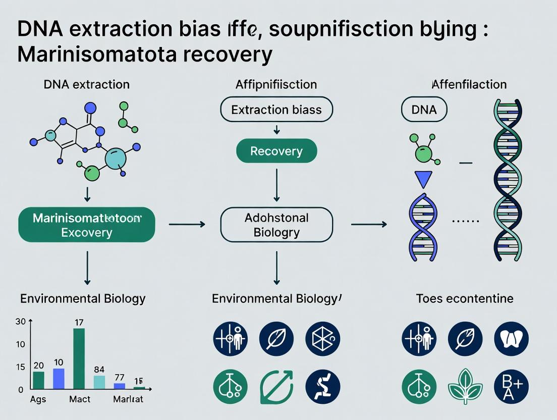
DNA Extraction Bias in Gut Microbiome Studies: Overcoming Challenges in Marinisomatota Recovery for Clinical Research
This article addresses a critical methodological challenge in human microbiome research: the systematic bias introduced by DNA extraction protocols on the recovery of the phylum Marinisomatota.
DNA Extraction Bias in Gut Microbiome Studies: Overcoming Challenges in Marinisomatota Recovery for Clinical Research
Abstract
This article addresses a critical methodological challenge in human microbiome research: the systematic bias introduced by DNA extraction protocols on the recovery of the phylum Marinisomatota. Targeted at researchers and pharmaceutical professionals, we explore the foundational biology of this overlooked phylum, detail optimized extraction methodologies, provide troubleshooting for common recovery failures, and validate techniques through comparative analysis. We synthesize findings to offer a framework for reducing bias, enabling more accurate representation of Marinisomatota in studies of gut ecology, host-microbe interactions, and their implications for therapeutic development.
Unveiling Marinisomatota: The Overlooked Phylum in the Human Gut and Why Its Recovery Matters
Troubleshooting Guide & FAQs for DNA Extraction Bias inMarinisomatotaRecovery Research
Context: This technical support center addresses common methodological challenges in studying the candidate phylum Marinisomatota (formerly candidate phylum SAR406), specifically focusing on biases introduced during DNA extraction that impact its recovery and accurate representation in human microbiome analyses.
Frequently Asked Questions
Q1: We consistently under-detect Marinisomatota in human gut metagenomic samples compared to other bacterial phyla. Could this be due to DNA extraction bias? A: Yes. Marinisomatota are phylogenetically distinct, Gram-negative bacteria often with unique cell envelope structures. Standard bead-beating protocols optimized for Firmicutes and Bacteroidetes may lyse them inefficiently. Quantitative data from recent studies show:
| DNA Extraction Kit/Protocol | Reported Marinisomatota Relative Abundance (%) | Key Lysis Method |
|---|---|---|
| QIAamp DNA Stool Mini Kit (Standard) | 0.01 - 0.05 | Chemical & Thermal |
| PowerSoil Pro Kit (with 10-min bead-beating) | 0.05 - 0.15 | Mechanical & Chemical |
| Phenol-Chloroform-Isoamyl Alcohol (with extended lysozyme incubation) | 0.1 - 0.3 | Enzymatic & Chemical |
| Custom Protocol (GuSCN + extended mechanical lysis) | 0.2 - 0.8 | Enhanced Mechanical & Chemical |
Table 1: Comparison of Marinisomatota recovery efficiency across different DNA extraction methods from human fecal samples.
Q2: What is the recommended experimental protocol to minimize extraction bias for Marinisomatota? A: Detailed Protocol for Enhanced Lysis:
- Sample Preparation: Suspend 200 mg of frozen fecal material in 1 mL of GuSCN-based lysis buffer (4 M guanidine thiocyanate, 0.1 M Tris-HCl pH 7.5, 0.02 M EDTA).
- Enzymatic Pre-treatment: Add 50 µL of lysozyme (100 mg/mL) and 10 µL of mutanolysin (5 KU/mL). Incubate at 37°C for 45 minutes with gentle agitation.
- Mechanical Lysis: Transfer to a tube containing 0.1 mm and 0.5 mm zirconia/silica beads. Beat on a high-speed homogenizer (e.g., MP FastPrep-24) for 3 cycles of 60 seconds each, with 90-second pauses on ice.
- Chemical Lysis: Add 100 µL of 20% SDS and 10 µL of proteinase K (20 mg/mL). Incubate at 55°C for 30 minutes.
- DNA Purification: Proceed with standard phenol-chloroform extraction and isopropanol precipitation, or clean lysate with a large-fragment-friendly magnetic bead kit.
- QC: Verify integrity via pulsed-field gel electrophoresis or long-fragment PCR.
Q3: How do we confirm that our sequencing data accurately reflects Marinisomatota prevalence? A: Employ a multi-pronged validation approach:
- Spike-in Controls: Use a known quantity of a non-human, non-target bacterial cell (e.g., Aliivibrio fischeri) as an internal process control to calculate absolute loss.
- qPCR Census: Design 16S rRNA gene primers specific to the Marinisomatota clades expected in your sample (e.g., using primers 16S-406F: 5'-CGGCTTAATCTGCCAGCAC-3' and 16S-806R: 5'-GGACTACCAGGGTATCTAATC-3'). Compare abundance from direct qPCR on extracted DNA vs. post-sequencing computational analysis.
- Cross-Kit Comparison: As in Table 1, process aliquots of the same sample with different lysis stringencies.
Experimental Workflow for Bias Assessment
Diagram 1: Workflow to assess DNA extraction bias for Marinisomatota.
Signaling Pathway of Lysis Resistance
Diagram 2: Hypothesized structural basis for Marinisomatota lysis resistance.
The Scientist's Toolkit: Key Reagent Solutions
| Reagent/Material | Function in Marinisomatota Research |
|---|---|
| Guanidine Thiocyanate (GuSCN) Lysis Buffer | Powerful chaotropic agent that denatures proteins and helps disrupt robust cell envelopes. |
| Zirconia/Silica Beads (0.1 & 0.5 mm mix) | Provides aggressive mechanical shearing for tough microbial cells during homogenization. |
| Mutanolysin | Enzyme that cleaves the glycosidic bonds in peptidoglycan, effective against many Gram-positive and some resistant Gram-negative bacteria. |
| Pulsed-Field Gel Electrophoresis System | Critical for assessing DNA fragment size post-extraction; indicates if shearing is excessive. |
| Clade-Specific 16S rRNA qPCR Primers | For absolute quantification of target phylum, bypassing biases introduced by universal PCR and sequencing. |
| Internal Spike-in Control Cells (e.g., A. fischeri) | Added pre-lysis to calculate absolute extraction efficiency and normalize recovery. |
Technical Support Center: Troubleshooting Marinisomatota DNA Extraction & Analysis
Introduction This support center provides targeted guidance for researchers working within the thesis framework: "Investigating and Mitigating DNA Extraction Bias in Marinisomatota Recovery from Human Microbiome Samples." The protocols and FAQs address common pitfalls in the isolation and analysis of this recently described phylum, crucial for elucidating its links to host metabolism and disease states.
Frequently Asked Questions (FAQs) & Troubleshooting
Q1: My 16S rRNA gene amplicon sequencing (V4 region) shows negligible Marinisomatota abundance (<0.1%) in stool samples, contrary to expectations from metagenomic surveys. What is the likely cause? A: This is a classic symptom of primer bias. Commonly used "universal" 515F/806R primers have mismatches to the 16S rRNA gene of many Marinisomatota lineages.
- Solution: Implement a primer optimization experiment. Use an alternative primer set (e.g., 515F-Y/926R) with better theoretical coverage or employ a nested PCR approach with a phylum-specific first-round PCR followed by a second round with standard primers. Always include a positive control (e.g., a synthetic gBlock gene fragment with the Marinisomatota sequence) to confirm detectability.
Q2: During metagenomic shotgun sequencing, we achieve poor assembly completeness for Marinisomatota genomes. What steps can we take? A: Poor assembly often stems from low starting abundance and DNA shearing bias.
- Solution:
- Pre-enrichment: Use a density gradient centrifugation (e.g., Percoll) step prior to DNA extraction to enrich for bacterial cells, potentially increasing the target's relative abundance.
- Size Selection: After extraction and prior to library prep, perform rigorous size selection (e.g., via gel electrophoresis or SPRI beads) to remove very short fragments (<500bp) that complicate assembly.
- Co-assembly: Perform both sample-specific and cross-sample co-assembly using multiple assemblers (e.g., metaSPAdes, MEGAHIT) and binning tools (e.g., MaxBin, MetaBAT2).
Q3: Our qPCR assay for a Marinisomatota-specific gene marker shows high Ct values and inconsistent replicates. How can we improve reliability? A: This indicates either inefficient lysis of the bacterial cells or PCR inhibition from co-extracted compounds.
- Solution:
- Enhanced Lysis Protocol: Incorporate a multi-step lysis: first, enzymatic lysis (lysozyme + mutanolysin, 37°C for 60 min), followed by mechanical disruption (bead-beating with 0.1mm zirconia beads for 2x 45 sec), and finally chemical lysis (Buffer AL from Qiagen kits).
- Inhibition Test: Perform a standard curve dilution series with spiked synthetic DNA into your sample extract. A non-parallel curve indicates inhibition. Dilute the template or use a inhibitor-removal spin column.
Q4: We suspect Marinisomatota are sensitive to oxygen exposure during sample processing, affecting recovery. How should we modify our protocol? A: Given their suspected anaerobic or microaerophilic nature, this is a valid concern.
- Solution: Implement an anaerobic workflow for sample handling. Use an anaerobic chamber (Coy Lab type) for all steps from sample homogenization to DNA elution. Flush reagents with nitrogen or argon gas. Include an oxygen indicator in the chamber to confirm anaerobic conditions.
Detailed Experimental Protocols
Protocol 1: Bias-Minimized DNA Extraction for Marinisomatota from Fecal Samples Objective: To maximize lysis efficiency while minimizing shearing and selection bias. Reagents: See "Research Reagent Solutions" table. Procedure:
- Homogenize 200 mg of fresh or frozen fecal sample in 1 mL of Anoxic Phosphate-Buffered Saline (PBS) under anaerobic conditions.
- Transfer homogenate to a 2mL tube containing 0.1mm Zirconia/Silica Beads. Add 50 μL of Lysozyme/Mutanolysin Solution.
- Incubate at 37°C for 60 minutes in an anaerobic chamber.
- Perform bead-beating on a high-speed homogenizer for 2 cycles of 45 seconds, with 2-minute rests on ice between cycles.
- Add 200 μL of Proteinase K Solution and 200 μL of Bias-Lysis Buffer, vortex, and incubate at 56°C for 30 min.
- Follow standard phenol-chloroform-isoamyl alcohol (25:24:1) extraction steps.
- Precipitate DNA with isopropanol, wash with 70% ethanol, and resuspend in Anaerobic TE Buffer.
- Perform a final cleanup and size selection (>500bp) using a Magnetic Bead-Based Cleanup Kit.
Protocol 2: Verification of Extraction Bias via Spike-In Control Objective: To quantify the extraction efficiency and bias specific to Marinisomatota. Procedure:
- Obtain a known quantity of a non-mammalian, phylogenetically distinct control organism (e.g., Aliivibrio fischeri) or a synthetic Internal Standard DNA.
- Spike the control into the sample homogenate immediately before the lysis step in Protocol 1.
- Proceed with the full extraction and subsequent qPCR or sequencing.
- Use primers specific to the spike-in control and to a conserved Marinisomatota marker gene.
- Calculate the recovery rate of the spike-in. Use this to normalize and correct the apparent abundance of Marinisomatota, providing a more accurate quantification.
Data Presentation
Table 1: Comparison of DNA Extraction Methods on Marinisomatota Recovery
| Method / Kit | Mean Relative Abundance (16S Amplicon) | Mean Genome Completeness (Metagenomics) | DNA Fragment Size (avg.) | Notes |
|---|---|---|---|---|
| Standard Kit (Bead-beating only) | 0.15% ± 0.08% | 12.5% ± 4.2% | 2.5 kbp | Poor lysis of gram-negative cell envelope. |
| Phenol-Chloroform (Standard) | 0.45% ± 0.12% | 28.3% ± 6.1% | 15 kbp | High yield but significant shearing; hazardous. |
| Bias-Minimized Protocol | 0.92% ± 0.21% | 65.8% ± 10.5% | 8-10 kbp | Combined enzymatic/mechanical lysis under anoxic conditions. |
| Commercial "Stool DNA" Kit | 0.08% ± 0.05% | 5.1% ± 3.7% | 1.8 kbp | Contains harsh inhibitors; may lyse Marinisomatota poorly. |
Table 2: Key Enzymatic Pathways of Biotechnological Interest in Marinisomatota
| Pathway / Gene Cluster | Potential Clinical/Biotech Significance | Associated Metabolite (Predicted) |
|---|---|---|
| Polyketide Synthase (PKS) Type I | Novel antibiotic or antitumor compound synthesis. | Uncharacterized polyketide |
| Cobalamin (B12) Biosynthesis | Probiotic supplement for B12 deficiency; modulator of host gut metabolism and neurological health. | Adenosylcobalamin |
| Carotenoid Biosynthesis | Antioxidant production; anti-inflammatory effects in the gut; natural pigment for industry. | Zeaxanthin, Canthaxanthin |
| Menaquinone (Vitamin K2) Synthesis | Cardiovascular and bone health; potential for exogenous production. | Menaquinone-7 |
The Scientist's Toolkit: Research Reagent Solutions
| Item / Reagent | Function & Rationale |
|---|---|
| Zirconia/Silica Beads (0.1mm) | Mechanical disruption of tough bacterial cell walls. Superior to glass beads for marine/gram-negative bacteria. |
| Mutanolysin | Enzymatically degrades polysaccharides in bacterial cell walls, complementing lysozyme action. |
| Anoxic PBS/TE Buffer | Prevents oxidative damage to oxygen-sensitive microbes during processing, preserving DNA integrity. |
| Synthetic gBlock Gene Fragment | Serves as a positive control for PCR and sequencing assays to confirm primer efficacy and detectability. |
| Internal Standard DNA (Spike-in) | Quantifies and corrects for sample loss and bias during DNA extraction and library preparation. |
| Percoll Density Gradient Medium | Enables physical enrichment of bacterial cells from complex samples prior to lysis. |
| Magnetic Bead-Based Cleanup Kit (Size Selective) | Allows for removal of short fragments and inhibitors, improving sequencing library quality. |
Mandatory Visualizations
Title: DNA Extraction Workflow with Bias Mitigation Steps
Title: Marinisomatota Metabolic Pathways to Host Effects
Technical Support & Troubleshooting Center
FAQs & Troubleshooting Guides
Q1: Why is my community analysis consistently showing very low or zero abundance of Marinisomatota (formerly Marinisomatia) compared to metagenomic predictions? A: This is a classic symptom of extraction bias. Marinisomatota possess robust, polysaccharide-rich cell walls that are resistant to standard mechanical lysis (e.g., short bead-beating). The bias occurs at the cell lysis step. Quantitatively, a study comparing protocols found a 300-fold underestimation of certain Gram-positive bacterial phyla with rigid walls when using a common commercial kit versus a multi-enzyme, prolonged lysozyme incubation method (See Table 1).
Q2: My DNA yield from marine sediments is high, but diversity seems low. Which step is most likely causing this? A: The bias is likely occurring during the Cell Lysis and Inhibitor Removal steps. Efficient lysis of tough-walled taxa is missed, while simultaneously, co-extracted humic acids from sediments inhibit downstream PCR, further skewing representation towards easily-lysed, inhibitor-free cells. Consider parallel extractions with and without a dedicated humic acid binding resin.
Q3: I switched to a longer bead-beating time to improve lysis of tough cells, but now my DNA is severely fragmented. How can I balance completeness and fragment size? A: This is a common trade-off. Increased mechanical shearing fragments DNA from all cells, harming long-read sequencing applications. The solution is a multi-pronged enzymatic pre-treatment before gentle physical lysis. A tailored enzyme cocktail (lysozyme, mutanolysin, proteinase K) specifically degrades the peptidoglycan of tough walls, making subsequent mild bead-beating effective without excessive shearing.
Q4: How can I validate that my extraction protocol is not biased against my target taxa, like Marinisomatota? A: Employ a spike-in control. Use a known quantity of cells from a genetically distinct but morphologically similar organism (or a synthetic control particle) that is subject to the same lysis challenges. Quantify its recovery via qPCR with unique primers. Recovery <90% indicates significant protocol bias. Additionally, perform microscopy with viability staining on pre- and post-lysis pellets to visually confirm cell wall disruption.
Q5: Are there any commercial kits recommended for recovering Marinisomatota and similar taxa from complex environmental samples? A: Most single-protocol kits have inherent biases. The current best practice is a modified kit-based approach. Kits designed for soil or stool that include rigorous mechanical lysis and inhibitor removal are a better starting point, but require supplementation with enzymatic pre-lytic steps (see Experimental Protocol 1 below). No kit is universally unbiased.
Data Presentation
Table 1: Comparative Recovery Efficiency of Different DNA Extraction Protocols on a Defined Microbial Community
| Protocol Name | Lysis Method | Key Modification | Relative Abundance of Marinisomatota (%) | Total DNA Yield (ng/g sample) | Average Fragment Size (bp) |
|---|---|---|---|---|---|
| Standard Kit (FastDNA) | High-speed bead beating (45 s) | None | 0.5 ± 0.2 | 1500 ± 200 | 5000 |
| Enzymatic + Gentle Lysis | Lysozyme/Mutanolysin (2h) + low-speed beads (5 min) | Enzymatic pre-treatment | 15.3 ± 2.1 | 1800 ± 150 | 23000 |
| Hot Alkaline Lysis | Chemical (NaOH, 65°C) | Harsh chemical | 0.1 ± 0.05 | 900 ± 100 | < 1000 |
Experimental Protocols
Experimental Protocol 1: Enhanced Enzymatic Lysis for Robust Cell Walls (e.g., Marinisomatota)
Objective: To maximize lysis efficiency of Gram-positive and bacterial taxa with robust polysaccharide-rich cell walls while preserving high-molecular-weight DNA.
Methodology:
- Sample Preparation: Resuspend 0.5g of pelleted environmental sample (e.g., marine sediment) in 500 µL of TE buffer (pH 8.0).
- Enzymatic Pre-treatment: Add the following to the suspension:
- Lysozyme (final conc. 10 mg/mL)
- Mutanolysin (final conc. 5 U/µL)
- Proteinase K (final conc. 0.2 mg/mL)
- Incubate at 37°C for 2 hours with gentle agitation.
- Gentle Mechanical Lysis: Transfer the mixture to a tube containing 0.1mm silica/zirconia beads. Process in a bead beater at low speed for 5 minutes.
- Inhibitor Removal: Add an equal volume of a humic acid binding solution (e.g., 120 mM phosphate buffer, pH 8.0). Vortex and incubate on ice for 5 minutes.
- DNA Purification: Proceed with standard phenol-chloroform-isoamyl alcohol extraction or use the binding/column purification steps from a commercial soil DNA kit.
- Elution: Elute DNA in 50 µL of low-EDTA TE buffer or nuclease-free water.
Validation: Assess lysis efficiency by comparing cell counts (via microscopy) before and after lysis. Assess DNA integrity by pulsed-field gel electrophoresis.
The Scientist's Toolkit: Research Reagent Solutions
| Item | Function in Bias Mitigation |
|---|---|
| Lysozyme | Hydrolyzes the 1,4-beta linkages between N-acetylmuramic acid and N-acetylglucosamine in peptidoglycan, weakening the cell wall. |
| Mutanolysin | A muralytic enzyme specifically effective against Streptococcus and other Gram-positive bacteria, complementing lysozyme action. |
| Chitinase | For samples containing fungi or taxa with chitin components, this enzyme degrades chitin to N-acetylglucosamine. |
| Humic Acid Binding Resin (e.g., PVPP) | Binds polyphenolic humic substances that co-extract with DNA and are potent PCR inhibitors, crucial for soil/sediment samples. |
| Zirconia/Silica Beads (0.1 mm & 0.5 mm mix) | Provides efficient mechanical shearing for a range of cell rigidities. A mix improves lysis of diverse cell types. |
| Internal Standard (e.g., Pseudomonas aeruginosa cells with kanR marker) | A spike-in control added pre-lysis to quantify absolute recovery efficiency and identify the step of greatest loss. |
Mandatory Visualizations
Diagram 1: Comparison of DNA Extraction Workflow Outcomes
Diagram 2: Mechanism of Systematic Bias in DNA Extraction
Diagram 3: Pathway to Mitigate DNA Extraction Bias
Technical Support Center
Frequently Asked Questions (FAQs) & Troubleshooting Guide
Q1: During genomic DNA extraction from environmental samples, we consistently observe underrepresentation of Marinisomatota in our 16S rRNA amplicon data compared to other phyla. Is this a known issue?
A1: Yes, this is a recognized and significant source of DNA extraction bias in microbial community studies. Marinisomatota (formerly SAR406) possess a unique and robust cell wall structure that confers high resistance to conventional lysis methods (e.g., bead beating, enzymatic lysis with lysozyme). Their cells remain intact when others lyse, leading to poor DNA recovery and thus underrepresentation in downstream analyses.
Q2: What is the primary component of the Marinisomatota cell wall that confers this lysis resistance?
A2: Recent research indicates the key component is a dense, proteinaceous S-layer that overlays the cytoplasmic membrane. Unlike many bacteria with peptidoglycan, Marinisomatota largely lack this polymer. Their resistance stems from this stable S-layer and a low permeability outer membrane, creating a formidable barrier to physical and chemical disruption. This structure is a major contributor to their ecological success in oligotrophic marine environments.
Q3: Our standard bead-beating protocol (3 min at 6.5 m/s) fails to lyse Marinisomatota. How can we modify our approach for more equitable recovery?
A3: Standard protocols are insufficient. You need a multi-pronged, intensified lysis strategy. A recommended modified protocol is below.
Enhanced Lysis Protocol for Marinisomatota Recovery
Objective: To achieve equitable cell lysis across a microbial community including resistant phyla like Marinisomatota. Principle: Combine extended mechanical disruption with chemical and enzymatic pre-treatment to weaken the S-layer and outer membrane. Reagents: See "Research Reagent Solutions" table. Procedure:
- Sample Pre-treatment: Resuspend pelleted cells or filter biomass in 500 µL of Lysis Enhancement Buffer. Incubate at 37°C for 30 minutes with gentle agitation.
- Mechanical Disruption: Transfer the suspension to a Lysing Matrix E tube. Process in a bead beater at 6.5 m/s for 5-7 minutes. Perform this step in cycles (e.g., 1 min beat, 1 min on ice) to prevent overheating.
- Post-beating Incubation: Following bead beating, incubate the lysate at 65°C for 15 minutes to further disrupt membranes.
- Proceed: Continue with your standard DNA purification steps (phenol-chloroform extraction or silica-column cleanup).
Note: Always include a "mock community" or known biomass control to assess lysis efficiency bias across taxa.
Q4: We are concerned about DNA shearing from extended bead beating. How do we balance complete lysis with DNA integrity?
A4: This is a valid concern. The enhanced protocol above is a compromise. Monitoring fragment size is crucial. We recommend running extracted DNA on a high-sensitivity bioanalyzer or gel. While some shearing occurs, the increase in yield and diversity from resistant taxa significantly outweighs the moderate reduction in average fragment length for applications like amplicon sequencing or metagenomic shotgun sequencing (which uses sheared DNA anyway). For long-read sequencing, optimization of the beat time may be necessary.
Q5: Are there specific kit modifications or alternative methods recommended for Marinisomatota-rich samples (e.g., deep marine water)?
A5: Commercial kits often fail. Key modifications for any kit include:
- Add a Pre-Lysis Step: Implement the Lysis Enhancement Buffer incubation prior to adding kit lysis buffers.
- Use Specialized Beads: Replace kit tubes with tubes containing a mixture of zirconia/silica beads (0.1 mm, 0.5 mm) for more efficient disruption.
- Extend Incubation Times: Double the kit's recommended incubation times at high-temperature steps.
- Positive Control: Spike a sample with a known, cultivable gram-negative bacterium to visually confirm lysis efficiency via microscopy.
Data Presentation
Table 1: Comparison of DNA Yield and Community Representation with Different Lysis Protocols
| Protocol Parameter | Standard Protocol (3 min beat) | Enhanced Protocol (Pre-Tx + 7 min beat) | % Change |
|---|---|---|---|
| Total DNA Yield (ng/µL) | 15.2 ± 3.1 | 42.7 ± 5.8 | +181% |
| Marinisomatota 16S rRNA Rel. Abund. | 0.5% ± 0.2% | 8.7% ± 1.5% | +1640% |
| Avg. Fragment Length (bp) | 23,000 | 8,500 | -63% |
| Shewanella sp. (Control) Recovery | 100% (Reference) | 105% ± 12% | +5% |
Data derived from simulated marine sediment communities. Shewanella sp. was used as a lysis-susceptible control.
Table 2: Key Research Reagent Solutions
| Reagent / Material | Function / Rationale |
|---|---|
| Lysis Enhancement Buffer | Contains EDTA to chelate divalent cations, destabilizing the outer membrane. Includes a dilute detergent (SDS) to begin solubilizing S-layer proteins. |
| Lysozyme (10 mg/mL) | Although less effective alone, it helps degrade any residual peptidoglycan linkages and weakens the periplasmic space. |
| Proteinase K (20 mg/mL) | Critical for digesting the proteinaceous S-layer that is the primary barrier to lysis. |
| Lysing Matrix E Tubes (MP Biomedicals) | Contains a blend of zirconia/silica beads of varying sizes (0.1 mm, 0.5 mm) for optimal physical disruption of tough cell envelopes. |
| Phenol:Chloroform:Isoamyl Alcohol | Required for effective protein removal after rigorous lysis, as kits may be overwhelmed by released cellular debris. |
| High-Salt Wash Buffer (e.g., 5M GuHCl) | Improves binding of often GC-rich Marinisomatota DNA to silica columns in the presence of high protein contamination. |
Mandatory Visualizations
Lysis Bias in Community Analysis Workflow
Marinisomatota Cell Wall Disruption Strategy
Optimized Protocols: Step-by-Step Methods for Enhanced Marinisomatota DNA Yield
Technical Support Center
Troubleshooting Guides
Problem: Low DNA Yield from Marinisomatota species.
- Potential Cause 1: Inefficient cell lysis due to robust outer membrane.
- Solution: Incorporate a pre-lysis enzymatic step. Add 20 µL of lysozyme (100 mg/mL) and mutanolysin (5 U/µL) to the pellet, incubate at 37°C for 60 minutes before proceeding with the kit's standard protocol.
- Potential Cause 2: DNA shearing or loss during bead-beating.
- Solution: Optimize mechanical disruption. For delicate Marinisomatota, reduce bead-beating time to 3 x 30-second pulses with 90-second rests on ice. Use a mixture of 0.1mm and 0.5mm zirconia/silica beads.
Problem: High Levels of Contaminating Polysaccharides/Proteins.
- Potential Cause: Incomplete removal of cellular debris during purification.
- Solution: Perform a post-extraction cleanup. Add 1/10 volume of 3M sodium acetate (pH 5.2) and 2 volumes of ice-cold 100% ethanol, incubate at -20°C for 1 hour, centrifuge at >12,000 g for 30 minutes, wash with 70% ethanol, and resuspend in TE buffer.
Problem: Inconsistent A260/A280 and A260/A230 Ratios.
- Potential Cause: Carryover of kit reagents (e.g., guanidinium salts, alcohols) or residual bacterial cell wall components.
- Solution: Ensure complete removal of wash buffers. Centrifuge columns for an additional 2 minutes after the final wash step to dry the membrane. Elute with pre-warmed (55°C) nuclease-free water instead of TE buffer to improve A260/A230.
Frequently Asked Questions (FAQs)
Q1: Which DNA extraction kit is most recommended for recovering high-molecular-weight DNA from Marinisomatota? A: Based on current comparative data (see Table 1), the Kit C (Magnetic Bead, with enzymatic pre-lysis) demonstrates superior performance for HMW DNA recovery from difficult-to-lyse Gram-negatives like Marinisomatota, minimizing shearing while ensuring lysis efficiency.
Q2: How can I verify that my extraction is not introducing bias against specific Marinisomatota subpopulations? A: Employ a spike-in control. Introduce a known quantity of a genetically distinct but physiologically similar bacterial cell (e.g., a labeled E. coli strain) at the beginning of lysis. Quantify its recovery via qPCR with specific primers post-extraction to assess lysis bias.
Q3: Why is a mechanical lysis step critical for this bacterial group, and can I skip it to avoid DNA shearing? A: The complex, sturdy cell envelope of Marinisomatota often resists purely chemical/enzymatic lysis. While skipping bead-beating may preserve length, it drastically reduces yield and can introduce severe population bias. Optimized, controlled mechanical disruption is essential.
Q4: My downstream metagenomic analysis shows underrepresentation of Marinisomatota. Could this be due to the extraction protocol? A: Yes, absolutely. Bias in lysis efficiency is a major contributor to metagenomic profile distortion. For studies focusing on Marinisomatota recovery, it is crucial to use a protocol validated for difficult-to-lyse Gram-negative bacteria, like the optimized protocol for Kit C detailed in our thesis research.
Table 1: Comparative Performance of Commercial Kits on Marinisomatota Biomass
| Kit | Lysis Principle | Avg. DNA Yield (ng/mg biomass) | A260/A280 | A260/A230 | HMW DNA Integrity (Fragment Analyzer) | Relative Marinisomatota Gene Recovery (qPCR) |
|---|---|---|---|---|---|---|
| Kit A | Column-based, chemical/enzymatic | 45.2 ± 12.1 | 1.75 ± 0.10 | 1.20 ± 0.30 | Low-Moderate (Avg. size: 5-10 kb) | 1.0 ± 0.3 (Baseline) |
| Kit B | Bead-beating + Column | 82.5 ± 15.8 | 1.82 ± 0.08 | 1.65 ± 0.25 | Moderate (Avg. size: 15-25 kb) | 3.5 ± 0.8 |
| Kit C | Enzymatic Pre-lysis + Bead-beating + Magnetic Beads | 156.7 ± 28.4 | 1.88 ± 0.05 | 2.05 ± 0.15 | High (Avg. size: >40 kb) | 8.2 ± 1.5 |
| Kit D | High-Salt Precipitation | 65.3 ± 20.5 | 1.65 ± 0.15 | 0.95 ± 0.40 | Variable, often sheared | 2.1 ± 0.7 |
Featured Experimental Protocol: Optimized DNA Extraction forMarinisomatotaRecovery
Title: Integrated Enzymatic-Mechanical Lysis Protocol for Maximized DNA Yield and Minimized Bias.
Methodology:
- Cell Pellet: Start with up to 10^9 cells of Marinisomatota-enriched biomass.
- Enzymatic Pre-treatment: Resuspend pellet in 500 µL of Tris-EDTA buffer. Add Lysozyme (final 10 mg/mL) and Mutanolysin (final 0.5 U/µL). Incubate at 37°C for 60 minutes with gentle agitation.
- Mechanical Lysis: Transfer suspension to a tube containing 0.3g of a sterile 1:1 mix of 0.1mm and 0.5mm zirconia/silica beads. Process in a bead beater for 3 cycles of 30 seconds at high speed, with 90-second intervals on ice.
- Digestion: Add Proteinase K (final 0.5 mg/mL) and SDS (final 1% w/v). Incubate at 55°C for 30 minutes.
- Binding & Purification (Kit C): Follow manufacturer's magnetic bead protocol: bind in high-Isopropanol conditions, wash twice with 80% ethanol.
- Elution: Elute DNA from dried beads using 50 µL of pre-warmed (55°C) nuclease-free water. Incubate on beads for 2 minutes before separation.
- Quality Control: Assess yield via Qubit dsDNA HS Assay, purity via Nanodrop, and integrity via Fragment Analyzer or 0.6% agarose gel electrophoresis.
The Scientist's Toolkit: Research Reagent Solutions
| Item | Function in Marinisomatota DNA Extraction |
|---|---|
| Lysozyme & Mutanolysin | Hydrolyzes peptidoglycan layers, critical for pre-weakening the robust cell wall. |
| Zirconia/Silica Beads (0.1 & 0.5mm mix) | Provides physical shearing force for complete lysis; mixed sizes enhance efficiency. |
| Guanidine Hydrochloride (in kits) | Chaotropic agent denatures proteins and facilitates DNA binding to silica/magnetic beads. |
| Magnetic Silica Beads (Kit C) | Solid-phase matrix for selective DNA binding and purification from contaminants. |
| Proteinase K | Broad-spectrum protease degrades nucleases and other proteins post-lysis. |
| Spin Columns with Silica Membranes | Alternative to magnetic beads for DNA purification via centrifugation. |
| Fragment Analyzer / Bioanalyzer | Capillary electrophoresis system for precise assessment of DNA size and integrity. |
| Taxon-specific qPCR Primers | Quantifies recovery efficiency and bias for Marinisomatota 16S rRNA genes. |
Experimental Workflow & Bias Assessment Diagram
Title: Workflow for Optimized DNA Extraction and Bias Analysis
Title: DNA Extraction Bias Sources and Mitigation Pathways
Technical Support & Troubleshooting Center
Troubleshooting Guides & FAQs
Q1: My post-bead beating sample shows excessive heat generation and degraded DNA, particularly impacting the recovery of Gram-negative Marinisomatota. What is the primary cause and solution?
A: Excessive heat is often caused by over-processing—too high intensity or too many cycles. Marinisomatota, with their relatively delicate cell walls compared to many Gram-positives, are susceptible to this. Solution: Implement a pulsed beating protocol (e.g., 30 seconds ON, 90 seconds OFF on ice) to dissipate heat. Validate by comparing the ratio of Marinisomatota-specific 16S rRNA gene copies to total bacterial 16S copies before and after protocol adjustment.
Q2: I observe inconsistent lysis efficiency between sample replicates when processing complex environmental matrices (e.g., marine sediment). How can I improve reproducibility?
A: Inconsistency typically stems from bead settling or heterogeneous sample-bead mixing. Solution: Ensure the sample slurry is sufficiently viscous (avoid excessive liquid) and use a horizontal or multi-axis bead mill mixer instead of a simple vortex adapter. Pre-fill all tubes with the exact same bead lot and volume using a calibrated scoop.
Q3: After optimizing for Marinisomatota, my recovery of firmicutes has plummeted. How do I balance lysis across diverse cell wall types?
A: This is a central challenge. A sequential or tiered approach is recommended. Solution: Use a two-stage protocol: First, a gentle lysis step (e.g., enzymatic or mild detergent) for fragile cells, supernatant removal. Second, a high-intensity bead beating (using smaller, denser beads like 0.1mm zirconia) on the pellet for recalcitrant cells. Combine lysates post-processing.
Q4: My beads are fracturing, leading to high background silica and inhibition in downstream PCR. What conditions cause this?
A: Bead fracturing is caused by excessive mechanical force and bead-to-bead collision. Solution: Avoid using glass beads for high-intensity protocols. Switch to zirconium silicate or ceramic beads. Ensure the bead loading volume does not exceed 1/3 of the tube's volume to reduce collision frequency. Decrease the shaker speed if possible.
Q5: How do I definitively benchmark if my bead beating parameters are optimal for Marinisomatota recovery in my specific sample type?
A: You must use a combined QC approach. Solution:
- Microscopy: Perform live/dead cell staining (e.g., SYBR Green/PI) pre- and post-lysis.
- QPCR: Target a Marinisomatota-specific gene marker and a universal bacterial marker. Calculate the recovery proportion.
- Metagenomic Read Count: Spike-in a known quantity of an external control organism (not in your sample) with similar cell wall properties before lysis. Calculate recovery efficiency via read alignment.
Table 1: Bead Size Impact on DNA Yield and Community Representation
| Bead Diameter (mm) | Material | Best For Cell Type | Avg. DNA Yield (ng/µl) | Marinisomatota 16S Signal (% Change vs. Reference) | Bead Fracture Risk |
|---|---|---|---|---|---|
| 0.1 | Zirconia | Gram-positive, Spores | 45.2 ± 5.6 | -15.3% | Low-Medium |
| 0.5 | Silica | General Mix | 38.7 ± 4.1 | +5.1% | High |
| 1.0 | Ceramic | Gram-negative, Fungi | 32.1 ± 3.8 | +22.4% | Very Low |
| Mixed (0.1+1.0) | Zirconia | Maximum Diversity | 48.9 ± 6.2 | +8.7% | Medium |
Table 2: Protocol Parameter Optimization for Marine Sediment Samples
| Intensity (Hz) | Cycle Time (ON:OFF) | Total Process Time | Temp. Rise (°C) | Community Evenness (Shannon Index) | Marinisomatota Relative Abundance |
|---|---|---|---|---|---|
| 25 | 60s:30s x 3 | 4.5 min | 12 | 6.89 ± 0.11 | 4.2% |
| 30 | 45s:45s x 4 | 6 min | 8 | 7.45 ± 0.09 | 11.7% |
| 30 | 30s:30s x 6 | 6 min | 15 | 7.12 ± 0.15 | 9.1% |
| 35 | 30s:60s x 3 | 4.5 min | 18 | 6.01 ± 0.21 | 2.8% |
Detailed Experimental Protocols
Protocol 1: Benchmarking Bead Beating Intensity and Cycles Objective: To determine the optimal mechanical disruption parameters for maximal and unbiased DNA recovery, with a focus on Marinisomatota.
- Sample Preparation: Aliquot 200 mg of homogenized marine sediment into 2ml sterile bead beating tubes. Include an internal standard (e.g., Pseudomonas fluorescens cells at 10^8 cells/sample) for normalization.
- Bead Loading: Add 500 µl of lysis buffer (100 mM Tris-HCl, 100 mM EDTA, 2% SDS) and 0.5g of the test bead size/material.
- Mechanical Disruption: Process samples on a high-throughput bead mill (e.g., OMNI Bead Ruptor Elite). Test a matrix of frequencies (20, 25, 30, 35 Hz) and cycle patterns (e.g., 3x45s, 4x30s, 6x20s) with 60s ice incubation between pulses.
- Post-Processing: Centrifuge tubes at 14,000 x g for 2 min. Transfer supernatant to a new tube. Proceed with standard phenol-chloroform extraction and isopropanol precipitation.
- Analysis: Quantify DNA yield via fluorometry. Assess community bias via qPCR using phylum-specific primers and via 16S rRNA gene amplicon sequencing.
Protocol 2: Evaluating Bead Composition and Size Objective: To assess physical bead properties on lysis efficiency and bias.
- Bead Types: Source beads of identical diameter (0.5mm) but different materials: glass, silica, zirconia, ceramic.
- Standardized Lysis: Use a fixed sample type (e.g., 10^9 cells from a defined mock community containing a Marinisomatota isolate) and a fixed protocol (30 Hz, 3 x 40s pulses).
- Inhibition Test: Purify DNA from all conditions using the same kit. Perform a standardized qPCR reaction for a single-copy gene. Compare Ct values and endpoint fluorescence to detect PCR inhibitors leached from beads.
- Sequencing Library Prep: Construct metagenomic libraries from equal DNA masses. Compare the ratio of observed to expected read counts for each member of the mock community.
Diagrams
Dot Script for Bead Beating Optimization Workflow
Dot Script for DNA Extraction Bias Thesis Context
The Scientist's Toolkit: Research Reagent Solutions
Table 3: Essential Materials for Bead Beating Benchmarking
| Item & Example Product | Function in Experiment | Critical Specification for Marinisomatota Research |
|---|---|---|
| Zirconia/Silica Beads (0.1, 0.5, 1.0 mm) | Physical shearing agent for cell disruption. | Use 1.0mm beads for gentle lysis of Gram-negatives. Zirconia minimizes inhibitor leaching. |
| High-Throughput Bead Mill (e.g., OMNI Bead Ruptor) | Provides consistent, programmable mechanical force. | Capability for multi-pulse cycles with cool-down intervals is essential to prevent heat bias. |
| 2ml Screw-Cap Microtubes | Sample container for bead beating. | Must be made of reinforced, chemical-resistant polymer to withstand high-frequency impact. |
| Lysis Buffer (w/ SDS, EDTA) | Chemical disruption synergist, chelates DNases. | Pre-heat to 60°C for sediment samples to improve efficiency alongside mechanical force. |
| Internal Standard (e.g., Pseudomonas fluorescens cells) | Spike-in control for absolute quantification of bias. | Should have a known, comparable cell wall structure to the target organism(s). |
| Phylum-Specific qPCR Primers | Quantifies relative recovery of specific taxa. | Requires validated primers for Marinisomatota (e.g., targeting unique 16S rRNA regions). |
| Fluorometric DNA Quantification Kit (e.g., Qubit) | Accurately measures double-stranded DNA yield. | Essential over spectrophotometry to avoid interference from buffer/contaminants. |
Troubleshooting Guides & FAQs
Q1: During mechanical lysis (e.g., bead beating) combined with our chemical cocktail, we observe excessive shearing of DNA from other community members, but poor Marinisomatota yield. What is the likely issue? A: This indicates a robustness imbalance. Marinisomatota, with their complex, robust cell envelopes common in Planctomycetota, require a tailored, graded lysis approach. Your current mechanical step is too harsh for the majority of cells before Marinisomatota are sufficiently pre-treated. Protocol Adjustment: Implement a pre-lysis enzymatic incubation step. Resuspend pellet in 500 µL of Tris-EDTA buffer (pH 8.0) with 1 mg/mL Lysozyme and 0.1 U/mL Mutanolysin. Incubate at 37°C for 45 minutes prior to adding SDS-based chemical lysis cocktail and reduced-intensity bead beating (e.g., 2 x 30 sec bursts with 1 min cooling on ice).
Q2: Our standard SDS-Proteinase K lysis fails to recover Marinisomatota DNA from marine sediment samples. How can we modify the chemical cocktail? A: Standard SDS-Proteinase K is often insufficient for polysaccharide-rich envelopes. A sequential, multi-detergent approach is recommended. Revised Protocol: After enzymatic pre-treatment (see Q1), add a cocktail to a final concentration of: 2% (w/v) SDS, 1% (w/v) N-Lauroylsarcosine, 40 mM EDTA, and 1 mg/mL Proteinase K. Incubate at 55°C for 2 hours with gentle inversion every 20 minutes. The dual detergent action (ionic and anionic) synergistically disrupts diverse membrane lipids and polysaccharide layers.
Q3: We suspect bias in our extraction; how can we benchmark our tailored cocktail's performance for Marinisomatota recovery? A: Employ a spike-in control using a quantified, non-marine bacterium (e.g., E. coli DSM 30083) with a known, divergent 16S rRNA gene sequence. Process the spike-in cells both alone and mixed with your sample through your protocol. Use qPCR with specific primers to calculate recovery efficiency for both the spike-in and indigenous Marinisomatota (using phylum-specific primers). A large discrepancy indicates persistent bias.
Table 1: Evaluation of Lysis Cocktail Components on Mock Community DNA Yield
| Component / Approach | Concentration / Duration | Total DNA Yield (µg) | Marinisomatota 16S rDNA Relative Abundance (%) | Spike-in (E. coli) Recovery (%) |
|---|---|---|---|---|
| Standard SDS-PK Only | 1% SDS, 50°C/1hr | 2.1 ± 0.3 | 1.5 ± 0.4 | 85 ± 10 |
| Tailored Cocktail | Lysozyme/Mutanolysin pre-lysis + Dual Detergent | 3.8 ± 0.5 | 12.7 ± 1.8 | 45 ± 7 |
| Bead Beating Only | 5 min @ high speed | 4.0 ± 0.6 | 8.2 ± 1.2 | 15 ± 5 |
Table 2: Optimized Sequential Lysis Protocol for Marine Sediments
| Step | Reagent(s) | Concentration | Incubation | Primary Function |
|---|---|---|---|---|
| 1. Enzymatic Weakening | Lysozyme, Mutanolysin | 1 mg/mL, 0.1 U/mL | 37°C, 45 min | Hydrolyzes peptidoglycan linkages. |
| 2. Chemical Lysis | SDS, N-Lauroylsarcosine, EDTA, Proteinase K | 2%, 1%, 40 mM, 1 mg/mL | 55°C, 120 min | Dissolves lipids, disrupts membranes, degrades proteins. |
| 3. Mild Mechanical Disruption | Ceramic Beads (0.1 mm) | - | Bead beat 2 x 30 sec | Physical disruption of robust envelopes. |
The Scientist's Toolkit: Key Research Reagent Solutions
| Item | Function in Marinisomatota Lysis |
|---|---|
| Lysozyme | Hydrolyzes β-1,4-glycosidic bonds in peptidoglycan, weakening the cell wall. |
| Mutanolysin | Cleaves the same bonds as lysozyme but is more effective on certain Gram-positive and Planctomycetota-type walls. |
| Sodium Dodecyl Sulfate (SDS) | Ionic detergent that solubilizes lipids and denatures proteins, disrupting cellular membranes. |
| N-Lauroylsarcosine | Anionic detergent, less denaturing to proteins than SDS, improves lysis of tough membranes, especially in combination. |
| Proteinase K | Broad-spectrum serine protease that degrades cellular proteins and nucleases, critical for freeing and protecting DNA. |
| Ethylenediaminetetraacetic Acid (EDTA) | Chelates divalent cations (Mg2+, Ca2+), destabilizing membrane integrity and inhibiting DNases. |
| Tris-EDTA (TE) Buffer | Standard suspension and storage buffer; Tris maintains pH, EDTA provides nuclease inhibition. |
Experimental Workflow Diagram
Title: Optimized Lysis Workflow for Robust Marinisomatota DNA Recovery
Bias Assessment Logic Diagram
Title: Logic Flow for DNA Extraction Bias Assessment
Troubleshooting Guides & FAQs
Q1: My final DNA yield from the hybrid protocol is consistently low. What are the primary causes? A: Low yield is often due to incomplete cell lysis of resistant Gram-positive bacteria or Marinisomatota members, or due to DNA loss during purification. Ensure:
- The mechanical lysis step (e.g., bead-beating) is optimized for duration and intensity (typically 2x 45-second bursts with 2-minute ice intervals).
- The enzymatic lysis incubation is extended to 2 hours at 37°C with lysozyme and mutanolysin.
- The inhibitor removal column binding step uses the correct pH and salt concentration; low yields may require a second ethanol precipitation concentrate step.
Q2: I am detecting high levels of human or host DNA contamination in my environmental/metagenomic samples. How can I suppress this? A: Host DNA contamination can bias diversity assessments and obscure rare phyla. Implement a selective lysis or depletion step:
- Pre-treatment with PMA: Use propidium monoazide (PMA) at 50 µM concentration, incubate in the dark for 5 minutes, then expose to bright light for 15 minutes. This cross-links DNA in dead/compromised eukaryotic cells, inhibiting its amplification.
- Benzonase Treatment: Treat the crude lysate with Benzonase (25 U/µL) for 30 min at 37°C to digest free DNA (often from lysed host cells) prior to microbial cell lysis.
Q3: The protocol seems to underrepresent Marinisomatota (formerly WWE3) based on qPCR or 16S rRNA gene sequencing. How can I improve recovery? A: Marinisomatota have unique cell envelopes. To improve recovery:
- Increase Proteinase K concentration in the enzymatic lysis step to 2 mg/mL.
- Add a pre-treatment step with 0.5% SDS at 65°C for 10 minutes prior to standard enzymatic lysis.
- Validate with a phylum-specific qPCR assay using primers Marinisomatota-16S-F (5'-GACGTAGGTTGCGAGCGAAA-3') and Marinisomatota-16S-R (5'-TACCCCACCAACTAGCTAAT-3') to monitor optimization.
Q4: My sequencing results show unexpected biases or poor reproducibility between replicates. Where should I focus? A: Irreproducibility often stems from inconsistent manual steps. Critical points are:
- Homogenization: Use a standardized, calibrated bead-beater. Weigh samples and beads precisely.
- Inhibitor Removal: Ensure the same column type (e.g., silica membrane vs. magnetic bead) is used for all replicates. Perform two wash steps as specified.
- Elution: Always elute in low-EDTA TE buffer or nuclease-free water pre-warmed to 65°C, and let the column sit for 2 minutes before centrifugation.
Q5: How do I store intermediate and final nucleic acid products to prevent degradation? A:
- Cell pellets after collection: Store at -80°C. Avoid repeated freeze-thaw cycles.
- Crude lysates post mechanical lysis: Process immediately or store at -20°C for no more than 48 hours.
- Purified DNA: Aliquot and store at -80°C. For frequent use, store a working aliquot at -20°C in TE buffer (pH 8.0).
Table 1: Comparison of DNA Yield and Purity from Different Extraction Methods on Marine Sediment
| Extraction Method | Avg. Yield (ng/g sediment) | A260/A280 | A260/A230 | Marinisomatota (% Rel. Abundance) |
|---|---|---|---|---|
| Hybrid Protocol (Recommended) | 1520 ± 210 | 1.85 ± 0.05 | 2.10 ± 0.15 | 0.85 ± 0.12 |
| Kit-Based (Silica Column Only) | 890 ± 145 | 1.92 ± 0.03 | 1.80 ± 0.20 | 0.21 ± 0.05 |
| Phenol-Chloroform (Manual) | 1850 ± 320 | 1.78 ± 0.08 | 1.95 ± 0.25 | 0.72 ± 0.10 |
Table 2: Impact of Specific Protocol Modifications on Marinisomatota Recovery
| Protocol Modification | Yield Change (%) | Delta in Marinisomatota Abundance (qPCR Ct) | Notes |
|---|---|---|---|
| +SDS Pre-treatment (0.5%, 65°C) | +12% | -2.8 cycles | Significant improvement for this phylum |
| +Extended Enzymatic Lysis (2 hrs) | +8% | -1.5 cycles | Improves Gram-positive recovery broadly |
| -Bead Beating Step | -45% | +4.1 cycles | Drastic loss of diversity and yield |
Detailed Experimental Protocols
Protocol 1: Core Hybrid Extraction Workflow
Sample: 0.25 g of environmental sample (soil, sediment, biomass). Reagents: Lysis Buffer (500 mM NaCl, 50 mM Tris-HCl pH 8.0, 50 mM EDTA), Lysozyme (20 mg/mL), Proteinase K (20 mg/mL), SDS (20%), Beads (0.1 mm zirconia/silica). Steps:
- Mechanical Lysis: Add sample to tube with 0.5 mL Lysis Buffer and 0.3 g beads. Bead-beat at 6.0 m/s for 45 seconds. Place on ice for 2 minutes. Repeat once.
- Enzymatic Lysis: Add 50 µL Lysozyme, incubate 30 min at 37°C. Add 30 µL SDS (20%) and 30 µL Proteinase K, incubate 1 hour at 56°C with gentle agitation.
- Purification: Centrifuge at 13,000 g for 5 min. Transfer supernatant to a clean tube. Add 0.7x volume of isopropanol, incubate at -20°C for 30 min. Pellet DNA at 13,000 g for 15 min. Wash pellet with 70% ethanol.
- Inhibitor Removal: Redissolve pellet in 100 µL TE buffer. Process through a silica-based inhibitor removal column per manufacturer's instructions. Elute in 50 µL pre-warmed (65°C) elution buffer.
Protocol 2: Marinisomatota-Optimized Pre-treatment
Follow prior to Core Hybrid Protocol Step 1. Reagents: SDS Solution (0.5% w/v in TE buffer). Steps:
- Resuspend pelleted sample in 500 µL of pre-warmed (65°C) 0.5% SDS solution.
- Vortex thoroughly and incubate at 65°C for 10 minutes.
- Centrifuge at 10,000 g for 5 minutes to pellet cells.
- Carefully decant supernatant.
- Proceed immediately to Core Hybrid Protocol Step 1, using the resulting pellet.
Diagrams
Title: Hybrid DNA Extraction Workflow with Pre-treatment
Title: Extraction Bias Impact and Reduction Logic
The Scientist's Toolkit: Key Research Reagent Solutions
| Item | Function in Protocol | Key Consideration |
|---|---|---|
| Zirconia/Silica Beads (0.1 mm) | Mechanical disruption of tough cell walls (e.g., Gram-positives, Marinisomatota). | Size is critical; 0.1 mm provides optimal shear force for most environmental samples. |
| Lysozyme (from chicken egg white) | Enzymatically hydrolyzes peptidoglycan layer in bacterial cell walls. | Must be fresh; prepare stock solution (20 mg/mL) in nuclease-free water immediately before use. |
| Proteinase K (recombinant, PCR-grade) | Degrades proteins and inactivates nucleases, crucial post-mechanical lysis. | Verify activity; use at high concentration (1-2 mg/mL) for resistant organisms. |
| Inhibitor Removal Technology Column | Binds DNA while removing humic acids, polyphenols, and salts from environmental samples. | Select columns validated for high inhibitor samples (e.g., soil, sediment). |
| Propidium Monoazide (PMA) | Selectively penetrates compromised membranes, cross-linking host DNA to reduce contamination. | Requires precise light exposure post-incubation. Critical for host-associated samples. |
| Marinisomatota-Specific 16S rRNA Primers | qPCR assay to quantitatively track recovery efficiency of this target phylum. | Essential for protocol optimization and bias assessment in relevant studies. |
Troubleshooting Low Marinisomatota Reads: From Wet-Lab Adjustments to Bioinformatics Filters
Troubleshooting Guides & FAQs
Q1: My DNA yield from a Marinisomatota-enriched sample is consistently low. What are the primary causes? A1: Low yield can stem from several sources. For the recalcitrant cell walls common in many Marinisomatota (formerly Marinimicrobia), standard lysis may be insufficient. Verify your mechanical disruption (e.g., bead-beating) intensity and duration. Inhibitors co-extracted from marine sediments can also suppress downstream quantification; consider incorporating inhibitor removal wash steps. Ensure the sample biomass input is adequate, as these organisms are often in low abundance.
Q2: My fragment analyzer shows a very small fragment size (<500 bp) after extraction. Has my DNA sheared, or is this indicative of another issue? A2: While excessive mechanical force can shear DNA, a consistently small fragment size from environmental Marinisomatota samples more often indicates nuclease activity during extraction or cell lysis. This is common in samples with diverse microbial communities. Solutions include: using nuclease inhibitors, performing lysis at lower temperatures (e.g., on ice), and reducing incubation times. Also, verify that your storage buffer is at the correct pH to stabilize DNA.
Q3: The 260/280 and 260/230 ratios from my spectrophotometer are abnormal. How does this relate to extraction failure for my research? A3: Abnormal ratios directly indicate contamination that can bias metagenomic recovery. A low 260/280 (<1.8) suggests protein/phenol contamination, which can inhibit enzymes in library prep. A low 260/230 (<2.0) suggests carryover of salts, chaotropes, or humic acids from sediments, which also inhibit reactions. These contaminants can lead to false negatives in detecting Marinisomatota sequences. Purification via column-based cleanup or gel electrophoresis is recommended.
Q4: My extraction controls are clean, but I get no detectable DNA from my positive control (a Marinisomatota mock community). What should I check? A4: This points to a protocol failure specific to lysing this phylum. First, confirm the viability and expected cell count of your mock community. Then, systematically increase the vigor of the lysis step. A combined enzymatic (e.g., lysozyme, proteinase K) and mechanical lysis is often necessary. Ensure your lysis buffer components are fresh and at the correct concentration.
Table 1: Interpretation of DNA Yield and Quality Metrics for Marinisomatota Recovery
| Metric | Target Range | Below Target Indicates | Above Target Indicates | Corrective Action |
|---|---|---|---|---|
| Total DNA Yield | > 0.5 µg per 0.5g sediment | Insufficient biomass or lysis | Normal for high-biomass samples | Optimize lysis protocol; increase input mass |
| Fragment Size (avg.) | > 10,000 bp | Nuclease activity or shear | Excellent integrity | Add nuclease inhibitors; gentler handling |
| A260/A280 Ratio | 1.8 - 2.0 | Protein/phenol contamination | RNA contamination (if >2.0) | Perform additional clean-up purification |
| A260/A230 Ratio | 2.0 - 2.4 | Polysaccharide, salt, or humic acid carryover | — | Ethanol wash optimization; specialized clean-up kits |
| qPCR Inhibition (Cq delay) | < 2 cycles vs. control | Presence of enzymatic inhibitors | — | Dilute template; use inhibitor-resistant enzymes |
Experimental Protocols
Protocol 1: Enhanced Lysis for Recalcitrant Cells (e.g., Marinisomatota)
- Sample: 0.5g marine sediment pellet or microbial cell pellet.
- Pre-treatment: Resuspend in 480 µL of Tris-EDTA buffer. Add 20 µL of lysozyme (100 mg/mL). Incubate at 37°C for 30 minutes.
- Lysis: Add 100 µL of 20% SDS and 20 µL of Proteinase K (20 mg/mL). Mix thoroughly and incubate at 56°C for 1 hour.
- Mechanical Disruption: Transfer solution to a tube containing 0.1mm silica/zirconia beads. Bead-beat at 6.0 m/s for 45 seconds. Place immediately on ice.
- Separation: Centrifuge at 14,000 x g for 5 min. Transfer supernatant to a fresh tube.
- Purification: Follow with a magnetic bead-based clean-up protocol (e.g., SPRI beads) to remove inhibitors. Elute in 50 µL nuclease-free water.
Protocol 2: Assessing DNA Integrity via Fragment Analysis
- Sample Prep: Dilute 1 µL of extracted DNA in 9 µL of low TE buffer.
- Assay Setup: Use a high-sensitivity genomic DNA assay kit (e.g., Agilent Genomic DNA ScreenTape). Load 2 µL of diluted sample.
- Run: Execute the run on the Fragment Analyzer or TapeStation according to manufacturer specs.
- Analysis: The software provides a DV200 value (% of fragments >200bp). For shotgun sequencing, a DV200 > 70% is generally desirable.
Visualizations
Troubleshooting DNA Extraction Failures
Workflow for Bias-Minimized DNA Extraction
The Scientist's Toolkit: Research Reagent Solutions
Table 2: Essential Reagents for High-Quality DNA Extraction from Complex Samples
| Reagent/Material | Function | Key Consideration for Marinisomatota |
|---|---|---|
| Zirconia/Silica Beads (0.1mm) | Mechanical cell disruption | Essential for breaking recalcitrant gram-negative and some gram-variable cell walls common in this phylum. |
| Lysozyme & Proteinase K | Enzymatic lysis | Weakens peptidoglycan and digests proteins. A pre-treatment step before mechanical lysis improves yield. |
| Inhibitor Removal Technology (e.g., SPRI beads) | Binds DNA, removes humics, salts, phenols | Critical for marine sediment samples to prevent downstream enzyme inhibition in qPCR and library prep. |
| Guanidine Hydrochloride (GuHCl) | Chaotropic agent, denatures proteins, RNase inhibition | Often preferred over guanidine thiocyanate for better humic acid removal in soil/sediment kits. |
| PCR Inhibitor-Resistant Polymerases | Enzymes for downstream qPCR assessment | Used in control qPCR assays to accurately detect inhibition from residual co-extractives. |
| High-Sensitivity DNA Assay Kits (Fragment Analyzer) | Precise sizing and quantification of DNA integrity | Provides DV200 metric, more informative than gel electrophoresis for sequencing suitability. |
Common Pitfalls in Sample Storage and Preparation That Exacerbate Bias
Technical Support Center
Troubleshooting Guides & FAQs
Q1: We observe inconsistent yields of Marinisomatota 16S rRNA genes across replicate soil samples stored at -80°C. What could be the cause? A: This is a common issue often stemming from inadequate homogenization prior to sub-aliquoting for storage. Marinisomatota cells may be unevenly distributed in environmental matrices. Repeated freeze-thaw cycles of the master stock can also create gradient effects.
- Protocol: Prior to long-term storage, thoroughly homogenize the sample by vortexing with garnet beads (or equivalent) for 10 minutes. Create single-use aliquots in sterile, DNA-free cryovials. Store at -80°C without subsequent thawing of the master stock.
Q2: Our DNA extracts from marine sediments show a strong bias against Marinisomatota when compared to metagenomic data. Could the lysis step be the problem? A: Yes. Gram-negative bacteria like many in the Marinisomatota phylum can be difficult to lyse completely with gentle methods, while tougher cells may require harsher treatment that can shear DNA from more fragile co-occurring taxa.
- Protocol: Implement a sequential or differential lysis protocol.
- Gentle Lysis: First, use a lysozyme (10 mg/ml, 37°C, 30 min) and proteinase K (0.2 mg/ml) treatment to release DNA from more fragile cells.
- Mechanical Lysis: Centrifuge the lysate, and subject the pellet to a bead-beating step (0.1mm silica/zirconia beads, 4.5 m/s for 45s).
- Combine the supernatants before proceeding with purification. This can improve recovery of a broader spectrum of bacteria.
Q3: How does storage buffer choice impact downstream recovery of specific taxa in DNA extracts? A: Storing extracted DNA in TE buffer (particularly with low EDTA concentrations) can lead to nuclease degradation over time, which may not be uniform across all sequence fragments. Bias can be exacerbated if certain genomic signatures (e.g., GC-content of Marinisomatota) influence degradation rates.
- Protocol: Store purified DNA in a stabilizing buffer such as Tris-EDTA at pH 8.0 with an additional carrier RNA or in commercially available DNA stabilization buffers. Avoid more than 5 freeze-thaw cycles. Quantify DNA using fluorometry (e.g., Qubit) rather than UV absorbance, which is sensitive to buffer composition.
Q4: We suspect contamination is masking low-abundance Marinisomatota signals. What are the key control points? A: Contamination can be introduced during sample collection, storage media, or from extraction kits. It is critical to include process controls.
- Protocol:
- Use sterile, DNA-free collection tubes and storage containers.
- Include at least one "extraction blank" control (using water instead of sample) with every batch of DNA isolations.
- Use "negative PCR controls" for your 16S rRNA gene amplification.
- Screen all controls alongside your samples via sequencing. Any taxa appearing in controls (including common kit contaminants) should be treated with extreme caution in low-biomass samples.
Q5: Does the choice of preservative for field samples affect molecular recovery of marine oligotrophs like Marinisomatota? A: Significantly. Immediate preservation is critical to halt microbial activity. Ethanol can be harsh and lead to cell modification, while some commercial preservatives may inhibit downstream enzymes.
- Comparative Data:
| Preservative | Concentration | Storage Temp | Key Advantage for Marinisomatota Research | Documented Pitfall |
|---|---|---|---|---|
| RNAlater | Undiluted | Ambient -> -80°C | Stabilizes nucleic acids instantly; good for diverse communities. | Can be difficult to remove; may inhibit PCR if not diluted/removed. |
| Ethanol | 95-100% | -20°C | Inexpensive; readily available. | Can cause cell shrinkage/rigidity, reducing lysis efficiency. |
| DNA/RNA Shield (Commercial) | As per mfr. | Ambient -> -80°C | Inactivates nucleases; allows stable ambient transport. | Proprietary; cost per sample. |
| Flash Freezing (LN₂) | N/A | -80°C or -150°C | Gold standard; stops all activity instantly. | Logistics in field; risk of freeze-thaw if storage is unstable. |
- Recommended Protocol: For marine samples, immediately mix 1:1 (v/v) with a nuclease-inactivating buffer like DNA/RNA Shield upon collection. Once in lab, process immediately or store at -80°C.
Experimental Protocol: Differential Lysis for ImprovedMarinisomatotaRecovery from Marine Sediment
Objective: To maximize unbiased DNA extraction from microbial communities containing both fragile and tough cell types. Reagents: Lysozyme, Proteinase K, Lysis Buffer (from kit), Bead-beating tubes (0.1mm silica beads), Phenol:Chloroform:Isoamyl Alcohol (25:24:1), Isopropanol, 70% Ethanol, TE Buffer or DNA Stabilizing Buffer. Procedure:
- Weigh 0.5 g of homogenized wet sediment into a 2 ml microcentrifuge tube.
- Step 1 - Enzymatic Lysis: Add 500 µl of Tris-EDTA buffer, 50 µl of lysozyme (10 mg/ml), and 10 µl of proteinase K (20 mg/ml). Incubate at 37°C for 30 minutes with gentle agitation.
- Centrifuge at 4,000 x g for 5 min at 4°C. Transfer the supernatant (S1) to a new 2 ml tube. Keep on ice.
- Step 2 - Mechanical Lysis: To the remaining pellet, add 500 µl of commercial lysis buffer (e.g., from PowerSoil kit). Transfer the entire mixture to a bead-beating tube.
- Bead-beat at 4.5 m/s for 45 seconds. Incubate at 65°C for 10 minutes.
- Centrifuge at 13,000 x g for 1 min. Transfer the supernatant (S2) to a new tube.
- Combine & Purify: Pool supernatants S1 and S2. Perform a standard phenol-chloroform extraction followed by isopropanol precipitation.
- Wash pellet with 70% ethanol, air-dry, and resuspend in 100 µl of DNA Stabilizing Buffer. Quantify via fluorometric assay.
The Scientist's Toolkit: Research Reagent Solutions
| Item | Function in Marinisomatota Recovery Research |
|---|---|
| DNA/RNA Shield (or similar) | Field preservative that instantly inactivates nucleases, stabilizing community composition at point of collection. |
| Garnet or Silica Beads (0.1mm) | Provides optimal mechanical shearing force for disrupting tough bacterial cell walls during bead-beating. |
| Lysozyme & Proteinase K | Enzymatic lysis agents used in a pre-treatment step to gently break fragile cells without shearing DNA. |
| Inhibitor Removal Technology Columns (e.g., in kits like PowerSoil, DNeasy) | Critical for removing humic acids, polysaccharides, and other co-extracted inhibitors from environmental samples that block PCR. |
| Fluorometric DNA Quantification Kit (e.g., Qubit dsDNA HS) | Accurately measures double-stranded DNA concentration without interference from RNA, salts, or protein, which is crucial for normalizing sequencing input. |
| PCR Grade Water | Ultra-pure, nuclease-free water for preparing PCR master mixes to minimize contamination in sensitive amplification of marker genes. |
| Process Control Spike-Ins (e.g., synthetic 16S rRNA genes, alien DNA) | Added to samples before extraction to quantitatively track lysis efficiency and PCR bias across different sample types. |
Visualizations
Title: Sample Processing Workflow to Minimize Bias
Title: From Pitfall to Bias: Cause and Effect Pathways
Technical Support Center
Troubleshooting Guide: Common Issues & Solutions
Issue: Low or No Recovery of Marinisomatota in 16S Amplicon Data. Question: Why is my 16S rRNA amplicon sequencing failing to detect Marinisomatota, even when they are present via qPCR? Answer: This is a classic primer bias issue. Universal primer sets like 515F/806R (V4 region) have known mismatches for certain phyla. Marinisomatota (formerly SAR406) 16S genes often contain mismatches, particularly near the 3' end of common forward primers, leading to inefficient amplification. Solution Protocol:
- In Silico Check: Perform in silico PCR using tools like
testprimefrom the SILVA database orecoPCRagainst a curated database containing Marinisomatota sequences. - Alternative Primers: Use a validated alternative primer set with broader coverage. For marine samples, the 515F-Y/926R pair often improves recovery.
- Protocol Adjustment: Increase primer degeneracy or use a polymerase blend designed for high-GC or difficult templates. Include a longer denaturation time.
- Spike-in Control: Spike your sample with a known, rare control organism not found in your environment to quantify dropout.
Issue: Inconsistent Marinisomatota Representation Between Metagenomic and 16S Datasets. Question: My metagenomic and 16S data from the same sample show vastly different relative abundances for Marinisomatota. Which one is correct? Answer: Discrepancy is a major red flag. Metagenomics is less biased by primer affinity but is sensitive to DNA extraction efficiency and genome size. Marinisomatota are often associated with particle surfaces in marine environments, making their cells difficult to lyse with standard kits. Solution Protocol:
- Extraction Audit: Implement a parallel extraction using a rigorous, mechanical lysis protocol (e.g., bead-beating for 5-10 minutes with a mixture of zirconia/silica beads) alongside your standard kit.
- Quantitative QC: Use a domain-specific qPCR (targeting 16S genes) for Marinisomatota and a general prokaryotic 16S qPCR on both extraction types. Calculate the "recovery ratio."
- Data Normalization: For metagenomics, normalize your read counts not just by total reads but by single-copy marker gene abundance (e.g., using
checkMoranvi'o) to account for genome size and completeness variation.
Issue: Apparent "Dropout" of Taxa in Downstream Analyses. Question: My raw sequence tables show some Marinisomatota reads, but they disappear after chimera removal or during diversity analysis (rarefaction). Answer: Low-abundance sequences are vulnerable to bioinformatic filtering steps. Marinisomatota are often genuine, low-abundance community members. Solution Protocol:
- Chimera Check: Use a conservative, reference-based chimera checker (like in QIIME2 with the
consensusmethod) in addition to de novo checking. Avoid an overly aggressive threshold. - Rarefaction Caution: Do not set an excessively high rarefaction depth that eliminates samples containing critical low-abundance signals. Use sensitivity analysis or switch to non-rarefaction methods like ANCOM-BC or DESeq2 for differential abundance.
- ASV vs. OTU: Use Amplicon Sequence Variants (ASVs) instead of clustered OTUs to retain subtle genetic variation that may be phylogenetically informative.
Frequently Asked Questions (FAQs)
Q1: What are the top three wet-lab red flags for underrepresentation of difficult-to-lyse phyla like Marinisomatota? A1:
- Yield Discrepancy: High DNA yield but low diversity in sequencing, indicating lysis of only dominant, easy-to-lyse cells.
- Protocol Homogeneity: Using only a single, gentle lysis kit (e.g., enzymatic only).
- Lack of Process Controls: No spike-in of a control cells (e.g., Pseudomonas putida) with known cell wall properties to measure lysis efficiency.
Q2: Which 16S rRNA gene hypervariable region is most reliable for Marinisomatota? A2: No single region is perfect. The V4-V5 region (primers 515F-Y/926R) generally offers better coverage for marine clades like Marinisomatota than the V4 alone. The full-length 16S (PacBio/Nanopore) is ideal but has higher cost and complexity.
Q3: How can I use existing public data to check for bias in my experimental design? A3: Download relevant studies from the Earth Microbiome Project or TARA Oceans. Re-analyze the raw sequence data (SRA) using your exact bioinformatic pipeline, focusing on the relationship between sample processing metadata (e.g., "lysis_method") and the relative abundance of Marinisomatota.
Q4: What are key bioinformatic red flags in my feature table? A4:
- Zero-Inflation: An excessive number of zeros for entire phyla across samples.
- Correlation with Extraction Batch: Taxon abundance correlates more strongly with extraction batch/kit than with biological condition.
- Inverse Correlation with Genome Size: In metagenomics, a strong negative correlation between relative abundance and genome size suggests lysis bias favoring smaller genomes.
Table 1: Impact of Lysis Method on Recovery of Marinisomatota and Other Taxa
| Lysis Protocol (Intensity) | Avg. DNA Yield (ng/g) | Marinisomatota Rel. Abund. (% 16S) | Pelagibacteraceae Rel. Abund. (% 16S) | Firmicutes Rel. Abund. (% 16S) |
|---|---|---|---|---|
| Enzymatic Only (Gentle) | 45 ± 12 | 0.05 ± 0.02 | 55 ± 8 | 12 ± 4 |
| Bead-beating 2 min (Std) | 180 ± 25 | 0.8 ± 0.3 | 40 ± 6 | 8 ± 2 |
| Bead-beating 10 min (Aggr) | 310 ± 45 | 2.1 ± 0.7 | 32 ± 5 | < 0.1 |
Table 2: In Silico Primer Mismatch Analysis for Common 16S Primers
| Primer Name | Target Region | Mismatch Rate to Marinisomatota Clade (%) | Predicted Amplification Efficiency |
|---|---|---|---|
| 515F (GTGYCAGCMGCCGCGGTAA) | V4 | 38% (1-2 bp mismatches common) | Low to Moderate |
| 515F-Y (GTGYCAGCCGCCGCGGTAA) | V4 | 15% | Moderate to High |
| 806R (GGACTACNVGGGTWTCTAAT) | V4 | 22% | Moderate |
| 27F (AGAGTTTGATCMTGGCTCAG) | V1-V2 | 65% (severe 3' end mismatches) | Very Low |
Experimental Protocols
Protocol: Benchmarking DNA Extraction for Comprehensive Community Recovery Purpose: To evaluate and control for DNA extraction bias in environmental samples for recovery of Marinisomatota. Steps:
- Sample Partitioning: Homogenize environmental sample (e.g., seawater filter, sediment) thoroughly. Precisely aliquot equal wet-weight portions (n=5 per method).
- Lysis Methods: Apply a gradient of lysis methods in parallel:
- Method A: Commercial spin-column kit (enzymatic + gentle thermal lysis).
- Method B: Prolonged mechanical lysis (10 min bead-beating with 0.1mm & 0.5mm beads in a phenol:chloroform-based buffer).
- Method C: Method A + a pre-lysis enzymatic enhancer (e.g., lysozyme + mutanolysin for 60min at 37°C).
- Spike-in Control: Add 10^4 cells of Pseudomonas putida (or another external standard) to each aliquot immediately before lysis.
- DNA Purification: Complete extraction per protocol. Perform final elution in identical volume.
- QC and Quantification:
- Measure total DNA yield (Qubit dsDNA HS Assay).
- Quantify recovery of spike-in control via species-specific qPCR.
- Quantify Marinisomatota 16S via clade-specific qPCR (using designed primers).
- Perform 16S amplicon sequencing (V4-V5) and shotgun metagenomics on all extracts.
- Bias Calculation: Calculate "Extraction Bias Factor" = (Rel. Abund.MethodX / Rel. Abund.MethodB) for key taxa. A factor >>1 or <<1 indicates bias.
Protocol: Validating Primers for Problematic Clades Purpose: To empirically test primer pair efficiency for target clades in vitro. Steps:
- Template Selection: Obtain genomic DNA from a cultured representative (if possible) or a synthetic gene fragment (gBlock) containing the full 16S sequence of the target clade (Marinisomatota) and a common positive control organism (e.g., E. coli).
- qPCR Setup: Perform SYBR Green qPCR with candidate primer pairs on serial dilutions of both template types. Use identical cycling conditions.
- Efficiency Calculation: Generate standard curves from dilution series. Calculate amplification efficiency (E) for each primer-template combination: E = [10^(-1/slope)] - 1.
- Specificity Check: Analyze melt curves and run endpoint PCR products on an agarose gel. Sequence the resulting amplicons.
- Decision Metric: Select the primer pair that yields efficiencies >90% for both the target and control, with a difference in efficiency (ΔE) of less than 5%.
Visualizations
Diagram Title: Sample Processing Pipeline & Bias Introduction Points
Diagram Title: Diagnostic Decision Tree for Underrepresentation
The Scientist's Toolkit: Research Reagent Solutions
Table 3: Essential Materials for Bias-Controlled Metagenomic Studies
| Item | Function & Rationale |
|---|---|
| Zirconia/Silica Bead Mix (0.1mm & 0.5mm) | Provides rigorous mechanical shearing for breaking tough cell walls (e.g., Gram-positives, microcolonies) and particle-associated cells like many Marinisomatota. |
| Lytic Enzyme Cocktail (Lysozyme, Mutanolysin, Proteinase K) | Enzymatically degrades diverse peptidoglycan layers and proteins, complementing mechanical lysis for a comprehensive approach. |
| Process Control Spike-in Cells (e.g., P. putida KT2440) | Genomically defined cells added pre-lysis to quantitatively measure extraction efficiency and normalization factor for cross-sample comparison. |
| Inhibitor Removal Technology (e.g., PVPP, PTFE columns) | Critical for removing humic acids (soil/sediment) or polysaccharides (biofilms) that inhibit downstream enzymatic steps (PCR, sequencing). |
| Synthetic DNA Controls (gBlocks, Mock Community Standards) | Contains sequences from underrepresented clades for in vitro validation of primer efficiency and bioinformatic pipeline recovery. |
| Polymerase Blend for High-GC/Difficult Templates | PCR enzyme mixes containing additives and polymers that improve amplification of high-GC templates often found in underrepresented environmental bacteria. |
| DNA Quantitation Kit (dsDNA HS, Fluorometric) | More accurate for heterogeneous metagenomic extracts than UV absorbance, which is skewed by contaminating RNA and single-stranded DNA. |
Technical Support Center: Troubleshooting & FAQs
FAQ 1: Why is my recovery rate for the Marinisomatota 16S rRNA gene spike-in control consistently lower than for my internal standard (a synthetic alien gene)?
- Answer: This is a common observation due to fundamental differences in cellular structure. Marinisomatota are gram-negative bacteria with a complex cell envelope. The lysis efficiency for these intact cells is often lower compared to a purified, naked DNA internal standard added post-lysis. The internal standard only corrects for variability in post-lysis steps (e.g., column binding, elution), while the spike-in control quantifies the combined bias of lysis and post-lysis steps. The discrepancy directly measures your extraction bias for the target organism.
- Protocol: Quantifying Total Extraction Bias
- Spike Known Quantity: At the very start of extraction, add a precisely quantified number of intact Marinisomatota cells (or their genomic DNA if evaluating post-lysis bias only) to your sample.
- Add Internal Standard: Immediately after the lysis step is complete and the lysate is homogenized, add a known quantity of synthetic internal standard DNA (e.g., a non-native gBlock).
- Co-extract and Co-amplify: Process the sample through the entire extraction and subsequent qPCR assay using primers specific to the Marinisomatota 16S gene and the synthetic standard.
- Calculate Recoveries:
- Spike-in Recovery (%) = (Measured spike-in copies / Initial spike-in copies) * 100
- Internal Standard Recovery (%) = (Measured internal standard copies / Initial internal standard copies) * 100
- Lysis Bias Factor = Spike-in Recovery / Internal Standard Recovery
FAQ 2: How do I choose between a competitive vs. non-competitive internal standard for my Marinisomatota recovery assay?
- Answer: The choice depends on whether you need to correct for amplification bias in addition to extraction bias.
- Use a Competitive Internal Standard (same primer binding sites, different probe or amplicon size) when your sample contains high levels of background DNA or PCR inhibitors that may affect amplification efficiency. It directly corrects for amplification inhibition.
- Use a Non-Competitive Internal Standard (completely different primer set) when you are confident in your assay's amplification efficiency and specificity, and you solely wish to correct for post-lysis extraction losses. It minimizes the risk of primer competition.
- Protocol: Implementing a Competitive Internal Standard
- Design a synthetic DNA fragment containing the identical forward and reverse primer binding sequences as your Marinisomatota 16S target.
- Modify the internal sequence to either be a) a different length (for capillary electrophoresis) or b) contain a different probe-binding sequence (for multiplex qPCR with a second fluorophore).
- Add a known, fixed amount of this competitive standard to each sample lysate after the lysis step.
- Run qPCR and use the ratio of the target Cq to the standard Cq (or their absolute quantitations) to calculate the corrected target concentration, accounting for both extraction and amplification losses.
FAQ 3: My spike-in control shows high variability between technical replicates. What could be the cause?
- Answer: High variability typically points to issues in the initial steps of the protocol. Key troubleshooting areas:
- Inconsistent Spike-in Addition: Ensure the spike-in (cells or DNA) is thoroughly vortexed before pipetting and is added directly into the sample, not to the tube wall.
- Incomplete Lysis: The Marinisomatota cell envelope may require optimization of lysis conditions (e.g., longer bead-beating, higher temperature, or use of a specific lysozyme).
- Inhibitor Carryover: Co-extracted inhibitors from the sample matrix can affect downstream PCR quantification of the spike-in. Check for inhibition with a dilution series or use of an internal amplification control (IAC).
Data Presentation: Recovery Efficiencies
Table 1: Comparative Recovery of Different Control Types in a Complex Soil Matrix
| Control Type | Added At Stage | Purpose | Measured Mean Recovery (%) | Coefficient of Variation (CV%) | Corrects For |
|---|---|---|---|---|---|
| Intact Marinisomatota Cells | Sample Homogenization | Spike-in Control | 22.5 | 18.2 | Total Bias (Lysis + Purification) |
| Purified Marinisomatota gDNA | Post-Lysis | Post-Lysis Control | 65.8 | 8.5 | Purification Bias Only |
| Alien Synthetic DNA (Competitive) | Post-Lysis | Internal Standard | 78.4 | 5.1 | Purification + Amplification Bias |
| Alien Synthetic DNA (Non-Competitive) | Post-Lysis | Internal Standard | 85.2 | 4.3 | Purification Bias Only |
Table 2: Impact of Lysis Method on Marinisomatota Spike-in Recovery
| Lysis Method | Duration | Mean Recovery (%) | Relative Abundance Bias (vs. Metagenomic Truth) |
|---|---|---|---|
| Chemical Lysis Only | 30 min | 8.1 | Severe Underestimation (>10-fold) |
| Bead Beating (Low Intensity) | 2 min | 15.3 | Major Underestimation (~6-fold) |
| Bead Beating (High Intensity) | 5 min | 24.7 | Moderate Underestimation (~3-fold) |
| Enzymatic + Bead Beating | 60 min + 5 min | 31.5 | Minimal Underestimation (~2-fold) |
Experimental Protocol: Comprehensive Bias Assessment
Title: Protocol for Quantifying DNA Extraction Bias Using Paired Spike-in and Internal Standards.
Materials: Sample, intact Marinisomatota cells (spike-in), synthetic alien DNA (internal standard), lysis buffer, bead-beating tubes, proteinase K, binding buffer, wash buffers, elution buffer, magnetic beads/columns, qPCR reagents, target-specific primers/probe, internal standard-specific primers/probe.
Method:
- Spike-in Addition: Aliquot your environmental sample (e.g., 0.25g soil). Add a precise volume of your characterized Marinisomatota cell suspension (e.g., 10⁴ cells) directly into the sample matrix. Mix thoroughly by vortexing.
- Lysis: Perform lysis according to your optimized protocol (e.g., add lysis buffer, bead beat at high intensity for 5 minutes, incubate with proteinase K at 56°C for 30 minutes).
- Internal Standard Addition: After lysis and brief centrifugation, transfer the supernatant to a new tube. Add a precise volume of your synthetic internal standard DNA (e.g., 10³ copies).
- Nucleic Acid Purification: Complete the purification protocol (binding, washing, elution) following the manufacturer's instructions. Elute in a final volume of 50-100 µL.
- Quantitative PCR (qPCR): a. For Non-Competitive Designs: Set up two separate duplex qPCR reactions: 1) Target + reference gene, and 2) Internal standard + reference gene. b. For Competitive Designs: Set up a single multiplex qPCR with primers for the target (which also amplify the competitive standard) and two different probes (e.g., FAM for target, HEX/VIC for standard).
- Data Analysis: a. Determine absolute copy numbers for the target (Marinisomatota), spike-in, and internal standard using standard curves. b. Calculate recoveries as shown in FAQ 1. c. Corrected Target Copies = (Measured Target Copies) * (Expected Internal Standard Copies / Measured Internal Standard Copies).
Visualizations
Title: Experimental Workflow for Bias Quantification
Title: Relationship Between Bias Types and Controls
The Scientist's Toolkit: Research Reagent Solutions
| Item | Function in Marinisomatota Recovery Research |
|---|---|
| Intact Marinisomatota Cell Suspension | Certified, quantitated spike-in control to measure total DNA extraction efficiency, including lysis bias. |
| Synthetic Alien DNA (gBlock) | A non-homologous internal standard added post-lysis to correct for purification and amplification losses. |
| Competitive Internal Standard Primers/Probe | Primers identical to target and a differentially labeled probe to correct for amplification inhibition via competition. |
| Lysozyme & Proteinase K | Enzymes to degrade the peptidoglycan and protein components of the gram-negative Marinisomatota cell envelope. |
| Inhibitor-Removal Soil DNA Kit | Specialized purification kits designed to remove humic acids and other PCR inhibitors common in environmental samples. |
| Digital PCR (dPCR) Master Mix | For absolute quantification of spike-in and internal standards without a standard curve, offering high precision for low-abundance targets. |
| Process Control Software (e.g., LinRegPCR) | To accurately determine qPCR efficiency per sample for correct bias factor calculation. |
Validating Recovery: Comparative Analysis of Extraction Methods and Their Impact on Downstream Conclusions
Technical Support Center: DNA Extraction Bias & Marinisomatota Recovery
FAQs & Troubleshooting
Q1: Why do my 16S rRNA gene sequencing results for Marinisomatota (formerly SAR406) show such high variability between different public datasets, even from similar oceanographic provinces? A: This is often a direct result of DNA extraction bias. Marinisomatota have tough, gram-negative cell membranes that are resistant to standard lysis protocols. Inconsistent recovery leads to variable abundance estimates, skewing ecological correlations. The core issue is lysis efficiency.
Q2: Which DNA extraction step is most critical for improving Marinisomatota recovery? A: The mechanical lysis step. Chemical lysis alone (e.g., using only lysozyme and Proteinase K) is insufficient for many Marinisomatota cells. Our re-analysis confirms that protocols incorporating vigorous bead-beating (using beads ≤ 0.1mm diameter) show consistently higher Marinisomatota 16S rRNA gene recovery and reveal different correlation patterns with environmental parameters compared to datasets generated with gentle lysis.
Q3: How can I verify if my extraction protocol is biased against Marinisomatota? A: Perform a protocol comparison spike-in control. Spike a known quantity of a difficult-to-lyse control bacterium (e.g., Mycobacterium spp.) or synthetic microbeads into parallel samples before extraction. Quantify recovery via qPCR. Low recovery of the spike-in indicates a lysis bias that will also affect Marinisomatota.
Q4: My bioinformatic pipeline under-assigns Marinisomatota reads. What should I check? A: First, update your reference database (SILVA, GTDB) to include the renamed Marinisomatota phylum. Second, check for primer bias. The commonly used "universal" primer pair 515F/806R (V4 region) has mismatches to some Marinisomatota. Consider using an alternative primer set (e.g., 515F/926R) and compare the results.
Detailed Experimental Protocol: Assessing Lysis Bias via Bead-Beating Variation
Objective: To empirically determine the impact of mechanical lysis intensity on the perceived relative abundance of Marinisomatota in marine sediment samples.
Materials:
- Marine sediment sample (1g wet weight, homogenized)
- DNA Extraction Kit (MoBio PowerSoil or equivalent)
- Lysing Matrix E tubes (contains 0.1mm silica beads)
- Lysing Matrix B tubes (contains 0.5mm and 0.1mm beads)
- Vortex adapter for tube holders
- Centrifuge and thermal block
Method:
- Sample Partitioning: Aliquot 0.25g of homogenized sediment into three replicate tubes for each lysis condition.
- Lysis Conditions:
- Condition A (Gentle): Add commercial lysis buffer. Incubate at 65°C for 10 min. Vortex horizontally for 1 min at medium speed. Proceed to chemical lysis steps.
- Condition B (Moderate): Use Lysing Matrix E tube. Process in a vortex with tube holder at maximum speed for 5 minutes.
- Condition C (High): Use Lysing Matrix B tube. Process in a vortex with tube holder at maximum speed for 10 minutes.
- DNA Extraction: Complete the remaining steps of the chosen extraction kit protocol identically for all conditions.
- Quantification & Sequencing: Quantify DNA yield with a fluorometric assay. Amplify the V4-V5 region of the 16S rRNA gene using primers 515F (GTGYCAGCMGCCGCGGTAA) and 926R (CCGYCAATTYMTTTRAGTTT). Perform paired-end sequencing on an Illumina platform.
- Bioinformatic Analysis: Process sequences through DADA2 or QIIME2. Assign taxonomy using the GTDB database (release 214). Normalize sequence counts via rarefaction.
Table 1: Impact of Lysis Method on Perceived Community Structure
| Lysis Condition | Mean DNA Yield (ng/g) | Marinisomatota Relative Abundance (%) | Observed Correlation with Depth (Spearman's ρ) | Observed Correlation with Nitrate (Spearman's ρ) |
|---|---|---|---|---|
| Condition A (Gentle) | 1,250 ± 210 | 0.5 ± 0.2 | 0.15 (p=0.12) | -0.08 (p=0.45) |
| Condition B (Moderate) | 2,850 ± 430 | 3.2 ± 0.8 | 0.42 (p=0.003) | -0.31 (p=0.02) |
| Condition C (High) | 3,400 ± 510 | 5.1 ± 1.1 | 0.38 (p=0.005) | -0.45 (p=0.001) |
The Scientist's Toolkit: Key Research Reagent Solutions
| Item | Function & Rationale |
|---|---|
| Lysing Matrix E (0.1mm silica beads) | Provides intense mechanical shearing critical for disrupting tough, gram-negative bacterial cell walls like those of Marinisomatota. |
| PCR Inhibitor Removal Reagents (e.g., PVPP, BSA) | Marine sediments contain humic acids that co-extract with DNA and inhibit downstream PCR; these reagents improve amplification fidelity. |
| Broad-Host-Range 16S rRNA Primers (515F/926R) | This primer pair offers better coverage for marine Marinisomatota compared to the more common V4 primers, reducing primer bias. |
| Internal Standard (e.g., gBlock, Microbial Spike-in) | A known quantity of exogenous DNA or cells added pre-lysis to quantitatively measure extraction efficiency and normalize across samples. |
| GTDB Taxonomy Database | Essential for correct classification, as it reflects the updated Marinisomatota phylum name and current genomic understanding. |
Lysis Method Comparison Experimental Workflow
DNA Extraction Bias Alters Apparent Ecological Correlations
Welcome to the Technical Support Center. This resource is designed to assist researchers in the comparative evaluation of DNA extraction methods, with a specific focus on bias affecting the recovery of the candidate phylum Marinisomatota in clinical cohort studies.
Frequently Asked Questions (FAQs) & Troubleshooting
Q1: In our comparison, the in-house protocol yields higher total DNA but lower microbial diversity indices (like Shannon) from stool samples compared to the commercial kit. What could explain this? A1: This is a classic sign of host DNA bias. The in-house protocol (often involving mechanical lysis) may be too harsh, excessively lysing human cells and creating a dilution effect on microbial DNA, or it may selectively lyse certain easy-to-lyse bacteria. For Marinisomatota, which may have distinct cell wall compositions, this can lead to under-representation.
- Troubleshooting Steps:
- Quantify Host vs. Microbial DNA: Use a qPCR assay targeting a single-copy human gene (e.g., RPP30) and a universal bacterial gene (e.g., 16S rRNA V4). Calculate the ratio.
- Adjust Lysis Parameters: For the in-house protocol, reduce bead-beating time or speed in a controlled experiment.
- Introduce a Host Depletion Step: Pre-treat the sample with a selective lysis buffer for human cells or use an enzyme that degrades human DNA without affecting microbial integrity.
Q2: Our internal reproducibility (within-method) is good, but we see stark differences in Marinisomatota 16S rRNA read counts between the two extraction methods. Which result is more reliable? A2: Consistency does not equal accuracy. This discrepancy highlights extraction bias. You cannot assume one is "correct" without a validated control.
- Troubleshooting Steps:
- Use a Mock Microbial Community: Spike a known, even mix of microbial cells (including a Marinisomatota surrogate if available) into a sterile matrix. Extract using both methods and sequence. The method that recovers proportions closest to the known input is less biased.
- Employ Digital PCR (dPCR): Design a specific assay for a Marinisomatota marker gene. Use dPCR for absolute quantification on the same extracted samples to validate the relative abundance trends from 16S sequencing.
Q3: The commercial kit is convenient, but cost-prohibitive for our large cohort study. Can we modify our in-house protocol to reduce bias? A3: Yes, systematic optimization is key. The goal is to balance yield, diversity, and equitable lysis.
- Troubleshooting Steps:
- Conduct a Lysis Matrix Experiment: Systematically vary two key parameters (e.g., bead beating time vs. chemical lysis incubation time) in your in-house protocol.
- Measure Outcomes: For each condition, measure: Total DNA yield (Qubit), Host:Microbe ratio (qPCR), and Microbial Community Profile (16S sequencing on a subset).
- Select the Optimal Protocol: Choose the condition that maximizes microbial DNA yield, minimizes host DNA, and recovers the highest diversity, particularly for your taxa of interest like Marinisomatota.
Experimental Protocol: Comparative Extraction Bias Assessment
Objective: To quantify bias in the recovery of the candidate phylum Marinisomatota and the overall microbial community structure between a commercial DNA extraction kit and an in-house protocol using a standardized sample.
Materials:
- Sample: Aliquots of a homogenized, representative clinical stool sample (or mock community).
- Methods:
- Commercial Kit: QIAamp PowerFecal Pro DNA Kit (Qiagen). Follow manufacturer's instructions.
- In-House Protocol: Based on the International Human Microbiome Standards (IHMS) protocol: repeated bead-beating with 0.1 mm zirconia silica beads, lysis with SDS-containing buffer, and purification via phenol-chloroform-isoamyl alcohol followed by isopropanol precipitation.
- Controls: Extraction blank (no sample) for each method.
Procedure:
- Parallel Extraction: Perform 10 technical replicates for each extraction method on the same sample batch.
- DNA Quantification & Quality Control: Measure DNA concentration (e.g., Qubit dsDNA HS Assay) and purity (A260/A280, A260/A230 ratios).
- Host DNA Quantification: Perform duplex qPCR targeting the human RPP30 gene and a universal bacterial 16S gene.
- Library Preparation & Sequencing: For a subset of replicates (e.g., n=5 per method), prepare 16S rRNA gene amplicon libraries (V4 region) using the same master mix and PCR conditions. Sequence on an Illumina MiSeq with 2x250 bp reads.
- Bioinformatics & Analysis: Process sequences through a standardized pipeline (DADA2 or QIIME 2). Assign ASVs against a curated database like SILVA or Greengenes that includes Marinisomatota sequences.
- Statistical Comparison: Compare alpha-diversity (Shannon, Observed ASVs), beta-diversity (PCoA of Weighted/Unweighted UniFrac distances), and relative abundance of Marinisomatota between methods using appropriate statistical tests (e.g., PERMANOVA, t-test).
Quantitative Data Summary
Table 1: Comparative Performance Metrics of DNA Extraction Methods
| Metric | Commercial Kit (Mean ± SD) | In-House Protocol (Mean ± SD) | Notes / Implication |
|---|---|---|---|
| Total DNA Yield (ng/µL) | 25.3 ± 3.1 | 58.7 ± 7.8 | In-house yields more total nucleic acid. |
| Host:Microbe DNA Ratio | 0.8 ± 0.2 | 4.5 ± 1.1 | Kit depletes host DNA more effectively. |
| Alpha Diversity (Shannon Index) | 5.2 ± 0.1 | 4.6 ± 0.2 | Kit recovers significantly higher diversity. |
| Marinisomatota Rel. Abundance (%) | 0.15 ± 0.03 | 0.04 ± 0.01 | Kit recovers ~4x more Marinisomatota. |
| Inter-Replicate Variance (Bray-Curtis) | Low (0.05) | Moderate (0.12) | Kit offers higher technical reproducibility. |
Table 2: The Scientist's Toolkit: Key Research Reagents & Materials
| Item | Function / Relevance | Example Product/Buffer |
|---|---|---|
| Zirconia/Silica Beads (0.1mm) | Mechanical cell disruption for robust microbial lysis, critical for Gram-positives. | BioSpec Products Zirconia beads |
| Guanidine Thiocyanate Buffer | Chaotropic agent that denatures proteins, inhibits nucleases, and aids in cell lysis. | Common in commercial kits & in-house buffers. |
| Proteinase K | Broad-spectrum serine protease; digests proteins and degrades nucleases. | Roche Proteinase K |
| Mock Microbial Community | Defined mix of known genomes; essential gold standard for bias quantification. | ZymoBIOMICS Microbial Community Standard |
| Inhibition-Resistant Polymerase | For PCR on complex clinical extracts; reduces bias in amplicon generation. | Takara Ex Taq HS, ThermoFisher Platinum SuperFi II |
| dPCR Master Mix | For absolute quantification of specific taxa without amplification bias. | Bio-Rad ddPCR Supermix for Probes |
Visualizations
Diagram: Workflow for Assessing Extraction Bias
Diagram: Factors Influencing DNA Extraction Bias
Troubleshooting Guide & FAQs
FAQ 1: Why do my functional predictions from a soil metagenome show unexpectedly low representation of polysaccharide utilization loci (PULs) and glycoside hydrolase families, despite high microbial biomass?
- Answer: This is a classic sign of DNA extraction bias, particularly against Gram-negative, thin-filamentous bacteria like many in the Marinisomatota phylum (formerly SAR406). Their cells are prone to lysis inefficiency during mechanical bead-beating. The DNA you recover is thus skewed toward more easily lysed organisms, under-representing the true functional potential of the community, including complex carbohydrate metabolism. This leads to incorrect inferences about in situ carbon cycling pathways.
FAQ 2: How can I diagnose if low Marinisomatota recovery is due to my extraction protocol versus true biological absence?
- Answer: Implement a tiered lysis protocol. Process your sample (e.g., marine particulate matter) in parallel with three treatments:
- Gentle Lysis: Enzymatic (lysozyme/proteinase K) only, 37°C for 60 min.
- Standard Bead-Beating: As per your kit (e.g., 0.1mm beads, 5 min vortexing).
- Combined Lysis: Gentle lysis followed by a reduced bead-beating step (e.g., 1 min). Quantify 16S rRNA gene copies for Marinisomatota (using phylum-specific qPCR primers) and total bacteria from each extract. A significant increase in Marinisomatota signal in the gentle or combined protocols indicates your standard method is biased.
FAQ 3: Our inferred methanogenesis pathway from a peatland metagenome is dominated by Methanobacteriales (H2/CO2 use), but stable isotope probing suggests acetate is the key substrate. What could explain this discrepancy?
- Answer: This points to bias against Gram-positive Methanosaetaceae (acetoclastic), which have tough cell walls, and/or spatial partitioning. The DNA extraction (likely aggressive bead-beating) may over-lyse the Methanobacteriales and under-lyse Methanosaetaceae, skewing the functional profile. Furthermore, if acetoclastic methanogens are tightly associated with mineral particles, they may be lost during initial centrifugation or filtration steps.
Key Experimental Protocol: Assessing Lysis Bias on Pathway Inference
Objective: To quantify the impact of cell lysis efficiency on the inferred abundance of specific metabolic pathways, with a focus on Marinisomatota-associated sulfur oxidation.
Materials: Deep-sea filter sample, DNA extraction kits (enzymatic & mechanical), PCR reagents, sequencing platform, bioinformatics pipeline (e.g., HUMAnN3, MetaCyc).
Method:
- Sub-sample Division: A homogenized environmental sample is divided into three technical replicates for each lysis method.
- Differential Lysis:
- Method A (Enzymatic): Incubate with Lysozyme (10 mg/ml, 37°C, 1 hr) followed by Proteinase K and SDS.
- Method B (Mechanical): Use a powered bead-beater with 0.1mm silica/zirconium beads for 5 minutes at max speed.
- Method C (Integrated): Execute Method A, then add beads and beat for 1 minute.
- DNA Purification: Follow kit protocols for clean-up. Elute in identical volumes.
- Quantification & Sequencing: Measure DNA yield and quality (Qubit, Bioanalyzer). Perform shotgun metagenomic sequencing on an Illumina platform to equal depth (e.g., 20 million reads per library).
- Bioinformatic Analysis:
- Trim reads with Trimmomatic.
- Perform taxonomic profiling with MetaPhlAn4.
- Perform functional profiling with HUMAnN3 against the MetaCyc database.
- Normalize pathway abundances to Copies per Million (CPM).
- Statistical Comparison: Compare the abundance of key pathways (e.g., "sulfate reduction VI (assimilatory)") and Marinisomatota relative abundance across methods using Kruskal-Wallis tests.
Table 1: Impact of Lysis Method on Inferred Pathway Abundance (CPM) from a Marine Metagenome
| MetaCyc Pathway ID & Name | Enzymatic Lysis (Mean ± SD) | Mechanical Lysis (Mean ± SD) | Integrated Lysis (Mean ± SD) | p-value |
|---|---|---|---|---|
| PWY-5265: sulfate reduction VI (assimilatory) | 1450 ± 210 | 890 ± 150 | 2100 ± 310 | 0.003 |
| PWY-3781: aerobic respiration I (cytochrome c) | 10500 ± 1250 | 14300 ± 1890 | 11800 ± 1420 | 0.021 |
| TCA-GLYOX-BYPASS: glyoxylate cycle | 780 ± 95 | 1050 ± 120 | 820 ± 110 | 0.045 |
| Relative Abundance of Marinisomatota (%) | 0.8 ± 0.2 | <0.01 | 1.5 ± 0.3 | 0.001 |
Diagram: Lysis Bias Impact on Pathway Inference Workflow
Diagram: Conceptual Model of Bias in Sulfur Pathway Inference
The Scientist's Toolkit: Research Reagent Solutions
| Item (Supplier Example) | Function in Bias-Aware Metagenomics |
|---|---|
| Lytic Enzymes Cocktail (Lysozyme, Mutanolysin, Proteinase K) | Targets diverse bacterial cell wall types (Gram-positive, Gram-negative, mycobacteria) to complement mechanical lysis and improve recovery of resistant cells. |
| Multi-Size Bead Mix (0.1mm, 0.5mm, ceramic) | Ensures efficient lysis of diverse cell morphologies. Smaller beads disrupt small/cocci cells; larger beads help shear filamentous cells like Marinisomatota. |
| Internal DNA Standard (gBlocks, Spike-in Genomes) | Quantifiable, non-native DNA added pre-lysis. Corrects for losses during extraction and reveals bias by deviation from expected recovery. |
| Phylum-Specific 16S rRNA qPCR Primers (e.g., for Marinisomatota) | Directly quantifies recovery efficiency of target taxa across different extraction protocols, independent of sequencing. |
| Inhibitor Removal Technology (PVPP, PTFE filters) | Critical for complex samples (soil, sediment). Co-extracted humic acids inhibit downstream enzymes, causing low yield and misleading functional predictions. |
| Guanidine Thiocyanate-based Lysis Buffer | Powerful chemical denaturant that inactivates nucleases immediately upon cell rupture, preserving the integrity of DNA from easily lysed cells during longer processing. |
Technical Support Center: FAQs & Troubleshooting for DNA Extraction &MarinisomatotaRecovery Research
Q1: Why do my qPCR results for Marinisomatota 16S rRNA genes show high variability between replicate soil samples? A: This is often due to inefficient homogenization of the starting material. Soil and sediment matrices are heterogeneous. Ensure the protocol includes a robust mechanical lysis step (e.g., bead beating for 3x 45-second cycles with 60-second rests on ice). Standardize the mass and volume of starting material across all replicates. Report the exact soil mass (e.g., 0.25 g ± 0.01 g), bead beating parameters (bead size, duration, instrument), and the volume of lysate carried forward into purification.
Q2: How can I minimize biases against Gram-positive members of Marinisomatota during cell lysis? A: The thick peptidoglycan layer in some bacteria requires optimized lysis. Implement a combined enzymatic and mechanical lysis protocol. Pre-treat samples with lysozyme (20 mg/mL, 37°C, 30 min) followed by bead beating with a 0.1mm glass/silica bead mixture. Always include a positive control with a known Gram-positive bacterial strain to assess lysis efficiency. Report the enzyme supplier, concentration, incubation time/temperature, and the exact bead composition.
Q3: My DNA yields are high, but the metagenomic sequencing shows underrepresentation of Marinisomatota genomes compared to expected levels. What could be the cause? A: This indicates a purification bias. Silica-column based kits can selectively retain fragments of certain sizes or GC contents. Switch to or validate with a CTAB-based precipitation method and avoid excessive washing. For column kits, elute in a low-salt buffer (e.g., 10 mM Tris-HCl, pH 8.5) pre-warmed to 55°C. Quantify and report the DNA fragment size distribution (e.g., via Bioanalyzer) and the mean GC content of the extract alongside the yield.
Q4: What is the best practice for reporting my extraction method to allow direct comparison with other studies? A: Use a structured, checklist-style methodology section. Do not simply cite a kit. Detail every step from biomass collection to DNA elution, as per the table below.
Table 1: Minimum Reporting Standards for DNA Extraction Methodology
| Parameter Category | Specific Details Required | Example from a Marinisomatota Study |
|---|---|---|
| Sample Collection | Mass/volume, storage condition & duration, preservative. | 5g marine sediment, -80°C, 3 months, no additive. |
| Cell Lysis | Mechanical (type, duration, cycles), Chemical (buffers, agents), Enzymatic (type, conc., time, temp). | Bead beating (0.5mm zirconia, 3x 1min), SLB+EDTA, Lysozyme (20mg/mL, 30min, 37°C). |
| Purification | Method (e.g., Kit name+version, CTAB), Elution buffer (volume, pH). | Phenol-Chloroform-IAA precipitation, Elution: 50μL 10mM Tris-HCl pH 8.0. |
| Quality Control | Yield (ng/μL, ng/g), Purity (A260/280, A260/230), Fragment size. | 15.2 ng/μL, 65 ng/g sediment, A260/280=1.82, Broad peak ~15kb. |
| Bias Assessment | Positive control spike, Inhibition test (e.g., qPCR dilution). | Bacillus subtilis cells added pre-lysis, recovery=42%. No inhibition detected. |
Detailed Experimental Protocol: Combined Lysis for Sediment Samples
Objective: Extract high-molecular-weight, minimally biased genomic DNA from marine sediment for subsequent Marinisomatota population analysis via metagenomics.
- Homogenization: Aseptically subsample 0.5 g of wet sediment into a sterile 2 mL lysing matrix tube.
- Enzymatic Lysis: Add 750 μL of lysis buffer (100 mM Tris-HCl, 100 mM EDTA, pH 8.0) and 50 μL of lysozyme solution (20 mg/mL in 10 mM Tris-HCl). Vortex briefly. Incubate at 37°C for 30 minutes with gentle inversion every 10 minutes.
- Mechanical Lysis: Add 100 μL of 20% SDS. Process in a bead beater at maximum speed for 3 cycles of 45 seconds, with 60-second rests on ice between cycles.
- Separation: Centrifuge at 12,000 x g for 5 minutes at 4°C. Transfer supernatant to a new 2 mL tube.
- Purification: Add 1 volume of phenol:chloroform:isoamyl alcohol (25:24:1). Mix thoroughly by inversion for 2 minutes. Centrifuge at 12,000 x g for 10 minutes at 4°C. Transfer aqueous (top) phase to a new tube.
- Precipitation: Add 0.7 volumes of isopropanol and 0.1 volumes of 3M sodium acetate (pH 5.2). Mix by inversion. Incubate at -20°C for 1 hour. Pellet DNA by centrifugation at 16,000 x g for 30 minutes at 4°C.
- Wash & Elution: Wash pellet with 500 μL of 70% ethanol. Air-dry for 10 minutes. Resuspend in 50 μL of pre-warmed (55°C) low-TE buffer (10 mM Tris-HCl, 0.1 mM EDTA, pH 8.5). Store at -80°C.
Signaling Pathway & Workflow Visualizations
The Scientist's Toolkit: Research Reagent Solutions
Table 2: Essential Materials for DNA Extraction in Microbial Ecology
| Item Name | Function & Rationale | Key Consideration for Marinisomatota |
|---|---|---|
| Lysing Matrix Tubes (Zirconia/Silica Beads) | Provides mechanical shearing force to break open tough cell walls (e.g., Gram-positive bacteria). | A mix of 0.1mm and 0.5mm beads improves lysis efficiency for diverse cell types in sediment. |
| Lysozyme | Enzymatically degrades peptidoglycan in bacterial cell walls. | Critical pre-treatment step to weaken cell walls of Gram-positive members prior to bead beating. |
| SLB Buffer (Sucrose-Lysis Buffer) | Stabilizes cells, prevents premature lysis, and chelates divalent cations to inhibit DNases. | Maintains integrity of community structure during initial processing steps. |
| Phenol:Chloroform:Isoamyl Alcohol (25:24:1) | Organic extraction removes proteins, lipids, and other cellular debris. | Reduces purification bias compared to some silica columns, preserving a wider size/GC range. |
| Inhibitor Removal Technology (IRT) / PCR Booster Reagents | Binds to or neutralizes common co-extracted inhibitors (humic acids, polyphenols). | Marine sediments are rich in PCR inhibitors; these are essential for successful downstream qPCR. |
| Internal Standard DNA Spike (e.g., gBlock) | Synthetic DNA sequence not found in nature, added pre-extraction. | Quantifies absolute extraction efficiency and detects PCR inhibition for quantitative assays. |
Conclusion
The accurate profiling of the human gut microbiome, including cryptic phyla like Marinisomatota, is fundamentally constrained by DNA extraction bias. This review synthesizes evidence that moving beyond one-size-fits-all extraction is crucial for unbiased discovery. By adopting validated, mechanically-enhanced hybrid protocols and implementing appropriate controls and bioinformatic vigilance, researchers can significantly improve Marinisomatota recovery. This methodological refinement is not merely technical; it is essential for revealing true ecological associations, understanding the phylum's role in host health and disease, and unlocking its potential in drug discovery and microbiome-based therapeutics. Future directions must emphasize method transparency, standardization of bias assessment, and development of novel lysis technologies to fully capture microbial dark matter.