Metagenomic Insights into Coastal Extracellular Enzymes: From Microbial Ecology to Biomedical Potential
This article explores the transformative role of metagenomics in deciphering the diversity, function, and dynamics of extracellular enzymes in coastal waters.
Metagenomic Insights into Coastal Extracellular Enzymes: From Microbial Ecology to Biomedical Potential
Abstract
This article explores the transformative role of metagenomics in deciphering the diversity, function, and dynamics of extracellular enzymes in coastal waters. Coastal ecosystems are hotspots of microbial activity where extracellular enzymes drive essential biogeochemical cycles by degrading complex organic matter. We examine how metagenomic and metatranscriptomic approaches are unraveling the vast genetic potential of uncultured microbial communities, revealing novel enzymes with implications for nutrient cycling, environmental monitoring, and drug discovery. The content covers foundational concepts of marine enzyme ecology, advanced methodological frameworks for functional profiling, strategies for overcoming analytical challenges, and comparative assessments of enzyme systems across diverse coastal habitats. For researchers and drug development professionals, this synthesis highlights how coastal metagenomics serves as a pipeline for discovering biologically active enzymes with therapeutic and industrial applications, from antibiotic resistance mechanisms to novel biocatalysts.
The Hidden World of Coastal Extracellular Enzymes: Fundamentals and Ecological Significance
Extracellular enzymes are fundamental functional components of marine ecosystems, initiating the critical first step in the biogeochemical cycling of organic matter by catalyzing the degradation of complex macromolecules into smaller, bioavailable substrates [1]. In marine environments, where an estimated 50% of surface primary production is processed through the microbial loop, these enzymes enable the transformation, repackaging, and respiration of organic compounds [1]. Most marine dissolved organic matter (DOM) exists as chemically complex polymers that are too large to cross cell membranes and must be hydrolyzed into molecules typically smaller than 600 Da by extracellular enzymes before microbial uptake can occur [1]. Measuring in situ seawater extracellular enzyme activity (EEA) thus provides fundamental information for understanding the organic carbon cycle and energy flow in the ocean [1]. The study of these enzymes, particularly through modern metagenomic approaches, is essential for elucidating the mechanisms underlying organic matter remineralization and the functional roles of marine microbial communities in coastal waters.
Quantitative Data on Marine Extracellular Enzymes
The activity and distribution of extracellular enzymes are key indicators of microbial functional diversity and biogeochemical processes. The tables below summarize core quantitative findings and major enzyme-producing taxa identified in marine environments.
Table 1: Key Hydrolytic Enzyme Activities in Chinese Marginal Seas (adapted from [1])
| Enzyme Type | Primary Substrate | Reported Contribution to Summed Hydrolysis Rates | Key Environmental Associations |
|---|---|---|---|
| Phosphatase | Organic Phosphorus | High | Nutrient acquisition, phosphate cycling |
| β-Glucosidase | Cellulose & β-linked polysaccharides | High | Carbon cycling, polysaccharide degradation |
| Protease | Proteins & Peptides | High | Nitrogen acquisition, protein degradation |
| Chitinase | Chitin | Variable (Substrate-dependent) | Degradation of crustacean/exoskeleton debris |
| Alginate Lyase | Alginate (Brown Algae) | Variable (Substrate-dependent) | Degradation of algal biomass |
Table 2: Major Marine Enzyme Classes and Their Industrial Relevance (adapted from [2] [3])
| Enzyme Class | Primary Function | Industrial/Biotechnological Application | Market Notes |
|---|---|---|---|
| Proteases | Hydrolyze peptide bonds in proteins | Detergents, leather processing, pharmaceuticals, food processing | Largest market share (42% in 2024) [3] |
| Lipases | Hydrolyze triglycerides into fatty acids and glycerol | Biofuels (biodiesel), nutraceuticals, food processing, diagnostics | Fastest-growing segment (CAGR 10.2%) [3] |
| Carbohydrases | Degrade complex carbohydrates (e.g., chitin, alginate, agar) | Biofuels, prebiotics, functional foods, cosmetics | Essential for marine polysaccharide processing [2] |
| Oxidoreductases | Catalyze redox reactions | Biosensors, bioremediation, chemical synthesis | Used in breaking down environmental pollutants [3] |
Table 3: Identified Marine Enzyme-Producing Microbial Clades (adapted from [1])
| Microbial Clade | Type | Examples of Enzymes Produced |
|---|---|---|
| Bacteroidetes | Bacteria | Proteases, polysaccharide-degrading enzymes (e.g., agarases) |
| Planctomycetes | Bacteria | - |
| Chloroflexi | Bacteria | - |
| Roseobacter | Bacteria (Alphaproteobacteria) | - |
| Alteromonas | Bacteria (Gammaproteobacteria) | - |
| Pseudoalteromonas | Bacteria (Gammaproteobacteria) | - |
| Streptomyces | Actinobacteria | Phospholipase C [2] |
| Aureobasidium pullulans | Yeast/Fungus | Proteases, Lipases [2] |
Experimental Protocols for Assessing Extracellular Enzyme Activity
This section provides a detailed methodology for measuring extracellular enzyme activity (EEA) in coastal water samples, a core technique for ecological studies and metagenomic validation.
Protocol: Sampling and Concentration of Extracellular Enzymes
Principle: To concentrate low-abundance extracellular enzymes from seawater for activity measurements, enabling the detection and quantification of hydrolysis rates on natural high-molecular-weight (HMW) polymers [1].
Materials:
- Water Sampling Bottles/Niskin Bottles: For collecting seawater samples.
- Prefiltration System: 20-μm pore-size filters (e.g., Millipore) to remove large particles and organisms.
- Tangential Flow Filtration (TFF) System: Equipped with 5000-Dalton molecular weight cut-off (MWCO) hollow-fiber or modified polyethersulfone membranes (e.g., Spectrum Laboratories) [1].
- Sterile Collection Vessels: For concentrated samples.
Procedure:
- Sample Collection: Collect surface water (e.g., from ~2 m depth) using a submersible pump or Niskin bottle [1].
- Prefiltration: Gently pass a known volume of seawater (e.g., 10 L) through a 20-μm filter to eliminate large particulates [1].
- Enzyme Concentration: Concentrate the prefiltered seawater (e.g., from 10 L to 50 mL) using the TFF system. Complete this process within 2 hours of sample collection to preserve enzyme activity [1].
- Fractionation (Optional): To separate dissolved from cell-associated enzymes, gently filter a portion of the concentrated sample (e.g., 25 mL) through a 0.22-μm syringe filter (e.g., Millipore Millex-GP) [1].
- Storage: Store processed samples at 4°C and begin enzyme assays within 1 hour in an onboard or nearby laboratory [1].
Protocol: Measuring Hydrolysis Rates Using Fluorogenic Substrates
Principle: The hydrolysis of a model substrate releases a fluorescent tag, the accumulation of which is measured over time to calculate enzyme activity. This method can be adapted for various enzyme classes.
Materials:
- Fluorogenic Substrates: e.g., 4-Methylumbelliferyl (MUF)- or 7-Amido-4-methylcoumarin (AMC)-linked substrate analogs (e.g., MUF-phosphate for phosphatases, MUF-β-D-glucoside for β-glucosidases) [1].
- Microplate Reader: Capable of measuring fluorescence (e.g., excitation/emission ~365/450 nm for MUF).
- Incubation Chamber: Temperature-controlled to maintain in situ or standardized conditions (e.g., 25°C) [1].
- Buffers and Stop Solutions: Appropriate buffers (e.g., Tris, PIPES) for pH control; a basic stop solution (e.g., 10 mM NaOH) can enhance fluorescence stability.
Procedure:
- Substrate Addition: Add a saturating concentration of the fluorogenic substrate to the concentrated seawater sample or its fractions in a multi-well plate or cuvette. Using high substrate concentrations ensures the measurement of potential enzyme activity [1].
- Incubation: Incubate the reaction mixture at a controlled temperature (e.g., 25°C or ambient sea surface temperature). To study temperature effects, a higher temperature (e.g., 35°C) can be used [1].
- Measurement: Monitor the increase in fluorescence at regular intervals over the course of the incubation (e.g., 30 minutes to several hours).
- Calculation: Calculate the hydrolysis rate from the slope of the fluorescence versus time curve, using a standard curve of the free fluorophore (e.g., MUF) for quantification. Report activity as moles of substrate hydrolyzed per unit volume per unit time (e.g., nmol L⁻¹ h⁻¹).
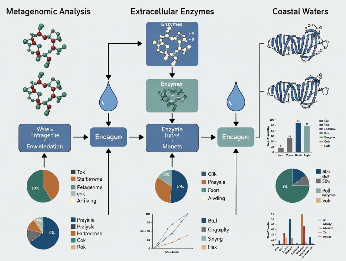
Visualization of Extracellular Enzyme Ecology
The following diagram illustrates the sources, pools, and ecological roles of extracellular enzymes in the marine environment, highlighting their connection to metagenomic analysis.
The Scientist's Toolkit: Research Reagent Solutions
This table outlines essential reagents, materials, and technologies for conducting research on extracellular enzymes in marine systems, with a focus on metagenomic-linked ecological studies.
Table 4: Essential Research Reagents and Materials for Marine EEA Studies
| Item | Specific Examples & Specifications | Primary Function in Research |
|---|---|---|
| Fluorogenic Substrates | 4-Nitrophenyl (pNP) or 4-Methylumbelliferyl (MUF)-linked analogs (e.g., MUF-phosphate, MUF-β-glucoside) [1] | Proxy substrates for measuring potential hydrolysis rates of specific enzyme classes (e.g., phosphatases, glucosidases). |
| Natural Polymer Substrates | Carboxymethyl cellulose (CMC), chitin, alginic acid, casein [1] | Measuring hydrolysis rates of environmentally relevant biopolymers to approximate in situ degradation. |
| Filtration Systems | 20-μm filters for pre-filtration; 0.22-μm polycarbonate membranes for separating cell-associated fractions; Tangential Flow Filtration (TFF) with 5-kDa membranes [1] | Concentrating dilute enzymes from large water volumes and separating dissolved from cell-associated enzyme fractions. |
| DNA Extraction Kits | Kits optimized for environmental samples (e.g., from filters); protocols including lysozyme and Proteinase K digestion [4] | Extracting high-quality microbial DNA from water or concentrated samples for subsequent metagenomic sequencing. |
| Metagenomic Sequencing Services/Platforms | Illumina NovaSeq (e.g., 2x151 bp chemistry) [4] | Determining the taxonomic and functional gene composition (e.g., CAZymes, peptidases) of the microbial community. |
| Bioinformatics Software & Databases | BBTools (BBDuk, bbmap), metaSPAdes assembler, Prodigal for gene prediction, NCBI protein database, KEGG [5] [4] | Processing raw sequencing data, assembling metagenomes, predicting genes, and annotating enzyme functions and pathways. |
Coastal waters are dynamic biochemical reactors where microbial communities play a pivotal role in nutrient cycling and organic matter degradation. Central to these processes are extracellular enzymes, including hydrolases, lipases, and phosphatases, which enable microorganisms to break down complex polymers into assimilable substrates. Metagenomic analysis of these enzymes provides a powerful lens for understanding microbial community function and ecological dynamics without the need for cultivation [6] [7]. This application note details the key methodologies and reagents for studying these critical enzyme classes within a metagenomic framework, providing researchers with standardized protocols for assessing microbial community functional potential in coastal ecosystems.
Key Enzyme Classes: Functions and Ecological Roles
Hydrolases and Lipases
Hydrolases catalyze the hydrolytic cleavage of ester bonds in the presence of water, and in low-water conditions can catalyze synthetic reactions like esterification and transesterification [6]. This enzyme class is characterized by a conserved catalytic triad of serine, aspartate (or glutamate), and histidine residues, with the catalytic serine embedded in the consensus motif Gly-X-Ser-X-Gly [6].
- Lipases vs. Esterases: While both are lipolytic enzymes, they differ in substrate specificity. Esterases hydrolyze short-chain fatty acid esters (<12 carbon atoms), while true lipases (EC 3.1.1.3) prefer long-chain fatty acid esters (≥12 carbon atoms) and often exhibit interfacial activation where a lid covering the active site opens at lipid interfaces [6].
- Bacterial lipolytic enzymes are classified into eight families (I-VIII) based on amino acid sequences and biological properties, with additional families discovered through metagenomic approaches [6].
Phosphatases
Phosphatases catalyze the liberation of orthophosphate from organophosphates through hydrolytic dephosphorylation [8]. They are crucial for phosphorus cycling in phosphorus-limited coastal environments [9].
- Classification: Based on optimum pH, phosphatases are categorized as alkaline phosphatases (AKP) or acid phosphatases (ACP). Based on substrate specificity, they include phosphomonoesterase, phosphodiesterase, and phosphotriesterase [8].
- Genetic Determinants: Key alkaline phosphatase encoding genes include
phoA(phosphomonoesterase in Bacteroidetes and Chloroflexi),phoDandphoX(target both phosphate monoesters and diesters in Proteobacteria, Actinobacteria, Bacteroidetes, and Cyanobacteria) [8].
Table 1: Key Enzyme Classes in Coastal Waters: Functions and Genetic Markers
| Enzyme Class | EC Number | Primary Function | Substrate Preference | Key Gene Markers |
|---|---|---|---|---|
| True Lipases | EC 3.1.1.3 | Hydrolysis of triacylglycerols | Long-chain fatty acid esters (≥12 C) | Families I-VIII (bacterial) |
| Esterases | EC 3.1.1.1 | Hydrolysis of carboxylic esters | Short-chain fatty acid esters (<12 C) | Families I-VIII (bacterial) |
| Alkaline Phosphatase | EC 3.1.3.1 | Organic phosphorus mineralization | Phosphate monoesters/diesters | phoA, phoD, phoX |
| Acid Phosphatase | EC 3.1.3.2 | Organic phosphorus mineralization | Phosphate monoesters | Various, less studied |
Quantitative Data on Environmental Responses
Environmental factors significantly influence enzyme activities and gene abundance in coastal waters. Microplastics and antibiotics pollution can alter microbial community structure and function.
- Microplastics Impact: A 60-day sediment simulation study showed that microplastics (PE, PP, PS, PVC, PET) significantly reduced total carbon (TC) and total nitrogen (TN) content, while enhancing alkaline phosphatase activity. They also inhibited ammonia assimilation and methane synthesis processes [10].
- Antibiotics Impact: Sulfamethoxazole (SMX) exposure increased phosphatase activity and elevated the abundance of antibiotic resistance genes (ARGs) including
sul1,sul2,dfrA, andermF[10]. - Phosphorus Source Influence: Microbial communities respond differently to phosphorus sources. Inorganic phosphates (IP) and cyclic-nucleoside-monophosphates (cNMP) supported the highest total organic carbon (TOC) removal efficiencies (64.8% and 52.3%, respectively). IP treatments encouraged Enterobacter, while cNMP treatments encouraged Aeromonas [8]. The abundance of
phoAandphoUgenes was higher in IP treatments, whereasphoDandphoXgenes dominated organophosphate (OP) treatments [8].
Table 2: Environmental Influences on Enzyme Activity and Microbial Community Structure
| Environmental Stressor | Impact on Enzyme Activity | Impact on Microbial Community/Genes | Experimental Conditions |
|---|---|---|---|
| Microplastics Mix (PE, PP, PS, PVC, PET) | Enhanced alkaline phosphatase activity; Reduced TC and TN | Inhibited ammonia assimilation & methane metabolism; Minimal impact on ARGs | Coastal sediments, 60-day exposure [10] |
| Antibiotic (Sulfamethoxazole) | Increased FDA hydrolase activity | Increased abundance of sul1, sul2, dfrA, ermF genes |
Coastal sediments, 60-day exposure [10] |
| Inorganic Phosphorus (IP) | Not specified | Higher abundance of phoA, phoU genes; Encouraged Enterobacter |
Activated sludge, 72h cultivation [8] |
| Organophosphorus (OP) | Not specified | Higher abundance of phoD, phoX genes |
Activated sludge, 72h cultivation [8] |
Experimental Protocols
Metagenomic DNA Extraction and Analysis from Coastal Sediments
Protocol Objective: To extract and analyze metagenomic DNA from coastal sediments for the identification of hydrolase, lipase, and phosphatase genes.
Materials & Reagents:
- Sediment samples from coastal regions (e.g., Liaodong Bay, Bohai Sea)
- DNA extraction kit (e.g., MO BIO PowerSoil DNA Isolation Kit)
- Phosphorus-free M9 medium for enrichment cultures [8]
- Phenotype Microarray plates (e.g., PM4A Microplate, Biolog Inc.) for testing phosphorus source utilization [8]
- PCR reagents and primers for target genes (e.g.,
phoD,phoX, lipase families) - High-throughput sequencing platform (e.g., Illumina)
Procedure:
- Sample Collection: Collect sediment cores from the desired coastal region. Store immediately at -80°C for DNA analysis or at 4°C for enrichment cultures.
- DNA Extraction: Extract total community DNA from 0.25-0.5 g of sediment using a commercial DNA isolation kit, following manufacturer's instructions.
- Enrichment Cultivation (Optional): For functional screening, inoculate sediment slurry into phosphorus-free M9 medium supplemented with different phosphorus sources (IP, NMP, cNMP, OP) as in PM4A Microplates. Incubate at in situ temperature (e.g., 30°C) for 3 days [8].
- Gene Amplification & Sequencing: Amplify target genes using degenerate primers. For lipases/esterases, target conserved regions around the catalytic triad. For phosphatases, use group-specific primers (e.g., for
phoD,phoX). - Sequencing & Bioinformatic Analysis: Perform high-throughput sequencing. Process reads via quality filtering, assembly/or binning, and predict open reading frames. Annotate genes against databases (e.g., NCBI NR, KEGG, COG) using BLAST-based searches.
- Quantitative Analysis: Quantify gene abundance via read mapping or perform qPCR for specific gene targets.
Measuring Alkaline Phosphatase Activity (APA) in Water and Sediment
Protocol Objective: To quantify alkaline phosphatase activity (APA) as a measure of microbial phosphorus acquisition effort.
Materials & Reagents:
- Artificial substrate: 4-Methylumbelliferyl phosphate (MUF-P) or p-Nitrophenyl phosphate (pNPP)
- Buffer: Tris-HCl (pH 8-9 for alkaline phosphatase)
- Fluorescence microplate reader or spectrophotometer
- Calibration standards (e.g., MUF or pNP)
Procedure:
- Sample Preparation: Filter water samples (0.2 µm) or create slurries with surface sediments and sterile-filtered water.
- Reaction Setup: Add artificial substrate (e.g., 200 µM MUF-P final concentration) to samples and controls (substrate blank, sample blank). Incubate in the dark at in situ temperature.
- Measurement: For MUF-P, measure fluorescence ( excitation ~365 nm, emission ~445 nm) at time zero and regularly over 1-3 hours. For pNPP, measure absorbance at 410 nm.
- Calculation: Calculate enzyme activity from the linear increase in product concentration over time, normalized to sample volume or chlorophyll-a content.
Visualizing the Workflow: From Sample to Functional Insight
The following diagram outlines the core metagenomic workflow for analyzing extracellular enzymes in coastal waters, from sample collection to data interpretation.
Metagenomic Analysis of Extracellular Enzymes
The Scientist's Toolkit: Research Reagent Solutions
Table 3: Essential Research Reagents for Metagenomic Enzyme Analysis
| Reagent / Material | Function / Application | Example Use Case |
|---|---|---|
| Phenotype Microarray (PM4A) | High-throughput profiling of microbial community utilization of 59 different phosphorus sources [8]. | Identifying preferential phosphorus sources (IP, cNMP, OP) and linking them to specific phosphatase gene abundance (phoA, phoX) [8]. |
| MUF/P substrates | Fluorogenic enzyme substrates (e.g., MUF-phosphate, MUF-acetate, MUF-fatty acid esters). | Quantifying hydrolytic enzyme activities (phosphatase, esterase) in environmental samples via fluorescence measurement [8]. |
| Phenol-Chloroform-Isoamyl Alcohol | Traditional method for high-quality DNA extraction from complex environmental matrices. | Extracting metagenomic DNA from coastal sediments for subsequent sequencing and functional gene analysis [10]. |
| Commercial DNA Extraction Kits (e.g., MO BIO PowerSoil) | Standardized protocol for efficient lysis and purification of community DNA from soils and sediments. | Obtaining high-quality, PCR-ready metagenomic DNA for amplicon or shotgun sequencing of hydrolase genes [10]. |
| Degenerate Primers | Amplification of diverse gene families (e.g., bacterial lipase families I-VIII) from metagenomic DNA. | Screening environmental DNA for novel lipolytic enzymes from uncultured microorganisms [6]. |
Spatial and Temporal Dynamics in Enzyme Distribution and Activity
This application note provides a detailed framework for investigating the spatial and temporal dynamics of extracellular enzyme activities in coastal marine environments, contextualized within a broader metagenomic analysis research thesis. Extracellular enzymes are functional components of marine microbial communities that catalyze the degradation of organic substrates, playing a critical role in nutrient remineralization and biogeochemical cycling [11]. In coastal waters, these enzymes exhibit significant variations across short temporal and spatial scales, directly influencing primary production and microbial loop dynamics [11] [12]. This document presents standardized protocols for assessing enzyme activities, data on observed dynamics, and essential methodological considerations for researchers investigating microbial ecology in coastal systems.
Temporal Dynamics of Extracellular Enzyme Activities
Observed Patterns of Variation
Temporal variability in extracellular enzyme activity occurs across multiple timescales, from diurnal to seasonal patterns. Research from the MICRO time series in Newport Pier, California, demonstrated that 34-48% of the variation in enzyme activity occurs at timescales shorter than 30 days [11]. Approximately 28-56% of the variance in related parameters including nutrient concentrations, chlorophyll levels, and ocean currents also occurs on these short timescales [11].
Diurnal fluctuations can be particularly dramatic, with studies in Mediterranean coastal waters showing that α- and β-glucosidase activities varied by 0-100% within 24-hour periods [12]. In contrast, aminopeptidase activities exhibited weaker diurnal variation but substantial day-to-day changes comparable in magnitude to seasonal variations [12].
Seasonal patterns are enzyme-specific, with β-glucosidase showing repeatable seasonal patterns correlated with spring phytoplankton blooms in the Southern California Bight [11]. These temporal dynamics reflect rapid responses of microbial communities to environmental triggers including phytoplankton blooms, upwelling events, wind patterns, and rainfall [11].
Key Environmental Correlations
Statistical analyses reveal significant relationships between enzyme activities and environmental parameters:
- Nutrient correlations: Most enzyme activities show weak but positive correlations with nutrient concentrations (r = 0.24-0.31) [11].
- Upwelling influence: Enzyme activities correlate with upwelling dynamics (r = 0.29-0.35) [11].
- Temperature effects: Seagrass coverage and aboveground biomass show significant positive correlations with temperature in coastal ecosystems [13].
- Oxygen and COD relationships: Seagrass density demonstrates significant positive correlation with dissolved oxygen (DO) but significant negative correlation with chemical oxygen demand (COD) [13].
Table 1: Temporal Variation Patterns in Coastal Enzyme Activities
| Enzyme | Short-term Variation (<30 days) | Diurnal Variation | Seasonal Pattern | Primary Correlates |
|---|---|---|---|---|
| β-glucosidase | 34-48% of total variation [11] | 0-100% fluctuation observed [12] | Elevated in spring blooms [11] | Phytoplankton blooms, upwelling [11] |
| Aminopeptidase | Similar magnitude to seasonal scale [12] | Weak diurnal variation [12] | Not specifically reported | Nutrient concentrations [11] |
| α-glucosidase | Not specifically quantified | 0-100% fluctuation observed [12] | Not specifically reported | Not specified in search results |
| Alkaline Phosphatase | Part of <30 day variation cohort [11] | Not specifically reported | Not specifically reported | Phosphate limitation [11] |
Spatial Distribution and Partitioning of Enzymes
Particle-Associated versus Free Enzymes
A crucial aspect of spatial distribution involves the partitioning of enzyme activities between particulate and dissolved phases. Research indicates distinct patterns across different enzyme types:
- Particle-dominated enzymes: For β-glucosidase and leucine aminopeptidase, most activity is bound to particles [11]. This localization potentially benefits enzyme producers by directly coupling hydrolysis with nutrient uptake [11].
- Freely dissolved enzymes: In contrast, 81.2% of alkaline phosphatase and 42.8% of N-acetyl-glucosaminidase activity occurs in the freely dissolved phase [11]. This distribution suggests that phosphorus release may occur throughout the water column rather than being concentrated on particles [11].
The proportion of enzymes in the dissolved phase can show extreme variability, with studies finding 0-100% of both α- and β-glucosidase in the dissolved phase within 24-hour periods [12]. Consistently high proportions of all three examined enzymes (α-glucosidase, β-glucosidase, and aminopeptidase) were found in the dissolved phase on seasonal scales [12].
Depth-Related Variations
Extracellular enzyme activities typically exhibit weak negative dependency with depth [12]. Activities are generally highest in surface waters where organic matter inputs from phytoplankton production and terrestrial sources are most abundant, gradually decreasing with depth due to reduced substrate availability and microbial biomass.
Experimental Protocols for Enzyme Activity Assessment
Sampling Methodology
Collection Protocol:
- Frequency: Sample up to three times per week to capture short-term variability [11].
- Timing: Collect between 08:00 and 09:00 local time to minimize diurnal variation effects [11].
- Location: Sample from surface waters (depth <0.5 m) using clean collection vessels [11].
- Replication: Collect two independent surface water samples on each date [11].
Sample Processing:
- Transport: Transport samples to laboratory at 15-20°C within 30 minutes of collection [11].
- Filtration: Process samples through sequential filtration:
- Bulk seawater: Unfiltered sample
- <2.7 μm fraction: Filtrate through GF/D filters
- <0.2 μm fraction: Filtrate through polyethersulfone syringe filters [11]
- Preservation: Freeze subsamples (-20°C) for subsequent nutrient analysis [11].
Fluorometric Enzyme Assays
Reaction Setup:
- Platform: Conduct assays in black 96-well microplates to minimize light scattering [11].
- Temperature: Run assays at 20°C to simulate environmental conditions [11].
- Duration: Incubate for 1.5 hours, ensuring fluorescence increase remains linear [11].
- Controls: Include appropriate controls:
- Sample blanks: 200 μL sample + 50 μL DI water
- Substrate blanks: 200 μL filtered, autoclaved seawater + 50 μL substrate solution [11]
Reaction Mixture:
- Add 50 μL substrate solution to 200 μL sample to initiate reaction [11].
- Include standards (10 μM 4-methyl-umbelliferone or 10 μM 7-amino-4-methylcoumarin) for fluorescence quantification and quenching correction [11].
- Gently tap microplates to mix solutions before reading [11].
Measurement Parameters:
- Read fluorescence at time zero and at 0.5-hour intervals [11].
- Use excitation/emission wavelengths of 360 nm/460 nm [11].
- Calculate enzyme activities based on product standard curves after accounting for quenching [11].
Table 2: Standardized Enzyme Assay Conditions
| Enzyme | Function | Substrate | Final Substrate Concentration | Fluorophore |
|---|---|---|---|---|
| Alkaline Phosphatase (AP) | Hydrolyzes phosphate monoesters | 4-MUB-phosphate | 200 μmol L⁻¹ | Methylumbelliferone (MUB) |
| β-glucosidase (BG) | Releases glucose from polysaccharides | 4-MUB-β-d-glucopyranoside | 40 μmol L⁻¹ | Methylumbelliferone (MUB) |
| Leucine Aminopeptidase (LAP) | Hydrolyzes polypeptides | l-leucine-AMC | 80 μmol L⁻¹ | 7-amido-4-methylcoumarin (AMC) |
| N-acetyl-glucosaminidase (NAG) | Releases N-acetyl-glucosamine from chitin | 4-MUB-N-acetyl-β-d-glucosaminide | 80 μmol L⁻¹ | Methylumbelliferone (MUB) |
The Scientist's Toolkit: Essential Research Reagents
Table 3: Key Research Reagents for Enzyme Activity Studies
| Reagent/Category | Specific Examples | Function/Application |
|---|---|---|
| Fluorogenic Substrates | 4-MUB-phosphate, 4-MUB-β-d-glucopyranoside, l-leucine-AMC, 4-MUB-N-acetyl-β-d-glucosaminide [11] | Enzyme activity measurement through fluorescent product generation |
| Filtration Materials | 2.7 μm GF/D filters, 0.2 μm polyethersulfone syringe filters [11] | Size fractionation of enzyme activities (particulate vs. dissolved) |
| Detection Instrumentation | Microplate reader (e.g., BioTek Synergy 4) [11] | Fluorometric measurement with 360 nm excitation/460 nm emission |
| Reference Standards | 4-methyl-umbelliferone (MUB), 7-amino-4-methylcoumarin (AMC) [11] | Quantification of reaction products and correction for fluorescence quenching |
| Sample Containers | Acid-washed polypropylene bottles, pre-rinsed scintillation vials [11] | Prevention of sample contamination during collection and processing |
Integrated Workflow for Spatiotemporal Enzyme Analysis
Data Interpretation and Integration with Metagenomic Analysis
Connecting Enzyme Activities to Microbial Community Dynamics
The spatial and temporal dynamics of extracellular enzymes provide crucial functional insights that complement metagenomic analyses of microbial community structure. Integrating these datasets enables researchers to:
- Link functional capacity with expression: Connect identified enzyme-coding genes in metagenomes with actual enzyme activities measured across temporal and spatial gradients [11].
- Identify key responding taxa: Correlate specific patterns of enzyme activity with shifts in microbial community composition from metagenomic data [11].
- Uncover regulatory mechanisms: Distinguish between changes in microbial abundance versus changes in per-cell enzyme production in response to environmental triggers [11].
Methodological Considerations
- Potential activity measurements: Note that standardized assays with artificial substrates measure potential enzyme activities rather than in situ rates, reflecting enzyme concentrations more than immediate environmental function [11].
- Substrate specificity limitations: Fluorogenic substrate analogs may not fully capture the diversity of natural substrates, potentially overlooking activities toward complex natural polymers [11].
- Integration challenges: Spatial and temporal mismatches between enzyme activity measurements (instantaneous) and metagenomic samples (snapshot in time) require careful experimental design to enable meaningful correlation analyses.
Linking Enzyme Profiles to Biogeochemical Cycling (Carbon, Nitrogen, Phosphorus)
Within marine ecosystems, microbial extracellular enzymes initiate the critical first step in the biogeochemical cycling of organic matter by hydrolyzing complex macromolecules into smaller, bioavailable substrates [1]. These enzymes are fundamental to the microbial loop, responsible for transforming an estimated 50% of surface water primary production [1]. In the context of metagenomic analysis of coastal waters, linking specific enzyme profiles to their biogeochemical functions provides a mechanistic understanding of organic matter processing. This application note details standardized protocols for measuring extracellular enzyme activity (EEA) and connecting these profiles to carbon (C), nitrogen (N), and phosphorus (P) cycling, enabling researchers to decipher the functional state of microbial communities.
Key Enzymes in Biogeochemical Cycling
The measurement of targeted enzyme activities provides a functional readout of microbial nutrient demands and their role in elemental cycling. The table below summarizes the key enzymes involved in the major biogeochemical pathways.
Table 1: Key Microbial Extracellular Enzymes and Their Biogeochemical Functions
| Element Cycle | Enzyme | Primary Function | Significance |
|---|---|---|---|
| Carbon | β-Glucosidase | Cleaves cellobiose to glucose [14] | Key step in cellulose degradation [1] |
| Phenol Oxidase (PHO) | Degrades recalcitrant aromatic compounds & lignin [14] | Regulates carbon storage via the "enzymic latch" mechanism [14] | |
| Nitrogen | Protease/Peptidase | Degrades proteins into amino acids [1] | Makes organic nitrogen bioavailable |
| Chitinase | Hydrolyzes chitin (N-acetylglucosamine polymer) [1] | Accesses nitrogen stored in fungal cell walls & exoskeletons | |
| Phosphorus | Phosphatase (e.g., PhoD) | Liberates inorganic phosphate from organic esters [14] [15] | Indicates phosphorus limitation; critical for P bioavailability [14] |
Experimental Protocols
Seawater Sampling and Fractionation
Objective: To collect and process water samples for the separation of dissolved and cell-associated enzyme fractions. Materials:
- Submersible pump or Niskin bottles
- Prefilters (20-μm pore size, Millipore)
- Polycarbonate membranes (0.22-μm pore size, Millipore)
- Tangential Flow Filtration (TFF) system with 5000-Dalton membranes (Spectrum Laboratories)
- Syringe filters (0.22-μm polypropylene, Millipore)
Procedure:
- Collection: Collect surface water (e.g., at ~2 m depth) using a submersible pump or Niskin bottles [1].
- Prefiltration: Pass ~10 L of seawater through a 20-μm filter to remove large particles and organisms [1].
- Concentration: Concentrate the prefiltered seawater from ~10 L to 50 mL using a TFF system. Complete the process within 2 hours to preserve enzyme integrity [1].
- Fractionation: To separate dissolved from cell-associated enzymes, gently filter 25 mL of the concentrated sample through a 0.22-μm syringe filter.
- The filtrate contains dissolved enzymes.
- The retentate on the filter contains cell-associated enzymes.
- Storage: Store samples for EEA measurement at 4°C and begin assays within 1 hour. For DNA analysis, preserve filters in liquid nitrogen and store at -80°C [1].
Measuring Extracellular Enzyme Activity (EEA)
Objective: To quantify the potential hydrolysis rates of various organic substrates using fluorogenic or chromogenic analogs. Materials:
- Substrate analogs: 4-Methylumbelliferyl (MUF)- or 4-Nitrophenyl (PNP)- labeled compounds (e.g., MUF-phosphate, PNP-β-D-glucoside, L-Leucine-7-amido-4-methylcoumarin)
- Incubation thermostat
- Microplate reader or spectrophotometer
- Buffers (e.g., TRIS, pH ~8 for seawater)
Procedure:
- Substrate Preparation: Prepare saturating concentrations of substrate analogs in an appropriate buffer. Using high substrate concentrations ensures the measurement of potential enzyme activity rather than in situ rates [1].
- Assay Setup: Combine sample (either dissolved or total fraction) with substrate solution in a microplate or test tube. Include controls with killed samples (e.g., addition of trichloroacetic acid) [1].
- Incubation: Incubate assays at in situ temperature (e.g., 25°C) or at elevated temperatures (e.g., 35°C) to test the effect of warming [1].
- Measurement:
- For fluorogenic substrates (MUF, AMC), measure fluorescence over time (e.g., excitation 365 nm, emission 455 nm).
- For chromogenic substrates (PNP), measure absorbance (e.g., 410 nm for p-nitrophenol).
- Calculation: Calculate hydrolysis rates from the linear increase in fluorescence or absorbance over time, using standard curves for the fluorescent or chromogenic product (e.g., MUF or p-nitrophenol). Rates are typically expressed as nmol L⁻¹ h⁻¹.
Metagenomic Analysis of Enzyme-Producing Taxa
Objective: To identify microbial clades with the genetic potential to produce target extracellular enzymes. Materials:
- DNA extraction kit
- PCR reagents
- Primers for functional genes (e.g., chiA for chitinase, phoD for phosphatase) and 16S rRNA genes
- High-throughput sequencing platform
Procedure:
- DNA Extraction: Extract genomic DNA from filters preserving the microbial community.
- Targeted Amplification/Sequencing: Amplify and sequence either:
- Bioinformatic Analysis:
- Process sequences (quality filtering, OTU/ASV picking).
- Assign taxonomy using reference databases (e.g., SILVA, Greengenes).
- Identify known enzyme-producing clades (e.g., Bacteroidetes, Planctomycetes, Chloroflexi, Roseobacter, Alteromonas, and Pseudoalteromonas) in your dataset [1].
- Integration: Statistically correlate the abundance of specific taxonomic groups or functional genes with measured EEA patterns to link genetic potential to ecosystem function.
Data Integration and Visualization
Integrating enzyme activity data with microbial community and environmental parameters reveals the functional state of the ecosystem. The following workflow diagram outlines the complete experimental pipeline from sampling to data integration.
Conceptual Framework for Data Interpretation
The integrated data can be interpreted through the framework of ecoenzymatic stoichiometry, which links extracellular enzyme activities to microbial resource allocation and nutrient limitation [14]. The following diagram illustrates the logical relationship between environmental conditions, microbial community response, and the resulting biogeochemical outcomes.
The Scientist's Toolkit: Research Reagent Solutions
Table 2: Essential Research Reagents and Materials for EEA Studies
| Item | Function/Application | Key Characteristics |
|---|---|---|
| Fluorogenic Substrates (e.g., MUF-/AMC-labeled) [1] | Quantifying hydrolysis rates of specific polymers (e.g., MUF-phosphate for phosphatase). | High sensitivity; allows measurement of low activity in dilute seawaters. |
| Chromogenic Substrates (e.g., PNP-labeled) | Alternative for activity measurement via absorbance. | Less sensitive than fluorogenic assays but widely used. |
| Tangential Flow Filtration (TFF) System [1] | Concentrating dilute extracellular enzymes from large water volumes (>10 L). | 5000-Dalton membranes; gentle on enzyme integrity. |
| Polycarbonate Membranes (0.22 μm) [1] | Fractionating cell-associated vs. dissolved enzymes; collecting biomass for DNA. | Low protein binding; sterile. |
| Primers for Functional Genes (e.g., phoD, chiA) [15] | Profiling microbial communities with genetic potential for enzyme production. | Targets genes encoding specific extracellular enzymes. |
This application note provides a standardized framework for linking microbial enzyme profiles to biogeochemical cycling in coastal waters. The detailed protocols for sample processing, activity measurements, and integrated metagenomic analysis empower researchers to move beyond correlative studies toward a mechanistic, function-based understanding of marine ecosystems. Applying these methods allows for the assessment of how environmental changes, such as nutrient inputs and warming, affect the fundamental microbial processes that drive carbon, nitrogen, and phosphorus transformations.
Within the framework of a broader thesis on the metagenomic analysis of extracellular enzymes in coastal waters, this application note addresses a fundamental aspect: identifying the dominant microbial taxa responsible for producing these crucial biocatalysts. In aquatic ecosystems, the initial step of organic matter degradation is primarily mediated by extracellular enzymes secreted by bacteria. Understanding the phylogenetic identity of these key enzyme producers is essential for deciphering microbial community function, ecological niche partitioning, and biogeochemical cycling in coastal environments. This document synthesizes recent research findings to delineate the principal enzyme-producing phyla, quantify their contributions, and provide standardized protocols for their study, serving as a resource for researchers and industrial applications in biotechnology and drug development.
Empirical studies from diverse coastal environments, including mudflats, seawater, and marine sediments, consistently identify three bacterial phyla as the dominant producers of industrial extracellular enzymes: Proteobacteria, Firmicutes, and Bacteroidetes [17] [18] [19]. The distribution and enzymatic strengths of these phyla are summarized in the table below.
Table 1: Dominant Enzyme-Producing Phyla in Coastal Marine Environments
| Phylum | Relative Abundance & Prevalence | Principal Enzyme Classes Produced | Notable Genera and Their Enzymatic Strengths |
|---|---|---|---|
| Proteobacteria | Often the most abundant phylum; frequently dominates cultured isolates and metagenomic sequences [17] [20]. | Peptidases, lipases, amylases [17] [21]. | Vibrio spp. (high lipase, amylase, protease) [17]; Pseudomonas, Shewanella (proteases, lipases) [17] [18]; Bacillus (proteases, amylases) [18]. |
| Firmicutes | Highly prevalent in culture-dependent studies from sediments and marine organisms [17] [22] [18]. | Proteases, amylases, phytases [22] [18]. | Bacillus spp. (dominant protease-producers) [18]; Solibacillus, Chryseomicrobium (amylase, lipase, protease) [17]. |
| Bacteroidetes | Major contributor in metagenomic studies; key in polysaccharide degradation [21] [20] [23]. | Carbohydrate-Active Enzymes (CAZymes), including those targeting laminarin, cellulose, and other complex polysaccharides [21] [23]. | Bacteroides, Alistipes, Prevotella (increased in specific metabolic niches) [24]; Tenacibaculum (amylase, lipase, protease) [17]. |
The quantitative output of these taxa is significant. For instance, one study screening 163 marine bacterial isolates found that 88.3% produced lipase, 68.7% produced amylase, and 68.7% produced protease [17] [19]. Furthermore, genetic analysis reveals that the gene pool for organic matter degradation is partitioned among these phyla: Bacteroidota are primary contributors to secretory CAZymes, while Gammaproteobacteria contribute more to secretory peptidases, and Alphaproteobacteria to specific transporters like the ATP-binding cassette (ABC) transporters [21].
Experimental Protocols for Identification and Characterization
A comprehensive understanding of enzyme-producing taxa requires integrating both culture-dependent and culture-independent methods. The following protocols detail standardized approaches for these analyses.
Protocol 1: Culture-Dependent Screening of Hydrolytic Enzyme-Producing Bacteria
This protocol is designed for the isolation and initial functional screening of culturable enzyme-producing bacteria from coastal sediment and water samples [17] [18].
Materials and Reagents
- Marine Agar 2216 & Marine Broth 2216: For general cultivation and growth of marine bacteria [17].
- Screening Agar Plates: Base agar prepared with artificial seawater, supplemented with specific substrates:
- Protease Screening: Add 1% casein and 2% gelatin. Protease activity is indicated by a clear halo zone around colonies [18].
- Amylase Screening: Add 1% soluble starch. Detect activity by flooding plates with iodine solution; a clear zone indicates starch hydrolysis [17].
- Lipase Screening: Use Spirit Blue Agar with lipid sources. Hydrolysis is indicated by a halo zone [17].
- Artificial Seawater: Synthetic sea salt dissolved at 3% (w/v) concentration [18].
- DNA Extraction Kit: e.g., DNeasy Blood and Tissue Kit for subsequent molecular identification [17].
Procedure
- Sample Collection: Aseptically collect coastal mud or water samples. Serially dilute samples (e.g., to 10⁻⁶) using sterile artificial seawater [18].
- Plating and Incubation: Spread 100 µL of each dilution onto the specialized screening agar plates. Incubate at temperatures relevant to the sample environment (e.g., 20°C or 35°C) until visible hydrolytic zones form [17] [18].
- Isolation and Purification: Select colonies surrounded by a hydrolytic zone and repeatedly streak them on fresh screening medium at least three times to obtain pure cultures [18].
- Enzyme Activity Quantification: Measure the hydrolytic zone diameter (in mm) and assign a score (e.g., 0: no zone; 3: zone ≥21 mm) to semi-quantify enzyme production strength [17].
- Molecular Identification:
- Extract genomic DNA from pure cultures.
- Amplify the 16S rRNA gene using universal primers (e.g., 27F: 5′-AGAGTTTGATCCTGGCTCAG-3′ and 1492R: 5′-GGTTACCTTGTTACGACTTC-3′) via colony PCR [17] [18].
- Sequence the amplicons and identify isolates by comparing sequences with databases like GenBank using the BLAST algorithm or the EzTaxon server [17].
Protocol 2: Culture-Independent Metagenomic Analysis of Enzyme Pathways
This protocol outlines the steps for assessing the functional potential of microbial communities via metagenomic sequencing, bypassing cultural biases [21] [20].
Materials and Reagents
- DNA Extraction Buffers: Lysis buffer containing Tris, EDTA, NaCl, and CTAB for efficient cell disruption from environmental samples [20].
- Phenol/Chloroform/Isoamyl Alcohol: For purification of metagenomic DNA.
- Illumina Sequencing Platform: e.g., HiSeq 2500 for high-throughput sequencing [20].
- Bioinformatics Software:
- SOAPnuke/MEGAHIT/IDBA: For sequence quality control and de novo assembly [20].
- MetaGeneMark: For predicting open reading frames (ORFs) from assembled contigs [20].
- DIAMOND/KEGG/CAZy: Tools and databases for functional annotation of predicted genes against KEGG, CAZy, and other specialized databases [21] [20].
Procedure
- Metagenomic DNA Extraction: From 10g of humus or sediment, extract DNA using a combination of chemical lysis (SDS, CTAB), enzymatic treatment (proteinase K), and organic purification (phenol-chloroform). Precipitate DNA with isopropanol, wash, and resuspend [20].
- Library Preparation and Sequencing: Prepare a paired-end sequencing library from the high-quality DNA and sequence on an Illumina platform to generate 150 bp reads [20].
- Bioinformatic Processing:
- Quality Control: Filter raw reads to remove adapters and low-quality sequences.
- Assembly: Assemble quality-filtered reads into contigs using assemblers like MEGAHIT with a range of k-mer sizes [20].
- Gene Prediction and Annotation: Predict ORFs from contigs. Functionally annotate the ORFs by alignment against public databases (KEGG, CAZy) to identify genes encoding extracellular enzymes (e.g., peptidases, CAZymes) and transporters (e.g., TonB-dependent transporters) [21] [20].
- Taxonomic Assignment: Assign taxonomy to contigs or ORFs by comparing them with reference databases to link enzymatic functions with phylogenetic identity [20].
The Scientist's Toolkit: Key Research Reagents
Table 2: Essential Reagents and Kits for Studying Enzyme-Producing Microbes
| Reagent / Kit Name | Function / Application | Key Features |
|---|---|---|
| Marine Agar/Broth 2216 | Cultivation of heterotrophic marine bacteria. | Standardized nutrient medium mimicking seawater. |
| DNeasy Blood & Tissue Kit | Extraction of high-quality genomic DNA from bacterial pure cultures. | Silica-membrane technology for purity and yield. |
| OMEGA Soil DNA Kit | Extraction of metagenomic DNA from complex environmental samples like sediment. | Effective for difficult-to-lyse cells and inhibitor removal. |
| MyTaq Mix | PCR amplification of 16S rRNA genes for phylogenetic identification. | Pre-mixed, optimized for robustness with complex templates. |
| Spirit Blue Agar / Starch Agar | Selective screening for lipolytic and amylolytic bacterial isolates. | Contains specific substrates for visual detection of enzyme activity. |
Integrated Workflow for Analysis of Enzyme-Producing Taxa
The following diagram illustrates the logical relationship and workflow between the two primary methodological approaches described in the protocols.
In coastal aquatic ecosystems, the microbial processing of organic matter is a fundamental driver of biogeochemical cycles. This process is initiated by extracellular enzymes produced by heterotrophic microbial communities, which hydrolyze complex organic polymers into smaller, assimilable molecules [21] [25]. The expression and activity of these enzymes are not static; they are dynamically regulated by key environmental drivers, including nutrient availability, temperature, and dissolved oxygen concentrations. Understanding these relationships is critical for predicting organic matter turnover and is a core component of metagenomic analyses of coastal waters. This Application Note details the experimental protocols for quantifying these relationships and their implications for microbial ecology and biogeochemical modeling.
Key Environmental Drivers and Quantitative Effects
The activity of microbial extracellular enzymes exhibits distinct and quantifiable responses to changes in the ambient environment. The table below summarizes the documented effects of specific environmental factors on key enzyme activities, serving as a reference for interpreting experimental results.
Table 1: Environmental Drivers of Extracellular Enzyme Activity (EEA) in Aquatic Systems
| Environmental Driver | Measured Effect on Enzyme Activity | Specific Enzymes / Systems Affected | Study Context |
|---|---|---|---|
| Temperature | Increase from 25°C to 35°C raised hydrolysis rates. | Polysaccharide hydrolases (e.g., for CMC, chitin, alginic acid) and protease. | Northern Chinese Marginal Seas [25] |
| Dissolved Organic Carbon (DOC) | Positive association with geographic distribution of EEA; higher concentrations correlated with higher inshore enzyme activity. | Phosphatase, β-glucosidase, protease. | Northern Chinese Marginal Seas; Neuse and Tar-Pamlico Rivers [26] [25] |
| Nutrient Availability | Microbial community nutrient demands influence enzymatic profiles; phosphorous limitation can stimulate phosphatase activity. | Phosphatase, peptidases, polysaccharide hydrolases. | Neuse and Tar-Pamlico Rivers [26] |
| Organic Matter Substrate Type | All tested substrates (polymers and oligomers) were hydrolyzed, but at different rates. Hydrolysis not strictly limited by molecule size. | Enzymes targeting CMC, chitin, alginic acid, casein, and their oligomers. | Northern Chinese Marginal Seas [25] |
| Salinity & Hydrology | Considerable spatiotemporal variability in EEA; hurricane-induced discharge led to persistent DOC maxima and stimulated bacterial production. | β-glucosidase, leucine aminopeptidase, phosphatase. | Neuse and Tar-Pamlico Rivers [26] |
Experimental Protocols
Protocol 1: Field Sampling and Metagenomic Analysis of Enzyme-Transporter Coupling
This protocol is designed to investigate the genetic potential for organic matter degradation in coastal bacterial communities, as revealed by metagenomic sequencing.
1. Sample Collection:
- Collect coastal water samples over a time-series (e.g., 22-day period) from multiple depths to capture temporal and spatial variability [21].
- Filter water samples through appropriate pore-size filters (e.g., 0.22 μm) to capture microbial biomass onto the filter for DNA extraction.
2. DNA Extraction and Metagenomic Sequencing:
- Perform genomic DNA extraction from the filters using a commercial soil or water DNA extraction kit.
- Prepare metagenomic libraries and sequence using an Illumina or similar high-throughput sequencing platform.
3. Bioinformatic Analysis:
- Assembly and Binning: Process raw sequencing reads to assemble contigs and bin them into Metagenome-Assembled Genomes (MAGs). A target of 163 MAGs, as in the cited study, provides robust data for correlation analysis [21].
- Gene Annotation: Annotate genes in the assembled contigs and MAGs against functional databases to identify key genes:
- Carbohydrate-Active Enzymes (CAZymes): Use dbCAN2 or similar tools.
- Peptidases: Use MEROPS database.
- Transporters: Identify genes for TonB-dependent transporters (TBDTs) and ATP-binding cassette (ABC) transporters.
- Statistical Correlation: At both the community-wide and MAG-specific levels, calculate correlation coefficients (e.g., Pearson's) between the abundance of extracellular enzyme genes (CAZymes, peptidases) and transporter genes (TBDTs, ABC transporters). This reveals potential coregulation and functional linkages [21].
Protocol 2: Measuring In-situ Extracellular Enzyme Activity (EEA)
This protocol measures the actual hydrolysis rates of organic matter, providing a ground-truthed measure of microbial functional response.
1. Water Sampling and Pre-processing:
- Collect surface water (e.g., from 2 m depth) using a submersible pump or Niskin bottles [26] [25].
- Pre-filter water through a 20-μm mesh to remove large particles and organisms.
- For total EEA, use the pre-filtered water directly. For dissolved EEA, further filter a portion through a 0.22-μm syringe filter to remove cells while retaining dissolved enzymes [25].
2. Enzyme Activity Assay via Substrate Hydrolysis:
- Substrate Preparation: Prepare a panel of fluorogenic or chromogenic substrate proxies. Common substrates include:
- 4-Methylumbelliferyl (MUF)- or 4-Nitrophenyl (PNP)- labeled derivatives (e.g., MUF-β-glucoside for β-glucosidase, MUF-phosphate for phosphatase, L-leucine-7-amido-4-methylcoumarin for protease) [26].
- Fluoresceinamine-labeled polysaccharides (e.g., carboxymethyl cellulose (CMC), chitin, alginic acid) and proteins (e.g., casein) to measure hydrolysis of real polymers [25].
- Incubation:
- Add substrates to seawater samples (total and dissolved) and incubate in the dark.
- Conduct assays at in-situ temperature (e.g., 25°C) and at a higher temperature (e.g., 35°C) to assess temperature sensitivity [25].
- Run controls with autoclaved or filtered sample to account for non-enzymatic hydrolysis.
- Measurement:
- For MUF/PNP substrates, measure fluorescence/absorbance at regular intervals using a plate reader. The increase in signal is proportional to hydrolysis rate.
- For polymer substrates, hydrolysis can be quantified by the increase in fluorescence as the labeled fragment is released, or by size-exclusion chromatography to detect the breakdown products [25].
3. Data Analysis:
- Calculate hydrolysis rates (nmol L⁻¹ h⁻¹ or μg L⁻¹ h⁻¹) from the linear increase of product over time.
- Correlate EEA rates with concurrently measured physicochemical parameters (temperature, DOC, nutrient concentrations, dissolved oxygen) to identify key environmental drivers [26] [25].
Visualization of Environmental-Microbial Interactions
The following diagram illustrates the logical and mechanistic relationships between environmental drivers, microbial genetic regulation, and the resulting biogeochemical outcomes in coastal waters.
Environmental Drivers Shape Microbial Enzyme Expression. This workflow diagrams how abiotic factors influence microbial genomics and metabolism, leading to biogeochemical outcomes like organic matter remineralization. Key interactions include the coupling between enzyme and transporter gene expression, a critical link identified via metagenomics [21].
The Scientist's Toolkit: Research Reagent Solutions
The following table lists essential materials and reagents required for the experimental protocols described in this note.
Table 2: Essential Research Reagents and Materials for EEA and Metagenomic Studies
| Reagent/Material | Function/Application | Key Considerations |
|---|---|---|
| Fluorogenic Substrate Proxies (e.g., MUF/AMC derivatives) | Quantifying hydrolysis rates of specific enzyme classes (e.g., glucosidases, phosphatases, peptidases) [26]. | Select substrates relevant to the organic matter pool (e.g., algal polysaccharides). Include a negative control. |
| Labeled Biopolymers (e.g., Fluoresceinamine-labeled CMC, Chitin) | Measuring hydrolysis rates of ecologically relevant polymers, not just proxies [25]. | Allows comparison of hydrolysis rates between polymers and their oligomers. |
| Tangential Flow Filtration (TFF) System | Concentrating extracellular enzymes from large water volumes (e.g., 10L to 50mL) to enable detection of activity on natural polymers [25]. | Use membranes with appropriate molecular weight cut-offs (e.g., 5 kDa). |
| Polycarbonate Membranes (0.22 μm) | Concentrating microbial biomass from water samples for subsequent DNA extraction and metagenomic analysis [21]. | Ensure sterile and nuclease-free conditions for DNA work. |
| DNA Extraction Kit | Isolating high-quality metagenomic DNA from environmental filters. | Optimized for environmental samples (soil, water) to overcome inhibitors. |
| Functional Annotation Databases (dbCAN2, MEROPS) | Bioinformatic annotation of CAZyme, peptidase, and transporter genes from metagenomic data [21]. | Use curated databases and set appropriate E-value cutoffs for homology searches. |
The expression and activity of microbial extracellular enzymes are powerfully shaped by the interplay of nutrients, temperature, and oxygen. Metagenomic approaches reveal the genetic potential and coupling of degradation pathways, while direct activity measurements capture the realized functional response of the community to environmental gradients. The protocols and data presented here provide a framework for researchers to systematically investigate these relationships, ultimately leading to a more predictive understanding of organic matter cycling in dynamic coastal waters.
Advanced Metagenomic Workflows: From Sampling to Functional Annotation
Sample Collection Strategies Across Coastal Gradients and Depths
The reliability of metagenomic data in coastal enzyme research is fundamentally constrained by the initial sample collection strategy. The dynamic interface of coastal environments, characterized by steep physical, chemical, and biological gradients, demands meticulous planning and execution of sampling protocols to ensure representative and uncontaminated samples. This document provides detailed application notes and protocols for designing and implementing sample collection strategies across coastal gradients and depths, specifically tailored for subsequent metagenomic analysis of extracellular enzymes. The objective is to equip researchers with standardized methodologies that enhance data comparability, minimize technical artifacts, and support robust ecological inferences regarding microbial community function in coastal waters.
Understanding the Sampling Environment
Coastal regions are transition zones where environmental parameters shift dramatically over small spatial and temporal scales. Recognizing these gradients is the first step in designing a statistically sound sampling plan.
Key Gradients and Their Implications
- Salinity Gradients: Salinity acts as a critical environmental filter on microbial communities [27] [28]. Cross-shore salinity gradients, often driven by riverine freshwater input, create distinct niches that host phylogenetically and functionally diverse microbial populations. Sampling must capture this variation to avoid biased functional profiles.
- Nutrient and Chemical Gradients: Land-based sources of pollution, including dissolved nitrogen, phosphorus, and silicate, can create strong water quality gradients that influence microbial metabolism and extracellular enzyme production [29]. As evidenced in Aua Reef, American Samoa, these gradients are measurable and can correlate with benthic community structure.
- Particulate and Organic Matter Gradients: The concentration of small microplastics (SMPs) and other particulates often decreases from the coastline toward the open ocean [30]. Similarly, the quality and quantity of organic matter, which drives extracellular enzyme activity (EEA), vary significantly with distance from shore and depth [31] [32].
Temporal Dynamics
Extracellular enzyme activities in coastal environments are highly dynamic. A multi-year time-series study in Southern California found that 34–48% of the variation in enzyme activity occurred at timescales of less than 30 days, influenced by short-term events like phytoplankton blooms, upwelling, and rainfall [33]. Sampling designs must therefore account for diel, tidal, and seasonal cycles to accurately capture the metabolic potential of the microbial community.
Sampling Strategies and Technologies
The choice of sampling technology is paramount for preserving the integrity of samples intended for sophisticated metagenomic analysis. The selection depends on the target sample type (water, sediment), depth, and required preservation state.
Water Sampling Techniques and Equipment
Table 1: Comparison of Seawater Sampling Technologies for Metagenomic Studies
| Technology/Sampler | Principle | Key Advantages | Key Limitations | Best Use Cases for Metagenomics |
|---|---|---|---|---|
| Niskin/Rosette Sampler [34] | Penetration form; cylindrical sampling chambers with end caps triggered at target depth. | Can house multiple chambers (e.g., 12-24); discrete depth sampling; standard for oceanography. | Potential for contamination between strata if valve closure is incomplete. | Collecting large-volume, discrete depth samples from the water column in coastal and offshore regions. |
| Gulper Sampler [34] | Negative pressure, plunger-based; spring-driven piston rapidly draws in water. | Rapid collection; adaptable for AUVs/ROVs; minimizes sample mixing. | Limited sample volume per deployment. | High-resolution spatial sampling from autonomous platforms; targeted sampling of transient features. |
| Gas-Tight Water Samplers [35] | Displacement-based collection with sophisticated sealing. | Eliminates gas exchange; preserves dissolved gases and volatile organics. | Complex operation; higher cost. | Studying anaerobic microbial processes or when preserving in-situ gas concentrations is critical. |
| Vacuum Chamber Samplers [34] | Pre-evacuated chambers open at depth; water is drawn in by pressure difference. | Simple mechanism. | Fixed, often small sample volume; volume is uncontrollable. | Small-volume water sampling for specific biomarker analysis. |
Sediment Sampling Techniques and Equipment
Sediment sampling requires specialized coring equipment to preserve the sediment-water interface and stratigraphic integrity, which is crucial for understanding depth-related microbial processes.
Table 2: Comparison of Sediment Coring Technologies for Metagenomic Studies
| Technology/Corer | Principle | Key Advantages | Key Limitations | Best Use Cases for Metagenomics |
|---|---|---|---|---|
| Multiple Corer (MUC) [35] | Gravity-assisted descent with hydraulic dampening. | Preserves the sediment-water interface; collects multiple, simultaneous, minimally disturbed cores. | Limited penetration depth (typically up to 60 cm). | Studying surface sediment processes, bioturbation, and the most recent depositional layer. |
| Giant Box Corer (GBC) [35] | Large-scale sampling platform with spring-loaded sealing. | Collects a large, undisturbed sediment volume (e.g., 50cm x 50cm surface area). | Significant disturbance during deployment and recovery; not suitable for fine-scale depth resolution. | When large sample volumes are needed for multiple analytical procedures (e.g., coupled metagenomics, enzyme assays, and chemistry). |
| Gravity Corer [35] | Precisely calculated weight for controlled penetration. | Achieves greater penetration depths; high recovery rates (90-98%). | Can compress sediment layers upon impact. | Sampling deeper sediment horizons to investigate historical microbial communities and paleo-metagenomics. |
Sampling Across a Coastal Gradient: A Practical Workflow
The following workflow diagram outlines a strategic approach to sampling across a coastal gradient, from inland waters to the outer shelf.
Strategic Coastal Sampling Workflow
Detailed Experimental Protocols
Protocol 1: Cross-Shore Water Column Profiling and Sampling
Application: Characterizing microbial community and extracellular enzyme potential across a salinity/nutrient gradient.
Materials:
- Research vessel or small boat
- CTD rosette system equipped with Niskin bottles (e.g., 5L or 10L)
- GPS unit
- Portable filtration rig
- Peristaltic pump and silicone tubing
- Sterile filtration units (0.22 µm pore size, polyethersulfone membrane)
- Cryovials for nucleic acid preservation
- Liquid nitrogen or -80°C dry shipper
Procedure:
- Transect Establishment: Based on remote sensing data of sea surface salinity [27] or known river plume pathways, establish 3-5 transect lines perpendicular to the coast, from estuarine or river-influenced waters to the outer shelf.
- Station Selection: Mark stations along each transect (e.g., at 1 km, 5 km, 10 km, and 20 km from shore) to capture the gradient.
- In-situ Profiling:
- At each station, lower the CTD rosette to within 5-10 meters of the seabed.
- Record continuous profiles of conductivity (salinity), temperature, depth, chlorophyll-a fluorescence, dissolved oxygen, and pH [29].
- Use this real-time data to identify specific sampling depths (e.g., surface, chlorophyll maximum, bottom waters).
- Discrete Water Sampling:
- Trigger Niskin bottles at the predetermined depths.
- Upon recovery, immediately transfer water from the Niskin bottles into pre-cleaned containers in a dedicated, contamination-controlled van or clean area on deck.
- Filtration for Metagenomics:
- For metagenomic analysis, process samples immediately.
- Filter a known volume of seawater (typically 1-4 L, depending on particulate load) through a 0.22 µm sterile filter using a peristaltic pump to capture microbial biomass [1].
- Using sterile forceps, carefully fold the filter and place it into a cryovial.
- Flash-freeze the filter in liquid nitrogen and store at -80°C until nucleic acid extraction.
Protocol 2: Multi-depth Sediment Coring and Sub-sampling
Application: Investigating the vertical stratification of microbial communities and extracellular enzymes in seafloor sediments.
Materials:
- Multiple Corer (MUC) or Gravity Corer, depending on depth requirement
- Polycarbonate or acrylic core tubes
- Core processing station (clean, level area)
- Nitrogen glove bag (for anoxic samples)
- Sterile syringes with trimmed ends
- Spatulas and scalpels
- Centrifuge tubes or vials for sub-samples
Procedure:
- Core Collection:
- Deploy the MUC/Gravity Corer at the designated station.
- Upon recovery, carefully extract the core tubes, ensuring they remain vertical. Cap the ends to prevent oxidation and disturbance.
- Core Description and Sectioning:
- At the processing station, remove the top cap and carefully note the appearance of the sediment-water interface.
- For metagenomic sub-sampling under anoxic conditions, place the entire core inside a nitrogen-filled glove bag.
- Using a sterile syringe with the end tip cut off, push the plunger in slightly and insert the syringe barrel into the sediment at the desired depth interval (e.g., 0-1 cm, 1-2 cm, 2-5 cm).
- Gently pull the plunger to extract a plug of sediment. Expel the sediment plug into a pre-labeled cryovial.
- Sample Preservation:
- For DNA analysis, immediately freeze the sediment sub-samples in liquid nitrogen and transfer to -80°C for long-term storage.
- Parallel sub-samples should be taken for measuring extracellular enzyme activity, which must be processed fresh or stored under specific conditions as required by the assay protocol [31] [32].
The Scientist's Toolkit: Essential Research Reagents and Materials
Table 3: Key Research Reagent Solutions for Field Sampling and Preservation
| Item/Category | Specific Examples | Function & Application Note |
|---|---|---|
| Filtration Membranes | Polyethersulfone (PES), Sterivex filter units, 0.22 µm pore size. | Sterile filtration of water samples to collect microbial biomass for DNA/RNA extraction. PES is preferred for low nucleic acid binding. |
| Nucleic Acid Preservation | RNAlater, DNA/RNA Shield, LifeGuard Soil Preservation Solution. | Chemically stabilizes nucleic acids immediately upon collection, preventing degradation during transport. Crucial for accurate metagenomic and metatranscriptomic results. |
| Sample Containers | Sterile polypropylene cryovials; Whirl-Pak bags. | Inert, leak-proof containers for storing filters, sediments, and water samples. Must be pre-cleaned and sterilized to prevent contamination. |
| Substrates for Enzyme Assays | 4-Methylumbelliferyl (MUF)-labeled substrates (e.g., MUF-phosphate, MUF-β-D-glucoside); 7-Amino-4-methylcoumarin (AMC)-labeled substrates (e.g., L-Leucine-AMC). | Fluorogenic model substrates used to measure potential extracellular enzyme activities (e.g., phosphatase, β-glucosidase, leucine-aminopeptidase) in water and sediment samples [33] [32]. |
| CTD Calibration Solutions | IAPSO Standard Seawater; pH buffer solutions (e.g., TRIS, AMP). | Used for the precise calibration of CTD sensors (conductivity, pH) before and after a research cruise to ensure data accuracy. |
Data Integration and Downstream Analytical Considerations
Effective sample collection is the foundation for meaningful metagenomic analysis. The relationship between field strategies and lab-based molecular workflows is illustrated below.
From Field Collection to Integrated Analysis
- Metadata is Paramount: Every sample must be linked to a comprehensive set of metadata, including GPS coordinates, depth, time/date of collection, and in-situ measurements (temperature, salinity, pH, dissolved oxygen). This environmental context is essential for interpreting metagenomic data.
- Integrated Analysis: The power of a well-designed sampling strategy is realized when metagenomic data (e.g., relative abundance of glycosyl hydrolase genes) is correlated with direct measurements of extracellular enzyme activity [1] [32] and environmental parameters (e.g., nutrient concentrations, salinity [28]). This integrated approach moves beyond correlation to provide mechanistic insights into the drivers of microbial biogeochemical cycles in coastal waters.
DNA Extraction and Metagenomic Library Preparation for Diverse Communities
Metagenomics has revolutionized the study of microbial communities, enabling researchers to analyze genetic material recovered directly from environmental samples. For research focused on extracellular enzymes in coastal waters, the quality of metagenomic data is profoundly influenced by the initial steps of DNA extraction and library preparation. These protocols must be optimized to effectively lyse diverse cell types, recover DNA from often low-biomass and inhibitor-rich aqueous environments, and construct libraries suitable for revealing functional potential, such as the genes encoding extracellular enzymes. This document provides detailed application notes and protocols to guide these critical processes.
DNA Extraction Method Comparison and Selection
The choice of DNA extraction method significantly impacts DNA yield, purity, and the representative nature of the subsequent metagenomic data. Different kits exhibit varying performance across sample types.
Table 1: Performance Comparison of Commercial DNA Isolation Kits for Different Sample Types [36]
| Kit Name | Short Name | Key Features | Recommended Sample Type | DNA Yield | Inhibitor Removal | Eukaryotic DNA Depletion |
|---|---|---|---|---|---|---|
| QIAamp PowerFecal Pro DNA Kit | PowerFecal | Bead beating, Inhibitor Removal Technology | Water, Sediment, Stool | High | Excellent | Moderate |
| DNeasy PowerSoil Pro Kit | PowerSoil | Bead beating, optimized for humic acid removal | Sediment, Soil | High | Excellent | Low |
| QIAamp DNA Microbiome Kit | Microbiome | Selective host DNA depletion (benzonase) | Host-associated (e.g., digestive tract) | Moderate | Good | Excellent |
| PureLink Microbiome DNA Purification Kit | PureLink | Mechanical & chemical lysis | Water, Sediment | Moderate | Good | Moderate |
Application Note: Depletion of Host and Extracellular DNA
Coastal water samples can contain extracellular DNA (eDNA) from lysed cells, which may not represent the active microbial community. Furthermore, samples like filter-feeder digestive tracts or particle-associated communities introduce high levels of non-target eukaryotic DNA. A method combining selective lysis and endonuclease digestion is highly effective for enriching for intracellular microbial DNA [37].
Protocol: Selective Lysis and Endonuclease Digestion for Water Filters [37]
- Sample Preparation: After filtering a water sample, resuspend the filter material in 1 mL of hypotonic lysis buffer (e.g., 10 mM Tris-HCl, 1 mM EDTA, pH 8.0).
- Eukaryotic Cell Lysis: Add a non-ionic detergent (e.g., 0.5% Tween-20) and incubate at 37°C for 30 minutes with gentle agitation to lyse eukaryotic cells without disrupting microbial cells.
- Endonuclease Digestion: Add MgCl₂ to a final concentration of 5 mM and a broad-spectrum endonuclease (e.g., Benzonase). Incubate at 37°C for 60 minutes to degrade extracellular DNA (both human and bacterial).
- Microbial Cell Pelletation: Centrifuge the sample at high speed (e.g., 14,000 x g for 10 minutes) to pellet intact microbial cells. Discard the supernatant containing digested DNA.
- DNA Extraction: Proceed with DNA extraction from the microbial cell pellet using a bead-beating kit (e.g., PowerFecal or PowerSoil) to ensure lysis of robust microbial cells.
Metagenomic Library Preparation Protocols
The construction of sequencing libraries is a critical step that influences gene detection and functional analysis.
Comparison of Library Preparation Kits
The choice of library prep kit can affect the number of genes detected and the overall community profile.
Table 2: Comparison of Metagenomic Library Preparation Protocols [38]
| Library Prep Kit | Fragmentation Method | Typical Insert Size | Relative Detected Gene Count | Key Characteristics |
|---|---|---|---|---|
| KAPA Hyper Prep Kit (KH) | Mechanical (e.g., sonication) | ~250 bp | Higher | Robust performance for metagenomic profiling. |
| TruePrep DNA Library Prep Kit V2 (TP) | Enzymatic (Tagmentation) | ~350 bp | Lower | Faster workflow; may have slightly lower gene detection. |
Protocol: Shotgun Metagenomic Library Preparation using the KAPA Hyper Prep Kit
This protocol is recommended for its high detected gene count and is suitable for functional metagenomics [38].
- DNA Fragmentation and Size Selection: Dilute 250 ng of high-quality metagenomic DNA in 50 µL of nuclease-free water. Fragment the DNA using a focused-ultrasonicator to a target peak of 250-300 bp. Clean up and size-select the fragments using solid-phase reversible immobilization (SPRI) beads.
- End Repair and A-Tailing: In a single step, combine the fragmented DNA with End Repair and A-Tailing Buffer and Enzyme. Incubate at 20-25°C for 30 minutes to create blunt-ended, 5'-phosphorylated fragments with a single 3'-dA overhang.
- Adapter Ligation: Add a unique dual-indexed adapter to the DNA fragments along with DNA Ligase. Incubate at 20-25°C for 15 minutes. Perform a post-ligation clean-up with SPRI beads to remove free adapters.
- Library Amplification: Amplify the ligated library via PCR (4-6 cycles) using high-fidelity DNA polymerase. Include Illumina P5 and P7 primer sequences.
- Final Library Purification and QC: Perform a final SPRI bead clean-up to purify the amplified library. Quantify the library using a fluorometric method (e.g., Qubit) and assess the size distribution using a bioanalyzer or tape station. Pool libraries at equimolar concentrations for sequencing.
The Scientist's Toolkit: Essential Research Reagents
Table 3: Key Research Reagent Solutions for Metagenomic Workflows [36] [37] [39]
| Item | Function | Example Product |
|---|---|---|
| Bead-Beating Kit | Mechanical cell lysis for robust Gram-positive bacteria. | DNeasy PowerSoil Pro Kit |
| Inhibitor Removal Resin | Binds humic acids and other PCR inhibitors from complex samples. | Included in PowerSoil/PowerFecal Kits |
| Broad-Spectrum Endonuclease | Degrades extracellular DNA to enrich for intracellular microbial DNA. | Benzonase |
| Size Selection Beads | Purifies and selects for DNA fragments of a specific size range post-fragmentation. | SPRI Beads |
| High-Fidelity DNA Polymerase | Amplifies library fragments with low error rates during PCR. | KAPA HiFi HotStart ReadyMix |
Workflow Visualization for Metagenomic Analysis
The following diagram outlines the complete workflow from sample collection to data analysis, highlighting critical decision points for studying extracellular enzymes.
Diagram 1: Metagenomic analysis workflow for coastal waters.
The DNA extraction step is a major source of bias and requires careful strategy selection based on the sample properties, as detailed below.
Diagram 2: DNA extraction kit selection guide.
Sequencing Platforms and Assembly Approaches for Enzyme Gene Discovery
This application note provides a structured framework for employing next-generation sequencing and genome-resolved metagenomics to discover novel enzyme genes from coastal aquatic environments. We present comparative performance data of sequencing platforms, detailed protocols for processing complex samples rich in extracellular DNA, and computational workflows for reconstructing microbial genomes from metagenomic data. The methods outlined herein are designed to maximize the recovery of coding sequences for biotechnologically relevant enzymes, including those involved in biodegradation and novel metabolic pathways, from the largely untapped microbial diversity of coastal waters.
Coastal waters represent a dynamic and complex microbial ecosystem with immense potential for the discovery of novel enzymatic activities. The metagenomic analysis of these environments, however, presents specific challenges, including high microbial diversity, the presence of closely related strains, and low microbial biomass relative to environmental DNA [37] [40]. Overcoming these hurdles requires a deliberate strategy in selecting sequencing technologies and assembly approaches. This document provides a detailed protocol for enzyme gene discovery, framed within a research project on extracellular enzymes, guiding the user from sample preparation to functional annotation.
Sequencing Platform Selection
The choice of sequencing platform profoundly impacts the depth of community analysis and the quality of genome reconstruction. Below, we compare the performance of second and third-generation sequencing platforms based on data from a benchmark study using complex synthetic microbial communities [41].
Table 1: Performance Comparison of Sequencing Platforms for Metagenomics
| Sequencing Platform | Technology Generation | Read Length | Key Strengths | Considerations for Enzyme Discovery |
|---|---|---|---|---|
| Illumina HiSeq 3000 | Second | Short | High accuracy, low error rate | Excellent for high-resolution taxonomic and functional profiling [41]. |
| MGI DNBSEQ-G400/T7 | Second | Short | Low indel error rates, cost-effective | Suitable as an alternative to Illumina for large-scale projects [41]. |
| ThermoFisher Ion S5/Proton | Second | Short | Fast run times | Lower percentage of uniquely mapped reads can impact quantification [41]. |
| PacBio Sequel II | Third | Long | Lowest substitution error rate, highly contiguous assemblies | Superior for de novo genome assembly; can fully reconstruct microbial genomes from mock communities [41]. |
| Oxford Nanopore MinION | Third | Long | Real-time sequencing, ultra-long reads | Higher error rate (~89% identity); hybrid assembly with short reads can improve accuracy [41]. |
Experimental Protocols
Sample Collection and DNA Extraction from Coastal Water
Objective: To obtain high-quality, high-molecular-weight (HMW) metagenomic DNA, enriched for intracellular microbial DNA, from coastal water samples.
Background: Coastal water samples can contain significant amounts of extracellular DNA (eDNA) from lysed cells and biofilms, which can bias metagenomic assemblies and functional profiles away from the viable microbial community [37]. The following protocol includes a step for the depletion of this extracellular DNA.
Reagents & Equipment:
- Sterile filtration system (e.g., peristaltic pump with 0.22 µm filter)
- Lysis Buffer A (Hypotonic): 10 mM Tris-HCl, 1 mM EDTA, pH 8.0
- Lysis Buffer B (Hypertonic): 10 mM Tris-HCl, 1 mM EDTA, 0.5 M NaCl, 10% Sucrose, pH 8.0
- Benzonase endonuclease or similar
- Proteinase K
- Phenol:Chloroform:Isoamyl Alcohol (25:24:1)
- Isopropanol and 70% Ethanol
- Commercial DNA extraction kit (e.g., DNeasy PowerWater Kit)
Procedure:
- Sample Collection and Concentration: Collect a large volume of coastal water (e.g., 1-10 L). Concentrate microbial cells by tangential flow filtration or by pumping water through a sterile 0.22 µm filter. The biomass retained on the filter constitutes the sample.
- Extracellular DNA Depletion: a. Resuspend the filter biomass in 10 mL of Lysis Buffer A. Incubate at room temperature for 15 minutes with gentle agitation. This hypotonic buffer selectively lyses eukaryotic cells and does not disrupt most bacterial cell walls [37]. b. Centrifuge at 5,000 x g for 10 minutes to pellet intact prokaryotic and fungal cells. Carefully discard the supernatant, which contains the lysed eukaryotic and extracellular DNA. c. Wash the pellet by resuspending in 10 mL of Lysis Buffer B. Centrifuge again at 5,000 x g for 10 minutes and discard the supernatant. d. Resuspend the final pellet in a nuclease digestion buffer (as recommended by the enzyme manufacturer). Add Benzonase endonuclease (e.g., 50 U/mL) and incubate at 37°C for 1-2 hours. This step degrades any remaining extracellular DNA, including bacterial extracellular DNA from biofilms [37].
- Microbial Cell Lysis and DNA Extraction: a. After nuclease digestion, pellet the cells again. Proceed with a standard DNA extraction protocol. This can involve a phenol:chloroform extraction followed by isopropanol precipitation, or the use of a commercial kit designed for environmental samples [42]. b. Validate the DNA quality and size using gel electrophoresis and a fluorometric assay (e.g., Qubit).
Library Preparation and Sequencing
Objective: To prepare sequencing libraries from the extracted metagenomic DNA for both short-read and long-read platforms to enable hybrid assembly.
Procedure:
- Short-Read Library (Illumina): a. Fragment HMW DNA via sonication (e.g., using a Covaris sonicator) to a target size of 150-800 bp [41]. b. Use a commercial library prep kit (e.g., Illumina DNA Prep) for end-repair, A-tailing, and adapter ligation. c. Perform a limited-cycle PCR to amplify the library if required. d. Quantify the library and sequence on an Illumina HiSeq 3000 or NovaSeq platform to a minimum depth of 20 million paired-end reads per sample.
- Long-Read Library (PacBio): a. For PacBio Sequel II, use the SMRTbell prep kit. The DNA is size-selected to maximize read length. b. The library is not amplified, avoiding PCR bias. c. Sequence using the Circular Consensus Sequencing (CCS) mode to generate highly accurate HiFi reads [41].
Bioinformatics Workflow for Genome-Resolved Metagenomics
Genome-resolved metagenomics moves beyond 16S rRNA amplicon sequencing, which has limited taxonomic and no functional resolution, to reconstruct entire genomes from metagenomic data, enabling direct discovery of complete enzyme coding sequences [43].
Pre-processing and Assembly
Objective: To generate high-quality, contiguous contigs from raw sequencing reads.
Software & Tools:
- Quality Control: FastQC, Trimmomatic, or Cutadapt for adapter removal and quality trimming.
- Short-Read Assembly: metaSPAdes [43] or MEGAHIT [43]. These are de Bruijn graph-based assemblers designed for complex metagenomic data.
- Long-Read Assembly: Flye or hifiasm-meta, which use the overlap-layout-consensus (OLC) model, ideal for long reads.
- Hybrid Assembly: OPERA-MS or MaSuRCA, which integrate short and long reads to produce more complete and accurate assemblies [41].
Procedure:
- Run quality control tools on raw reads to remove adapters and low-quality bases.
- Perform multiple assembly strategies in parallel:
- Assemble quality-filtered short reads using
metaSPAdes. - Assemble long reads using
Flye. - Combine short and long reads for a hybrid assembly using
OPERA-MS.
- Assemble quality-filtered short reads using
- Compare assembly metrics (N50, number of contigs, total assembly size) to select the best assembly for downstream binning.
Binning, Refinement, and Annotation
Objective: To reconstruct individual microbial genomes (Metagenome-Assembled Genomes, MAGs) from the assembled contigs and annotate their gene content.
Software & Tools:
- Binning: MetaBAT2, MaxBin2. These tools group contigs into "bins" (draft genomes) based on sequence composition (k-mer frequencies) and abundance across multiple samples [43].
- Bin Refinement: MetaWRAP. This tool uses a consensus approach to refine bins from multiple tools, removing contaminants and improving bin quality.
- Taxonomic Classification: GTDB-Tk to assign taxonomy to the refined MAGs.
- Functional Annotation: Prokka for rapid gene calling and annotation, or DRAM (Distilled and Refined Annotation of Metabolism) for detailed metabolic pathway annotation, which is crucial for enzyme discovery.
Procedure:
- Map the quality-controlled reads back to the assembly to generate coverage profiles for each contig.
- Run at least two binning tools (e.g., MetaBAT2 and MaxBin2) on the assembly and coverage files.
- Use the
bin_refinementmodule in MetaWRAP to consolidate the results and generate a refined set of high-quality bins (MAGs). - Assess MAG quality (completeness and contamination) with CheckM.
- Annotate high-quality MAGs using
DRAMto identify genes, including those encoding for putative enzymes, and to distill metabolic pathways.
The Scientist's Toolkit: Research Reagent Solutions
Table 2: Essential Reagents and Kits for Metagenomic Enzyme Discovery
| Item | Function/Application | Example Product/Source |
|---|---|---|
| Sterile Filtration System | Concentrating microbial cells from large water volumes. | 0.22 µm Sterivex filter units with a peristaltic pump. |
| Extracellular DNA Depletion Kit | Selective lysis of human/eukaryotic cells and digestion of free DNA to enrich for intracellular microbial DNA. | Hypotonic lysis buffers and Benzonase endonuclease [37]. |
| HMW DNA Extraction Kit | Extracting long, intact DNA fragments suitable for long-read sequencing. | DNeasy PowerWater Kit, MagAttract HMW DNA Kit. |
| Short-Read Library Prep Kit | Preparing sequencing libraries for Illumina platforms. | Illumina DNA Prep Kit. |
| Long-Read Library Prep Kit | Preparing SMRTbell libraries for PacBio sequencing. | SMRTbell Prep Kit 3.0. |
| Metagenomic Assembly Software | Piecing together short reads into contigs. | metaSPAdes [42], MEGAHIT [43]. |
| Binning Software | Grouping contigs into draft genomes (MAGs). | MetaBAT2 [43], MaxBin2. |
| Functional Annotation Pipeline | Predicting genes and assigning functional terms to MAGs. | DRAM, Prokka. |
Functional Annotation Using CAZy, KEGG, and Custom Databases
Metagenomic analysis has revolutionized our understanding of microbial communities in coastal waters, revealing an immense diversity of organisms and functions. A critical step in extracting biological meaning from metagenomic sequence data is functional annotation, which assigns putative functions to predicted genes. This process enables researchers to move beyond taxonomic census to infer the functional potential and biogeochemical roles of microbial assemblages. For researchers studying extracellular enzymes in coastal ecosystems, three annotation approaches are particularly powerful: the Carbohydrate-Active enZYmes (CAZy) database, the Kyoto Encyclopedia of Genes and Genomes (KEGG), and custom databases tailored to specific research questions. This protocol details their integrated application for investigating the enzymatic machinery that drives carbon cycling in dynamic coastal environments.
Database Fundamentals and Applications
The CAZy Database for Carbohydrate-Active Enzymes
The CAZy database provides a sequence-based family classification of enzymes that synthesize and degrade complex carbohydrates, which is crucial for understanding the breakdown of organic matter in marine systems [44] [45].
Scope and Coverage: CAZy classifies enzymes into families of structurally related catalytic and carbohydrate-binding modules. The core enzyme classes include [46] [45]:
- Glycoside Hydrolases (GHs): Catalyze the hydrolysis of glycosidic bonds.
- GlycosylTransferases (GTs): Form glycosidic bonds.
- Polysaccharide Lyases (PLs): Non-hydrolytically cleave glycosidic bonds.
- Carbohydrate Esterases (CEs): Hydrolyze carbohydrate esters.
- Auxiliary Activities (AAs): Redox enzymes that work in conjunction with CAZymes, including lytic polysaccharide monooxygenases and lignin-modifying enzymes [47]. Additionally, Carbohydrate-Binding Modules (CBMs) are non-catalytic domains that facilitate binding to carbohydrates [45].
Relevance to Coastal Waters: In marine metagenomics, CAZy annotation helps trace the processing of phytoplankton-derived polysaccharides such as cellulose, chitin, and complex heteropolysaccharides. For instance, a metagenomic study of seasonal change in Sendai Bay, Japan, demonstrated that functional gene composition, including carbohydrate-active enzymes, varied with chlorophyll a concentration and water temperature [48].
The KEGG Resource for Metabolic Reconstruction
KEGG is an integrated database resource for linking genomic information to higher-level biological functions [49] [50].
Core Components: The most utilized components for functional annotation are:
- KEGG Orthology (KO): A database of ortholog groups that serves as a functional node in pathway maps.
- KEGG PATHWAY: Manually drawn maps of metabolic and signaling pathways.
- KEGG ENZYME: An implementation of the Enzyme Nomenclature (EC number system) [50].
Functional Insights: KEGG annotation allows researchers to place genes and metabolites within the context of entire metabolic pathways. This is invaluable for constructing a system-level view of microbial metabolism in coastal waters, such as understanding how communities shift their metabolic strategies between bloom and non-bloom periods [48] [51]. KEGG Mapper tools are then used to project annotated genes onto pathway maps for visual interpretation [49].
Custom Databases for Targeted Analysis
While comprehensive databases like CAZy and KEGG are indispensable, custom databases are often necessary to address specific ecological questions or to study gene families not well-represented in general resources.
- Applications:
- Antibiotic Resistance: The Comprehensive Antibiotic Resistance Database (CARD) can be used to annotate antimicrobial resistance genes (ARGs), which are a growing concern in coastal environments influenced by anthropogenic activity [52] [46].
- Virulence Factors: The Virulence Factor Database (VFDB) helps identify genes involved in pathogenicity.
- Specialized Metabolites: Tools like antiSMASH identify biosynthetic gene clusters for secondary metabolites [46].
- Taxonomic Profiling: Custom databases built from curated genomic sequences can improve the resolution and accuracy of taxonomic assignments.
Integrated Experimental Protocol for Coastal Water Metagenomics
This section provides a step-by-step protocol for the functional annotation of metagenomes from coastal water samples, with a focus on extracellular enzymes.
Sample Collection and Metagenomic Sequencing
Materials:
- Niskin bottle or similar sampling device
- Peristaltic pump and filtration apparatus
- Series of membrane filters (e.g., 20-μm, 5-μm, 0.8-μm, and 0.2-μm pore sizes) to separate different size fractions [48].
- DNA extraction kit (e.g., PowerWater DNA Isolation Kit)
- High-throughput sequencer (e.g., Illumina NovaSeq, Ion Torrent PGM)
Procedure:
- Collect surface water samples (e.g., ~1m depth) from coastal monitoring sites.
- Pre-filter water through a 100-μm or 20-μm mesh to remove large eukaryotes and debris [48].
- Sequentially filter known volumes of water through a series of filters to capture microbial cells. For the analysis of particle-associated microbes, collect the 0.8–5-μm size fraction on a 0.8-μm filter [48].
- Store filters at –80°C until DNA extraction.
- Extract total genomic DNA from the filters according to the manufacturer's protocol.
- Prepare metagenomic libraries and perform whole-metagenome shotgun sequencing on your platform of choice.
Bioinformatic Processing and Functional Annotation
Computational Resources:
Procedure:
- Quality Control and Preprocessing:
- Remove adapter sequences and low-quality bases using Trimmomatic or similar tools.
- Remove reads mapping to host genomes (e.g., human) or sequencing vectors (e.g., PhiX) using bowtie2 [53].
- Gene Prediction and Quantification:
- Functional Annotation:
- CAZy Annotation: Compare predicted protein sequences against the dbCAN hidden Markov model (HMM) database or run HMMER searches against CAZy family HMMs to assign sequences to CAZy families [45].
- KEGG Annotation: Use tools like BlastKOALA or KEGG Mapper to annotate sequences with KO identifiers, which can then be mapped to pathways and modules [49] [51].
- Custom Database Annotation: Perform BLASTp or HMM searches against your custom database (e.g., CARD, VFDB) using an appropriate e-value cutoff (e.g., 1e-10) [48] [46].
- Taxonomic Annotation:
- Use Kraken2 or a similar k-mer-based classifier with a standardized database (e.g., PlusPF) to assign taxonomic labels to reads or contigs [53].
- Quality Control and Preprocessing:
The following workflow diagram summarizes the key steps in this protocol:
Table 1: Key research reagents, databases, and software tools for metagenomic functional annotation.
| Item Name | Function/Application | Specifications/Notes |
|---|---|---|
| PowerWater DNA Isolation Kit | DNA extraction from water filters | Optimized for low-biomass environmental samples; critical for success [48]. |
| Ion PGM Sequencing 400 Kit | Metagenomic library sequencing | For use with Ion Torrent PGM system; other kits available for Illumina platforms [48]. |
| CAZy Database | Annotation of carbohydrate-active enzymes | Manually curated; classifies enzymes into GH, GT, PL, CE, AA, and CBM families [44] [45]. |
| KEGG Database | Pathway mapping and metabolic reconstruction | Requires institutional subscription; BlastKOALA is a common annotation tool [49] [50]. |
| CARD Database | Annotation of antibiotic resistance genes (ARGs) | Uses Resistance Gene Identifier (RGI) tool for prediction; relevant for pollution monitoring [52] [46]. |
| Trimmomatic | Read quality control and adapter removal | Handles format; crucial for accurate downstream assembly and annotation [53]. |
| MetaGeneMark | Gene prediction from metagenomic contigs | Predicts open reading frames (ORFs); Prodigal is a common alternative [48] [53]. |
| Kraken2 | Taxonomic classification of sequences | k-mer-based; requires a pre-built database (e.g., PlusPF) [53]. |
Data Interpretation and Analysis
Normalization and Quantitative Analysis
Functional annotation generates count data (number of reads or genes assigned to a function) that must be normalized before comparative analysis.
Normalization Methods:
- Reads Per Kilobase Million (RPKM) or Transcripts Per Million (TPM): Normalize for sequencing depth and gene length.
- Cumulative Sum Scaling (CSS) or Other Compositional Methods: Account for the compositional nature of the data.
Comparative Analysis:
Case Study: Seasonal Dynamics in Sendai Bay
A metagenomic study in Sendai Bay, Japan, provides an excellent model for data interpretation [48]. Researchers collected 22 metagenomes over 14 months and found:
- The functional gene composition of the prokaryotic community varied seasonally and correlated with chlorophyll a, water temperature, and salinity.
- Spring bloom periods were associated with increased abundances of genes involved in amino acid metabolism, likely due to the processing of nitrogen-rich organic matter from phytoplankton.
- Post-bloom periods showed an increase in genes related to signal transduction and cellular communication, potentially associated with a particle-attached lifestyle on degrading organic aggregates.
- Conversely, genes for carbon metabolism were more abundant in the low-chlorophyll period, suggesting a shift in metabolic strategies.
Table 2: Example functional signatures in coastal metagenomes from a seasonal study [48].
| Environmental Condition | Enriched Functional Groups | Proposed Ecological Interpretation |
|---|---|---|
| Spring Phytoplankton Bloom | Amino acid metabolism pathways; specific CAZymes for algal polysaccharides. | High production of organic matter stimulates pathways for protein and complex carbohydrate degradation. |
| Mid- to Post-Bloom Period | Signal transduction, cellular communication; particle-associated taxa. | Microbial colonization and social interactions on sinking particles and marine snow. |
| Low Chlorophyll a Period | Central carbon metabolism (e.g., TCA cycle); CAZymes for generic carbon substrates. | Community shifts towards free-living oligotrophs utilizing a diffuse pool of dissolved organic carbon. |
Troubleshooting and Common Pitfalls
- Low Annotation Rates: A significant proportion of predicted genes often have no known function. This "microbial dark matter" can be mitigated by using multiple databases and sensitive HMM-based searches.
- Database-Specific Biases: Be aware that KEGG is heavily curated but can have incomplete coverage for non-model microbes, while CAZy is highly specialized. Results should be interpreted within the constraints of the database used.
- False Positives in Custom Databases: When building custom databases, ensure high-quality, non-redundant sequences and use conservative e-value cutoffs during BLAST searches to minimize false annotations.
- Integration of Multi-Omics Data: For a more complete picture, correlate metagenomic functional potential with metatranscriptomic (gene expression) and metabolomic (metabolite pools) data where possible.
The integrated application of CAZy, KEGG, and custom databases provides a powerful framework for deciphering the functional capabilities of microbial communities in coastal waters. The detailed protocols outlined here—from sample collection to bioinformatic analysis and data interpretation—offer a roadmap for researchers to investigate the extracellular enzymatic processes that underpin carbon and nutrient cycling in these critical ecosystems. This approach enables the generation of testable hypotheses about the relationship between microbial community structure, function, and environmental drivers in the dynamic coastal ocean.
{# The Application of Metagenome-Assembled Genomes (MAGs) for Linking Enzymes to Taxa in Coastal Waters}
Authors
Microbiology Research Group
This application note provides a detailed protocol for using metagenome-assembled genomes (MAGs) to link extracellular enzymes to their microbial taxa of origin in coastal waters. We outline a robust pipeline—from sample collection to functional annotation—and demonstrate its efficacy through a case study on organic matter degradation, where MAGs revealed a significant positive correlation between TonB-dependent transporters and extracellular enzymes in Bacteroidota. The protocol includes standardized methods for DNA extraction, metagenomic sequencing, genome binning, and enzyme annotation, supported by quantitative data and workflow diagrams to facilitate implementation.
Coastal waters are microbial hotspots that drive critical biogeochemical cycles through the action of extracellular enzymes, which break down complex organic molecules. A principal challenge in marine microbial ecology has been connecting these enzymatic functions to the specific uncultured microorganisms that produce them. Metagenome-assembled genomes (MAGs) overcome this limitation by enabling the genome-resolved study of uncultured microorganisms directly from environmental samples [54]. This genome-resolved approach allows researchers to move beyond community-level functional profiles and directly link key metabolic processes, such as the degradation of polymers like polyhydroxybutyrate (PHB) or the expression of carbohydrate-active enzymes (CAZymes), to specific, often novel, microbial lineages [55] [56]. This application note details a standardized protocol for generating and analyzing MAGs to elucidate the taxonomic origins and ecological roles of extracellular enzymes in coastal marine environments.
Experimental Design and Workflow
The following workflow is designed to maximize the recovery of high-quality MAGs from complex coastal water samples, enabling robust linkage between enzymatic functions and microbial taxa.
Sample Collection and Preservation
- Site Selection: Choose sites that represent the ecological gradient of interest (e.g., from freshwater-influenced estuaries to marine-dominated coastal waters). A spatiotemporal series, as implemented in the northern Gulf of Mexico study, is ideal for capturing community dynamics [57].
- Collection and Filtration: Collect water samples using sterile equipment. Filter a sufficient volume of water (e.g., 1-10 L, depending on biomass) through a series of sterivex filters (e.g., 0.22 µm pore size) to capture microbial cells [4].
- Preservation: Immediately after filtration, preserve the filters in DNA/RNA stabilization buffer (e.g., RNAlater) and flash-freeze in liquid nitrogen. Store at -80°C until DNA extraction. Avoid repeated freeze-thaw cycles to prevent DNA shearing [54].
DNA Extraction and Quality Control
- Extraction Protocol: Use a protocol designed to maximize yield and molecular weight from filters. A common method includes a lysozyme incubation (50 mg/mL, 37°C for 30 min) followed by overnight digestion with Proteinase K (1 mg/mL) and SDS buffer (10%) at 55°C [4].
- Purification: Precipitate DNA with isopropanol and purify using a commercial genomic DNA Clean and Concentrator kit [4].
- Quality Control: Assess DNA quality and quantity using a fluorometer (e.g., Qubit) and spectrophotometer (e.g., Nanodrop). High-quality DNA should have A260/A280 ratios between 1.6-2.0 and A260/A230 ratios between 2.0-2.2 [4].
Library Preparation and Sequencing
- Library Prep: Prepare sequencing libraries using a commercial kit (e.g., Illumina TruSeq Nano DNA with dual barcoded indexes) [58].
- Sequencing Platform: Perform sequencing on an Illumina platform (e.g., NovaSeq 6000 or HiSeqX) to generate 150 bp paired-end reads. A sequencing depth of 20-40 million reads per sample is a typical target [57] [58].
Bioinformatic Processing for MAG Generation
The following table summarizes the key software and parameters for a successful MAG pipeline.
Table 1: Standard Bioinformatic Tools and Parameters for MAG Generation
| Pipeline Step | Recommended Tool (Version) | Key Parameters / Purpose |
|---|---|---|
| Read QC & Trimming | Kneaddata (v0.7.7) or BBTools (bbduk v38.94) | Remove adapters, quality trim (Phred score >3), min length 51 bp [58] [4]. |
| Metagenomic Assembly | metaSPAdes (v3.15.5) | Standard k-mer sets for complex communities; remove contigs <200 bp [57] [58]. |
| Binning | metaWRAP (v1.3) or DASTool | Uses multiple binning tools (e.g., MaxBin2, CONCOCT) and reconciles outputs for higher quality [57] [58]. |
| MAG Quality Control | CheckM | Assess completeness (>75%) and contamination (<5-10%) [57] [58]. |
| Taxonomic Classification | GTDB-Tk | Accurate taxonomic assignment using the Genome Taxonomy Database [58]. |
| Functional Annotation | Prokka (v1.14.5) | Rapid gene calling and annotation [58]. |
| Enzyme-Specific Annotation | dbCAN2, MEROPS, REBEAN | Annotate CAZymes, peptidases, and other enzymes from reads/contigs [55] [59]. |
Advanced Binning: Subtractive Iterative Assembly (SIA)
For particularly complex communities, standard assembly and binning may miss rare or low-abundance populations. The SIA approach can recover additional MAGs [57].
- Perform an initial assembly and binning round (the "Full assembly").
- Map all reads to the recovered MAGs and remove the mapped reads from the dataset.
- From the remaining unmapped reads, randomly subset a fraction (e.g., 1%, 5%, 10%) and perform a new round of assembly and binning.
- Repeat this process iteratively (e.g., 6 rounds) with increasing subset sizes. Finally, combine and dereplicate MAGs from all rounds.
This method was pivotal in recovering 28% of the 1,313 MAGs in a Gulf of Mexico study, including unique members of the SAR11 and Asgardarchaeota groups [57].
The Scientist's Toolkit: Key Research Reagents and Solutions
Table 2: Essential Research Reagents and Kits for MAG-based Studies
| Item | Function / Application | Example Product / Component |
|---|---|---|
| Sterivex Syringe Filters | On-site concentration of microbial cells from large water volumes. | Millipore Sterivex-GP 0.22 µm [4]. |
| Nucleic Acid Preservation Buffer | Stabilizes DNA/RNA immediately after filtration, preventing degradation. | RNAlater, OMNIgene.GUT [54]. |
| DNA Extraction Kit | High-yield, high-molecular-weight DNA extraction from environmental filters. | QIAamp PowerFecal Pro DNA Kit [58]. |
| DNA Clean-up Kit | Final purification and concentration of extracted DNA. | Zymo Research Genomic DNA Clean & Concentrator [4]. |
| Library Preparation Kit | Preparation of Illumina-compatible sequencing libraries. | Illumina TruSeq Nano DNA LT Kit [58]. |
| Enzyme Annotation Database | Functional annotation of enzymatic potential in MAGs/contigs. | dbCAN2 (CAZymes), MEROPS (peptidases) [55]. |
Case Study: Linking Extracellular Enzymes to Taxa in Coastal Waters
A recent study employed MAGs to investigate the genetic coupling between extracellular enzymes and substrate uptake systems in coastal bacteria [55]. The research generated 163 bacterial and archaeal MAGs from a 22-day time-series.
Quantitative Findings: Metagenomic analysis revealed that the gene pool for organic matter degradation was primarily contributed by three bacterial classes:
- Bacteroidota: Primary contributors to secretory carbohydrate-active enzymes (CAZymes).
- Gammaproteobacteria: Major contributors to secretory peptidases and TonB-dependent transporters (TBDTs).
- Alphaproteobacteria: Primary contributors to ATP-binding cassette (ABC) transporters [55].
Correlation Analysis: At the community level, the abundance of TBDT genes was more positively correlated with extracellular enzymes than ABC transporters. A deeper, MAG-level analysis revealed taxon-specific strategies:
- Bacteroidota MAGs showed a significant positive correlation between TBDTs and extracellular enzymes.
- Gammaproteobacteria and Alphaproteobacteria MAGs showed weak or no significant correlations [55].
Ecological Interpretation: This suggests a tight functional linkage and potential coregulation in Bacteroidota, where the same organism is responsible for both cleaving large polymers and importing the breakdown products. This machinery facilitates ecological niche partitioning, with different taxa employing distinct strategies for organic matter assimilation [55].
Workflow and Data Interpretation Diagrams
Diagram 1: From Sample to Functional MAGs
This diagram outlines the complete experimental and computational workflow for linking enzymes to taxa using MAGs.
Diagram 2: Enzyme-Transporter Correlation Analysis
This diagram illustrates the logical process and key finding from the case study on correlating enzymes and transporters within MAGs.
Troubleshooting and Technical Notes
- Low MAG Completeness: If MAGs are consistently fragmented, consider increasing sequencing depth or employing the SIA method to reduce competition from dominant taxa during assembly [57].
- Enzyme Annotation of Novel Sequences: For discovering novel enzymes that lack homology to reference databases, leverage new language model-based tools like REBEAN, which annotates enzymatic potential directly from reads without assembly [59].
- Contamination Check: Rigorously assess MAG contamination with CheckM. MAGs with >5-10% contamination should be viewed with caution for functional interpretations.
The protocol outlined here provides a comprehensive roadmap for employing MAGs to decisively link extracellular enzymes to microbial taxa in coastal waters. By integrating robust experimental methods with advanced bioinformatic pipelines, including iterative binning strategies, researchers can illuminate the functional roles of uncultured microbes. This approach is indispensable for building predictive models of microbial community dynamics and their impact on coastal biogeochemical cycling.
Applications in Monitoring Environmental Health and Ecosystem Function
Metagenomic analysis has revolutionized our ability to monitor environmental health by providing a comprehensive, non-targeted view of microbial community structure and function directly from environmental samples. In coastal ecosystems, microbial communities are fundamental drivers of biogeochemical cycles, and their functional capacity, particularly through the production of extracellular enzymes, serves as a critical indicator of ecosystem status and function. These enzymes are the initial agents in the breakdown of complex organic matter, directly influencing nutrient availability and carbon cycling [60]. The integration of advanced molecular techniques with robust quantitative frameworks now allows researchers to move beyond mere compositional snapshots to obtain absolute quantitative data on gene abundances, offering unprecedented insights into microbial processes and their responses to environmental change.
Key Quantitative Findings in Coastal Ecosystems
Recent metagenomic studies in coastal waters have yielded concrete data on the functional genes governing organic matter decomposition. The tables below summarize core findings regarding the distribution of these genes across major bacterial taxa and their quantitative relationships.
Table 1: Primary Contributors to Organic Matter Degradation Gene Pools in Coastal Bacterioplankton [60]
| Major Bacterial Class | Primary Functional Gene Contribution | Implication for Niche Partitioning |
|---|---|---|
| Bacteroidota | Secretory Carbohydrate-Active Enzymes (CAZymes) | Specialists in the initial degradation of complex polysaccharides. |
| Gammaproteobacteria | Secretory Peptidases & TonB-Dependent Transporters (TBDTs) | Key players in protein degradation and substrate uptake. |
| Alphaproteobacteria | ATP-Binding Cassette (ABC) Transporters | Major contributors to the uptake of a broad range of substrates. |
Table 2: Correlation Analysis of Transporter and Extracellular Enzyme Gene Abundance [60]
| Analysis Level | Taxonomic Group | Correlation between TBDTs and Extracellular Enzymes | Ecological Interpretation |
|---|---|---|---|
| Community-Level | Whole Community | Strong Positive Correlation | Suggests a community-wide genetic coupling of degradation and uptake. |
| MAG-Level | Bacteroidota MAGs | Significant Positive Correlation | Indicates a potential coregulation or functional linkage in these taxa. |
| MAG-Level | Gammaproteobacteria MAGs | Weak or No Significant Correlation | Suggests distinct genetic strategies for carbon metabolism. |
| MAG-Level | Alphaproteobacteria MAGs | Weak or No Significant Correlation | Suggests distinct genetic strategies for carbon metabolism |
The data in Table 1 demonstrates clear functional partitioning among dominant bacterial classes, which was observed to shift over a 22-day sampling period, indicating dynamic microbial responses to changing organic matter pools [60]. Furthermore, as shown in Table 2, the positive correlation between TonB-dependent transporter (TBDT) genes and extracellular enzymes at the community level, particularly within Bacteroidota, highlights a tight functional linkage between the machinery for breaking down and taking up organic substrates, a key adaptation for marine heterotrophic prokaryotes [60].
Beyond core carbon cycling, metagenomics effectively profiles genes indicative of anthropogenic pressure. For instance, studies in the Yellow Sea and Yangtze River Delta have identified multidrug resistance genes as the most abundant type of antibiotic resistance gene (ARG) in these coastal waters [61]. The abundance and distribution of these ARGs were strongly influenced by environmental factors such as temperature, dissolved oxygen, pH, and depth, and were linked to potential sources including agricultural runoff, wastewater, and oil pollution [61].
Experimental Protocols for Quantitative Metagenomics
The transition from relative to absolute quantification in metagenomics is crucial for calculating gene removal rates in engineered systems and environmental exposure doses. The following protocol, benchmarked for wastewater surveillance and adaptable to coastal water samples, details the steps for quantitative metagenomic analysis [62].
Protocol: Absolute Quantification of Genes via Metagenomics with Internal Standards
Principle: This protocol uses synthetic DNA sequences (meta sequins) spiked into environmental DNA extracts as internal standards. These sequins exhibit no homology to natural sequences and are present in a ladder of known concentrations, enabling the calculation of absolute gene copy numbers in the original sample [62].
Limits of Quantification and Detection: When employing a mean sequencing depth of 94 Giga base pairs (Gb), the following limits were established for wastewater samples [62]:
- Limit of Quantification (LoQ): ~1.3 x 10³ gene copies per μL DNA extract.
- Limit of Detection (LoD): ~1 gene copy per μL DNA extract.
Materials & Reagents:
- Environmental Samples: Water samples filtered onto 0.45-μm mixed cellulose-ester filters.
- FastDNA Spin Kit for Soil (MP Biomedicals): For mechanical and chemical lysis.
- ZymoBIOMICS DNA Clean & Concentrator Kit: For DNA purification.
- Meta Sequins (Mixture A, Garvan Institute): Synthetic DNA standards.
- Qubit Fluorometer with dsDNA HS Assay Kit: For DNA quantification.
- Illumina Sequencing Platform: For deep sequencing (~100 Gb/sample recommended).
Procedure:
- Sample Collection and DNA Extraction:
- Collect water samples (e.g., 50-500 mL, depending on turbidity) and vacuum-filter onto 0.45-μm filters.
- Fix filters with ethanol and store at -20°C.
- Extract genomic DNA using a commercial kit for soil (e.g., FastDNA Spin Kit) that includes a bead-beating step for efficient cell lysis across diverse microbial taxa [62] [63].
- Purify the DNA extract to remove PCR inhibitors (e.g., humic acids) and quantify using a fluorescence-based method [62].
Spike-In of Internal Standards:
- Resuspend the lyophilized meta sequin standards to a concentration of 2 ng/μL using molecular grade water.
- Spike the meta sequin mixture into the replicate environmental DNA extracts at a defined mass-to-mass percentage (m/m%). A logarithmic dilution series is recommended to validate linearity [62].
Library Preparation and Sequencing:
Bioinformatic Processing and Quantification:
- Process raw sequencing reads (quality filtering, adapter removal) using tools like Trimmomatic [60].
- Assemble reads into contigs using assemblers such as MEGAHIT [60].
- Bin contigs into Metagenome-Assembled Genomes (MAGs) using software like MetaBAT 2 and assess quality with CheckM2 [60].
- Annotate genes against functional databases (e.g., CAZy, KEGG) using DIAMOND or similar tools [60].
- Quantify gene abundances by counting aligned reads. Use the known concentration of the spiked-in meta sequins to create a standard curve, which allows for the conversion of read counts for target genes (e.g., CAZymes, ARGs) into absolute volumetric concentrations (e.g., gene copies per μL of DNA extract or per liter of water) [62].
The Scientist's Toolkit: Essential Research Reagents and Materials
The following table catalogs key reagents and kits critical for executing the metagenomic workflows described in this application note.
Table 3: Essential Research Reagents and Kits for Metagenomic Analysis of Environmental Samples
| Item Name | Function / Application | Reference / Source |
|---|---|---|
| FastDNA Spin Kit for Soil | Efficient lysis and DNA extraction from complex environmental matrices like soil, sediment, and filtered biomass. | MP Biomedicals [62] [63] |
| DNeasy PowerSoil Kit | Another robust kit for DNA extraction from soil and water filters, known for effective removal of inhibitors. | Qiagen [63] |
| ZymoBIOMICS DNA Clean & Concentrator | Post-extraction purification of DNA to remove humic substances and other contaminants that inhibit downstream reactions. | Zymo Research [62] |
| Meta Sequins | Synthetic DNA internal standards for absolute quantification and quality control in metagenomic sequencing. | Garvan Institute [62] |
| Qubit dsDNA HS Assay Kit | Highly specific fluorescent quantification of double-stranded DNA, superior to UV-spectrophotometry for environmental samples. | Invitrogen [62] |
Workflow Visualization of a Coastal Metagenomic Study
The diagram below illustrates the integrated workflow from sample collection to data interpretation for a metagenomic study of extracellular enzymes in coastal waters.
Coastal Metagenomic Study Workflow
This workflow integrates both laboratory and computational phases. The wet lab phase begins with the collection of coastal water, followed by concentration of microbial biomass via filtration. The use of internal DNA standards (meta sequins) is a critical step for transitioning from relative to absolute quantification [62]. The computational phase involves assembling and annotating the sequenced DNA to identify key functional genes like CAZymes and antibiotic resistance genes (ARGs) [60] [61]. The final output is a quantitative model that links gene abundances to environmental parameters and potential pollution sources, providing a comprehensive picture of ecosystem health and function.
Overcoming Analytical Challenges in Marine Enzyme Metagenomics
Addressing Low Abundance and Sequence Diversity in Enzyme Families
The metagenomic analysis of extracellular enzymes in coastal waters is crucial for understanding microbial contributions to global biogeochemical cycles, such as the degradation of complex organic matter. However, researchers frequently encounter a significant methodological challenge: the low abundance and high sequence diversity of key enzyme families within microbial communities [65]. These characteristics often place target genes below reliable detection thresholds for conventional, similarity-based metagenomic tools, creating a blind spot in functional potential assessments.
This application note details a robust protocol that leverages a novel language model-based approach, REBEAN (Read Embedding-Based Enzyme ANnotator), to overcome these limitations [59]. The method enables sensitive, reference-free annotation of enzymatic functions directly from unassembled metagenomic reads, thereby bypassing the bottlenecks associated with gene calling, assembly, and alignment to reference databases.
Workflow for Detecting Low-Abundance Enzymes
The following diagram illustrates the comprehensive workflow for annotating enzymatic functions in metagenomic data, from sample collection to functional interpretation.
Figure 1. A workflow for the annotation of enzymatic functions in metagenomic data. The process begins with sample collection and proceeds through sequencing and quality control [66]. The core analytical step involves embedding reads using the REMME model, followed by enzymatic function prediction with the REBEAN classifier, which assigns Enzyme Commission (EC) classes [59].
Key Research Reagents and Computational Tools
Successful implementation of this protocol requires the following key reagents, software, and datasets.
Table 1: Essential Research Reagents and Computational Tools
| Category | Item | Function/Specification |
|---|---|---|
| Sample Collection | 0.2 µm Polycarbonate Membrane Filter | Concentration of microbial biomass from large water volumes (e.g., 30L) [65]. |
| Nucleic Acid Extraction | AllPrep DNA/RNA/miRNA Universal Kit | Simultaneous co-extraction of DNA and RNA for multi-omic analyses [65]. |
| Bead Beater with Ceramic Beads | Mechanical lysis for robust cell disruption, including tough fungal cell walls [65]. | |
| Sequencing | Illumina NovaSeq 6000 | High-throughput sequencing platform; S4 flow cell for PE 150 bp reads is recommended [65]. |
| Quality Control | Trimmomatic or BBDuk | Removal of adapter sequences and low-quality bases from raw sequencing reads [66] [65]. |
| Computational Model | REMME (Read EMbedder for Metagenomic Exploration) | Foundational DNA language model for generating numeric embeddings of metagenomic reads [59]. |
| REBEAN (Read Embedding-Based Enzyme ANnotator) | Fine-tuned classifier predicting first-level EC numbers from REMME read embeddings [59]. |
Detailed Experimental Protocols
Sample Collection and Nucleic Acid Extraction from Coastal Waters
This procedure is critical for obtaining high-quality genetic material representative of the native microbial community.
- Collection: Using a CTD rosette equipped with Niskin bottles, collect a sufficient volume of seawater (e.g., 30 L) from the target depth in coastal waters [65].
- Filtration: Rapidly filter the water through a 0.2 µm polycarbonate membrane filter using a peristaltic or diaphragm pump to capture microbial biomass. Processing time should be minimized to reduce RNA degradation [65].
- Preservation: Immediately flash-freeze the filter in liquid nitrogen and store at -80°C until extraction.
- Lysis and Extraction:
- Aseptically cut the filter into small pieces and place them in a tube containing ceramic beads and lysis buffer (e.g., Buffer RLT Plus from the AllPrep kit) [65].
- Perform bead beating for two cycles of 40 seconds each, with the samples cooled in liquid nitrogen between cycles to prevent overheating.
- Centrifuge the lysate and use the supernatant with the AllPrep kit or equivalent, following the manufacturer's instructions for the simultaneous isolation of DNA and RNA [65].
- Quality Assessment: Quantify nucleic acid yield using a fluorometer (e.g., Qubit) and assess RNA integrity using an instrument such as a TapeStation [65].
Sequencing Library Preparation and Data Generation
This protocol covers the preparation of metagenomic libraries for Illumina sequencing.
- Library Construction: For metagenomic DNA, use a library prep kit such as the KAPA HyperPrep Kit, following the manufacturer's protocol [65].
- Sequencing: Pool libraries and sequence on an Illumina NovaSeq 6000 system using an S4 flow cell for 150 bp paired-end reads, aiming for a minimum of 60 million read pairs per sample to ensure sufficient depth for detecting low-abundance genes [65].
Computational Analysis for Enzyme Annotation
The core analytical workflow for detecting diverse and low-abundance enzymes directly from reads.
Quality Control and Preprocessing:
Functional Annotation with REBEAN:
- Input: Provide the preprocessed, unassembled metagenomic reads in FASTQ format as input.
- Embedding: Process the reads through the pre-trained REMME model. This foundational DNA language model converts nucleotide sequences into informative numeric vectors (embeddings) that capture contextual patterns [59].
- Classification: Feed the read embeddings generated by REMME into the REBEAN classifier. REBEAN is a fine-tuned model that predicts the first-level Enzyme Commission (EC) class for each read based on its embedding [59].
- Output: The primary output is a table of read IDs and their predicted EC classes (e.g., EC 1: Oxidoreductases, EC 3: Hydrolases). This enables subsequent analysis of the functional potential encoded in the metagenome, even from low-abundance organisms.
Comparative Analysis of Metagenomic Annotation Approaches
The table below contrasts the traditional reference-based method with the language model-based approach described in this protocol.
Table 2: Comparison of Metagenomic Enzyme Annotation Methodologies
| Feature | Traditional Reference-Based Assembly | REBEAN (Read Embedding-Based) |
|---|---|---|
| Core Principle | Relies on alignment to curated reference sequences and genome assembly [59]. | Uses a DNA language model to understand sequence context and predict function [59]. |
| Dependency | Requires a comprehensive, pre-existing reference database. | Reference-free; does not depend on sequence homology [59]. |
| Sensitivity to Novelty | Low; struggles with genes that are divergent from known references [59]. | High; can annotate previously unexplored and "orphan" sequences [59]. |
| Handling of Low-Abundance Genes | Poor; requires sufficient coverage for assembly and gene calling. | Good; functions at the read level, bypassing the need for assembly [59]. |
| Key Advantage | Well-established and provides direct links to known proteins. | Unlocks the functional "dark matter" of metagenomes by discovering novel enzymes [59]. |
Technical Notes
- Data Quality is Paramount: The accuracy of the REBEAN model is contingent on high-quality input data. Strict quality control is non-negotiable [66] [59].
- Interpretation of Results: Predictions from REBEAN, especially for novel sequences, should be considered putative. Where possible, correlate findings with metatranscriptomic data to confirm expression and infer ecological activity [65].
- Expanding Functional Classes: The current implementation of REBEAN predicts first-level EC classes. For more granular annotation (e.g., to the fourth level), the model would need to be retrained or supplemented with other tools.
- Addressing Zeros in Data: Be aware that the absence of a gene signal (a zero) in metagenomic data can mean either true biological absence or a failure of detection due to low abundance or methodological limitations [67].
Statistical Frameworks for Differentiating True Enzyme Signals from Background
The metagenomic analysis of extracellular enzymes in coastal waters provides crucial insights into microbial community function and biogeochemical cycling [4]. However, accurately differentiating true enzymatic signals from background noise presents significant analytical challenges that require robust statistical frameworks. This protocol details methodologies for identifying genuine enzyme functions in metagenomic data, addressing issues of signal contamination from extracellular DNA and analytical artifacts that can compromise data interpretation [37] [68]. These approaches are particularly relevant for studying coastal environments like the Gulf of Mexico and Newport Beach, where human activities and complex hydrodynamics create dynamic microbial reservoirs [69] [4]. The statistical frameworks described herein enable researchers to resolve true metabolic potential from background interference, supporting advanced investigations into microbial ecology, antibiotic resistance gene dynamics, and hydrocarbon biodegradation pathways in coastal ecosystems.
Statistical Foundations for Enzyme Signal Detection
Probability Distribution Framework for Enzyme Annotation
The core statistical approach for differentiating true enzyme signals involves modeling the probability distribution of enzyme annotation counts across reference metagenomes. For a given enzyme g in metagenome m, the observed count y_{gm} is assumed to follow a Poisson or multinomial distribution, with the expected count per million annotations calculated as:
λ{gm} = (y{gm}/N_m) × 10^6
where N_m represents the total number of enzymes annotated in the entire metagenome m [69]. This parameter λ_{gm} is not treated as constant but rather as being sampled from a distribution that captures natural environmental variability. Enzymes exhibiting annotation frequencies significantly different from the reference distribution are flagged as having atypical behavior worthy of further investigation [69]. This method successfully identified enzymes involved in petroleum biodegradation in Gulf of Mexico samples when compared against worldwide marine water references, demonstrating its utility for detecting environmentally relevant enzymatic activities [69].
Extracellular Enzyme Activity (EEA) Data Curation
For fluorometric EEA assays using MUF-substrates, a specialized statistical framework addresses outliers and ensures measurements occur during the linear phase of enzyme saturation [68]. The method employs standardized residuals to identify and remove outliers caused by optical artifacts:
Standardized Residuals = (Distance to Regression Line - Mean Distance to Regression Line) / (Standard Deviation of Distances to Regression Line)
The default implementation uses a maximum standardized residual threshold of 1.5, though this can be optimized for specific datasets to retain the maximum number of valid samples [68]. After outlier removal, samples are evaluated based on the coefficient of determination (R²) of the linear regression, with a default minimum threshold of 0.9, and a requirement of at least 5 remaining time-point measurements to ensure reliable slope estimation [68].
Table 1: Statistical Thresholds for EEA Data Curation
| Parameter | Default Value | Purpose | Effect of Adjustment |
|---|---|---|---|
| Maximum Standardized Residual | 1.5 | Identify optical artifact outliers | Higher values retain more data but may include artifacts |
| Minimum R² | 0.9 | Ensure linear phase measurements | Lower values accept non-linear progress curves |
| Minimum Time Points | 5 | Ensure reliable regression | Higher values increase reliability but exclude sparse data |
Experimental Protocols
Reference-Based Metabolic Potential Inference
Purpose: To identify enzymes with statistically significant overrepresentation in target samples compared to an environmental reference.
Materials:
- Target metagenomes (e.g., coastal water samples)
- Reference metagenomes from similar biome (e.g., 19 GEOTRACES ocean samples [69])
- Computing infrastructure with MG-RAST suite or equivalent annotation pipeline [69]
- Statistical computing environment (R/Python)
Procedure:
- Sequence Processing: Quality filter raw reads using fastp or BBDuk (v38.94+) to remove adapters and low-quality sequences [69] [4].
- Functional Annotation: Annotate gene products against databases using MG-RAST with threshold of 45% sequence identity and e-value ≤ 1E−04, classifying by Enzymatic Commission (EC) number [69].
- Reference Distribution Construction: For each enzyme, determine probability distribution of normalized counts (per million annotations) across reference metagenomes.
- Statistical Testing: Compare target sample enzyme frequencies against reference distributions using appropriate statistical tests (e.g., Z-test for Poisson distributions).
- Multiple Testing Correction: Apply false discovery rate (FDR) correction to account for multiple comparisons across thousands of enzymes.
- Pathway Reconstruction: Use significantly overrepresented enzymes (FDR < 0.05) for metabolic reconstruction in platforms like KEGG [69].
EEA Data Curation Protocol
Purpose: To identify and remove outliers in extracellular enzyme activity measurements while ensuring linear phase analysis.
Materials:
- Fluorometric plate reader capable of kinetic measurements
- MUF-substrate solutions
- Soil or environmental samples
- R statistical environment with custom curation function [68]
Procedure:
- Data Collection: Measure fluorescence at discrete time points (minimum 5) to create progress curves for each sample.
- Standard Curve Generation: Include MUF standards in each run to convert fluorescence to concentration.
- Initial Linear Regression: Fit linear model to time versus product concentration for each sample.
- Residual Calculation: Compute standardized residuals for each time point measurement.
- Outlier Removal: Exclude measurements with standardized residuals exceeding threshold (default: 1.5).
- Secondary Regression: Fit new linear model to remaining data points.
- Quality Assessment: Discard samples with R² below threshold (default: 0.9) or with insufficient remaining time points (default: 5).
- Activity Calculation: Calculate EEA as slope of regression line divided by sample mass and time [68].
Visualization and Workflows
Statistical Framework for Enzyme Signal Differentiation
EEA Data Curation Workflow
Research Reagent Solutions
Table 2: Essential Research Reagents for Metagenomic Enzyme Analysis
| Reagent/Resource | Function/Purpose | Application Notes |
|---|---|---|
| MUF-substrates | Fluorogenic model substrates for hydrolytic enzymes | Cleaves to release fluorescent MUF; used for EEA assays [68] |
| MG-RAST suite | Metagenomic analysis pipeline | Functional annotation with EC number classification [69] |
| Remazol Brilliant Blue R dye | Covalent labeling of substrates | Enables quantitative dye-release assays for enzymatic activity [70] |
| Lysozyme & Proteinase K | Cell lysis and DNA liberation | Essential for DNA extraction from environmental samples [4] |
| Sterivex syringe filters | Microbial biomass collection | Concentration of cells from large water volumes [4] |
| metaSPAdes | Metagenomic assembly | Contig assembly from mixed microbial sequences [69] [4] |
| Zymo DNA Clean & Concentrator | DNA purification | Removal of inhibitors for high-quality sequencing libraries [4] |
| R Statistical Environment | Data curation and analysis | Implementation of EEA curation algorithms [68] |
Application to Coastal Water Research
In coastal water metagenomics, these statistical frameworks enable the detection of meaningful enzymatic patterns against high background noise. The reference-based approach identified hydrocarbon degradation enzymes in Gulf of Mexico waters, correlating with petroleum industry activities [69]. For the Newport Beach time series, similar methods could elucidate dynamics of antibiotic resistance genes and their carriers across seasonal cycles [4]. The EEA curation framework ensures that measured activities reflect genuine biological processes rather than analytical artifacts, which is particularly important when assessing nutrient cycling in coastal sediments and water columns [68]. By applying these statistical frameworks, researchers can confidently identify true enzymatic signals that reflect microbial community responses to environmental perturbations, anthropogenic influence, and natural temporal dynamics in coastal ecosystems.
Optimizing Gene Calling and Annotation for Short and Fragmented Reads
Metagenomic analysis of microbial communities in coastal waters provides unparalleled insights into the processes governing carbon and nutrient cycling. A pivotal step in this analysis is the accurate identification and functional annotation of genes from sequencing data. However, the inherent complexity of these communities, combined with the short and fragmented nature of reads produced by high-throughput sequencing, makes gene calling particularly challenging. This application note details optimized protocols for gene calling and annotation, specifically designed for fragmented metagenomic data, framed within research investigating extracellular enzymes in coastal bacterial communities [21] [71].
Core Challenges in Metagenomic Gene Calling
Gene prediction in metagenomes is more complex than in isolated genomes for several reasons [72]:
- Read Length: Many sequencing technologies produce very short reads, leading to incomplete genes where one or both ends exceed the fragment boundaries.
- Unknown Source Genomes: The genomic origin of most fragments is unknown or novel, complicating the use of species-specific statistical models for gene prediction.
- Fragmented Assemblies: In complex communities with highly uneven species distribution, it is often impossible to reliably assemble short reads into long contigs, necessitating gene prediction directly from the reads or short contigs [72].
Optimized Workflow for Gene Calling and Annotation
The following integrated workflow is designed to maximize the accuracy of gene discovery and functional interpretation from fragmented metagenomic data.
Workflow Diagram
The diagram below outlines the core steps and decision points in the optimized gene calling pipeline.
Step 1: Data Preprocessing
Purpose: To ensure the accuracy of downstream analyses by removing low-quality sequences and contaminating DNA.
- Quality Control (QC): Use tools like Trimmomatic or custom scripts to remove sequencing adapters and filter out low-quality reads [73].
- Host DNA Removal: When working with samples from host-associated environments (e.g., marine organisms), it is critical to map reads to the host genome and remove those that align to eliminate host contamination [73].
Step 2: Assembly and Binning
Purpose: To reconstruct genomic fragments from short reads and group them into putative genomes.
- Assembly: For complex metagenomes, use assemblers like metaSPAdes or MEGAHIT. metaSPAdes is noted for its high sensitivity in complex communities, while MEGAHIT offers speed advantages for large datasets [73].
- Binning: Tools like MAXBIN can group assembled contigs into Metagenome-Assembled Genomes (MAGs) based on sequence composition and abundance. This step is crucial for linking genes to specific taxonomic groups, as demonstrated in the coastal water study that reconstructed 163 bacterial and archaeal MAGs to explore taxon-specific metabolic strategies [21] [71].
Step 3: Gene Prediction
Purpose: To identify coding sequences within assembled contigs or directly from unassembled reads.
Given the high fragmentation, a dual-strategy is recommended:
- Prediction on Assembled Contigs: For well-assembled contigs and high-quality MAGs, use tools like Prodigal (optimized for prokaryotic genes) or MetaGeneMark (which also has some compatibility with eukaryotic genes) [73].
- Direct Prediction on Reads/Fragments: For communities where assembly is poor, bypass assembly and use tools designed for short, unassembled reads. Meta-MFDL is a powerful method that fuses multiple features (monocodon usage, monoamino acid usage, ORF length coverage, and Z-curve features) and uses a deep learning model for classification, showing superior performance on short fragments [72]. Other tools include FragGeneScan, which accounts for sequencing errors.
Step 4: Functional Annotation
Purpose: To assign biological functions to the predicted genes.
- Database Alignment: Compare predicted protein sequences against functional databases using fast alignment tools like DIAMOND (a faster alternative to BLAST) or BLAST+ [73].
- Key Databases:
- KEGG: Provides comprehensive metabolic pathway information [73].
- CAZy (Carbohydrate-Active enZYmes): Essential for annotating enzymes involved in carbohydrate degradation, highly relevant for studies on extracellular enzyme systems in carbon cycling [21] [71].
- eggNOG: Provides information on orthologous groups and functional annotation [73].
- Quantitative Analysis: Tools like HUMAnN can be used for quantitative analysis of pathway abundance [73].
The Scientist's Toolkit: Research Reagent Solutions
The table below summarizes key reagents, databases, and software tools essential for conducting metagenomic gene annotation.
Table 1: Essential Research Reagents and Resources for Metagenomic Gene Annotation
| Item Name | Type | Primary Function |
|---|---|---|
| Prodigal | Software | Predicts protein-coding genes in prokaryotic sequences from assembled contigs [73]. |
| Meta-MFDL | Software | Predicts genes directly from short metagenomic reads by fusing multiple features with deep learning [72]. |
| DIAMOND | Software | A high-speed alignment tool for comparing predicted protein sequences against functional databases [73]. |
| KEGG Database | Database | A resource for assigning genes to metabolic pathways and understanding higher-order functionality [73]. |
| CAZy Database | Database | A specialist database for annotating carbohydrate-active enzymes, key for studying polysaccharide degradation [21]. |
| MEGAHIT | Software | A metagenome assembler designed for efficient assembly of large and complex datasets [73]. |
| eggNOG Database | Database | A database of orthologous groups and functional annotation for comprehensive gene function analysis [73]. |
Application in Coastal Water Extracellular Enzyme Research
This optimized pipeline directly enables the study of genetic mechanisms behind organic matter cycling in marine environments. For example, a recent metagenomic analysis of coastal waters employed these strategies to investigate the coupling between TonB-dependent transporters (TBDTs) and extracellular enzymes [21] [71]. The study revealed:
- Niche Partitioning: Different bacterial taxa contributed uniquely to the gene pool: Bacteroidota were primary contributors to secretory carbohydrate-active enzymes (CAZymes), while Gammaproteobacteria contributed more to secretory peptidases and TBDTs [71].
- Genetic Coupling: At the community level, the abundance of TBDT genes was more positively correlated with extracellular enzymes than with other transporters like ABC transporters. This correlation was particularly strong in Bacteroidota MAGs, suggesting a potential functional linkage in their strategy for organic matter assimilation [21] [71].
Quantitative Comparison of Gene Prediction Tools
Selecting the appropriate gene prediction tool is critical and depends on the nature of your data. The following table summarizes the performance of different tools as reported in independent benchmarks.
Table 2: Performance Comparison of Gene Prediction Tools on Benchmark Datasets
| Tool | Methodology | Recommended Use Case | Reported Performance (Accuracy) |
|---|---|---|---|
| Meta-MFDL | Deep learning fusion of multiple features (MCU, MAU, ORF coverage, Z-curve) | Short, unassembled reads (120bp & 700bp) | Powerful performance in 10-fold CV and independent tests [72] |
| Prodigal | Prokaryotic dynamic programming | Assembled contigs from prokaryotic communities | High accuracy for complete genes in assembled genomes [73] |
| FragGeneScan | HMM accounting for sequencing errors | Short, unassembled reads, especially with errors | Effective for predicting genes directly from reads [72] |
| MetaGeneMark | HMM for gene prediction | Assembled contigs (prokaryotic & some eukaryotic) | Good performance on metagenomic contigs [73] |
Accurate gene calling and annotation from short, fragmented metagenomic reads is a non-trivial but manageable challenge. By implementing a tailored workflow that includes rigorous preprocessing, strategic assembly and binning, and—most critically—the application of specialized gene prediction tools like Meta-MFDL for short fragments, researchers can reliably extract meaningful biological insights. This optimized protocol is particularly powerful for dissecting the complex functional dynamics of microbial communities, such as those in coastal waters driving carbon cycling through extracellular enzyme activity.
Distinguishing Intracellular vs. Truly Extracellular Enzyme Genes
In the field of marine microbial ecology, accurately distinguishing the genes encoding truly extracellular enzymes from those for intracellular enzymes is pivotal for understanding organic matter cycling in coastal waters. Extracellular enzymes are secreted by microbes to break down large, complex organic polymers in the environment into smaller, assimilable molecules [60]. These enzymes are the initial and rate-limiting step in the microbial loop, driving the remineralization of carbon and nutrients [60]. Metagenomic analyses reveal that the genetic machinery for these enzymes is predominantly contributed by specific bacterial classes, with Bacteroidota being key contributors to secretory carbohydrate-active enzymes (CAZymes) and Gammaproteobacteria to secretory peptidases [60]. However, metagenomic DNA extracts from environmental samples contain a mixture of DNA from living cells (intracellular DNA), dead cells, and even freely associated extracellular DNA (eDNA) released into the environment [74]. This mixture poses a significant challenge, as the presence of extracellular DNA can lead to the misassignment of a dormant or historical genetic signal to a active microbial host, thereby obscuring the true in situ functional potential of the living microbial community [74]. This application note provides detailed protocols for the physical separation and metagenomic analysis of intracellular and extracellular DNA fractions, enabling researchers to accurately link extracellular enzyme genes to their active microbial hosts within coastal marine environments.
Physical Separation of DNA Fractions
The foundational step for distinguishing gene origins is the physical separation of intracellular DNA (iDNA) from extracellular DNA (eDNA) prior to cell lysis and DNA extraction. The following protocol, adapted from sediment studies and applicable to water column samples, ensures targeted analysis of the microbial active fraction [74].
Protocol for Separating eDNA and iDNA from Coastal Water Samples
Principle: Extracellular DNA (eDNA) is first isolated from a water sample through a series of washing and centrifugation steps designed to preserve cell integrity. Subsequently, the pelleted cells are lysed to isolate intracellular DNA (iDNA) [74].
Materials:
- Coastal water sample (e.g., from a phytoplankton bloom)
- Sterile, DNA-free phosphate buffer (e.g., 0.1 M, pH 7.4)
- Benchtop centrifuge capable of cooling and precise speed control
- Sterile centrifuge tubes
- Lysis buffer (e.g., containing SDS or guanidine salts) [75]
- DNA purification kit (silica membrane-based or particle-based) [75]
Procedure:
- Sample Collection: Collect coastal water samples in sterile containers. Process immediately or store at 4°C for a short period.
- Initial Centrifugation: Centrifuge a known volume of water (e.g., 50-100 mL) at 5,000 × g for 5 minutes at 4°C to gently pellet microbial cells without lysing them.
- eDNA Extraction: a. Carefully transfer the supernatant to a new sterile tube. This supernatant contains the eDNA fraction. b. To isolate DNA from this fraction, use a commercial DNA purification system designed for low-biomass or aqueous samples, following the manufacturer's instructions [75].
- Cell Washing (Critical Step): a. Resuspend the cell pellet gently in a pre-chilled, sterile phosphate buffer. b. Centrifuge again at 5,000 × g for 5 minutes at 4°C. c. Discard the supernatant. This wash step removes residual adsorbed eDNA from the cell pellet [74]. d. Repeat the wash step once more to ensure purity.
- iDNA Extraction: a. After the final wash, proceed to lyse the washed cell pellet using a standard genomic DNA extraction protocol. b. This can involve chemical methods (e.g., detergents, chaotropes), enzymatic methods (e.g., lysozyme, proteinase K), or physical methods (e.g., bead beating) depending on the microbial community [75]. c. Purify the resulting lysate using a genomic DNA isolation kit. Silica-based chemistries are highly efficient for this purpose [75].
Troubleshooting Note: Validation via qPCR of control genes (e.g., 16S rRNA) is recommended. The iDNA fraction should show significantly higher gene copy numbers than the eDNA fraction, confirming successful separation [74].
Metagenomic Analysis and Host Assignment
Once separated and purified, the iDNA and eDNA fractions are subjected to shotgun metagenomic sequencing. The iDNA metagenome represents the genetic potential of the intact microbial community at the time of sampling and should be used for host assignment.
Workflow for Bioinformatic Analysis
The following workflow outlines the key steps for processing metagenomic data to link extracellular enzyme genes to their microbial hosts.
Detailed Protocol for Host Assignment
Principle: Host assignment is most accurately achieved by reconstructing Metagenome-Assembled Genomes (MAGs) from the iDNA sequence data. Genes for extracellular enzymes (e.g., secretory CAZymes, peptidases) identified within a MAG are assigned to that host organism [60].
Materials:
- High-quality computing cluster or server
- Bioinformatic workflows (e.g., Gene Surfing snakemake pipeline) [76]
- Software: Fastp, MEGAHIT, MetaBAT 2, Prokka, DIAMOND, MMseqs2 [60] [76]
Procedure:
- Quality Control: Use tools like Fastp to remove adapter sequences and low-quality reads from the raw iDNA sequencing data [76].
- Metagenome Assembly: Assemble the quality-filtered reads into contigs using an assembler like MEGAHIT [76].
- Binning: Group contigs into putative MAGs based on sequence composition and abundance coverage across samples using software such as MetaBAT 2. Assess MAG quality (completeness, contamination) with CheckM2 [60].
- Gene Prediction and Annotation: Predict open reading frames on the assembled contigs/MAGs using Prokka. Annotate genes against functional databases (e.g., CAZy for carbohydrates, MEROPS for peptidases) using homology search tools like DIAMOND [60] [77].
- Identify Extracellular Enzyme Genes: Select candidate enzymes based on the presence of signal peptide sequences for secretion (e.g., via SignalP).
- Gene-to-Host Linkage: Assign extracellular enzyme genes to hosts by their co-localization within a MAG. For genes not binned, genomic context (e.g., proximity to transporter genes on the contig) can provide supportive evidence [60].
Key Insight: Studies show that the correlation between extracellular enzymes and TonB-dependent transporters (TBDTs) is particularly strong in Bacteroidota MAGs, revealing a genetically coupled strategy for polysaccharide uptake [60] [78]. This coupling can serve as additional genetic evidence for a true extracellular enzyme system.
Key Experimental Considerations and Validation
Research Reagent Solutions
Table 1: Essential Reagents for Differentiating Intracellular and Extracellular Enzyme Genes
| Reagent / Tool | Function / Description | Application Note |
|---|---|---|
| Sterile Phosphate Buffer | Washing buffer for removing adsorbed eDNA from cell pellets without causing lysis. | Critical for achieving high-purity iDNA fraction. Must be nuclease-free [74]. |
| Silica-Membrane DNA Kits | DNA binding and purification; efficient for both high-quality gDNA (iDNA) and eDNA. | Choose kits designed for environmental samples to co-purify inhibitors [75]. |
| Lysis Enhancers (e.g., Lysozyme, Proteinase K) | Enzymatic disruption of diverse cell walls in complex microbial communities. | Essential for comprehensive iDNA recovery from Gram-positive bacteria and fungi [75]. |
| Bioinformatic Workflow (e.g., Gene Surfing) | Integrated pipeline for QC, assembly, binning, and annotation. | Ensures reproducibility and scalability in metagenomic analysis [76]. |
| Functional Databases (e.g., CAZy, MEROPS) | Curated databases for annotating carbohydrate-active enzymes and peptidases. | Fundamental for accurate identification of target extracellular enzyme families [60]. |
Quantitative Expectations and Validation
Table 2: Expected Quantitative Outcomes from DNA Fractionation
| Parameter | Intracellular DNA (iDNA) | Extracellular DNA (eDNA) |
|---|---|---|
| 16S rRNA Gene Copies (qPCR) | High abundance [74] | Low abundance; typically 1-2 orders of magnitude lower than iDNA [74] |
| Total ARG Relative Abundance (Metagenomics) | Higher abundance and diversity [74] | Significantly lower relative abundance [74] |
| Community Representation | Represents the viable microbial community at sampling time. | Can skew community profile due to persistent DNA from dead cells [74]. |
| Utility for Host Assignment | High-fidelity; suitable for MAG construction and reliable gene-to-host linkage [60]. | Low-fidelity; not suitable for host assignment as it is dissociated from its source organism [74]. |
Validation Strategies:
- qPCR Spike-In Controls: During method development, spike a known amount of control DNA (e.g., from a non-marine organism) into the sample post-collection. After fractionation, quantify the recovery in each fraction to assess cross-contamination [74].
- Taxonomic Correlation: Analyze the correlation between the abundance of specific taxonomic groups in the iDNA fraction and the abundance of extracellular enzyme genes. A strong positive correlation in the iDNA fraction, absent in the eDNA fraction, supports a valid functional link [60].
The precise distinction between intracellular and extracellular enzyme genes is not merely a technical exercise but a prerequisite for accurately interpreting microbial community function in coastal waters. The combined experimental and bioinformatic protocol outlined here—centered on the physical separation of iDNA and its subsequent analysis via MAG construction—provides a robust framework. This approach moves beyond correlative inferences to enable the genetic linkage of extracellular enzymes, such as those targeting polysaccharides, to their specific bacterial hosts, like Bacteroidota. By applying these detailed protocols, researchers can dramatically reduce the noise introduced by extracellular DNA, leading to a more accurate understanding of the microbial actors and processes that govern carbon and nutrient cycling in dynamic coastal marine ecosystems.
Quality Control and Contamination Removal in Complex Metagenomes
Metagenomic analysis of extracellular enzymes in coastal waters provides powerful insights into microbial community function and biogeochemical cycling [79]. However, the accuracy of this research is critically dependent on effective quality control and contamination removal strategies. The complex nature of coastal samples, which often contain low microbial biomass mixed with diverse environmental contaminants, presents unique challenges for distinguishing true biological signals from contamination [80]. This application note outlines integrated experimental and computational protocols for contamination management throughout the metagenomic workflow, specifically tailored for coastal water extracellular enzyme research.
Experimental Design and Sampling Protocols
Contamination-Aware Sampling Strategies
Sampling for coastal metagenomics requires meticulous attention to contamination prevention from the initial collection point. The low-biomass nature of many aquatic environments means even minimal contamination can disproportionately impact results [80].
Key Considerations for Coastal Water Sampling:
- Sample Volume: Filter large volumes (5-15L) through pre-combusted glass fiber filters (GF/F, 0.7µm) to concentrate sufficient particulate organic matter (POM) for analysis [79].
- Equipment Decontamination: Implement rigorous decontamination protocols for all sampling equipment using 80% ethanol followed by nucleic acid-degrading solutions (e.g., sodium hypochlorite, UV-C exposure) to remove both viable cells and cell-free DNA [80].
- Personal Protective Equipment (PPE): Utilize appropriate PPE including gloves, goggles, and coveralls to minimize contamination from human operators. Avoid breathing directly on samples during collection [80].
Detailed Sampling Protocol:
- Prepare sampling equipment by autoclaving and UV-C treatment
- Decontaminate Niskin bottles with ethanol and DNA removal solution
- Collect water from multiple depths (surface, DCM, mesopelagic, bottom)
- Transfer water to acid-washed carboys using decontaminated silicone tubing
- Process samples immediately for enzyme assays and microbial composition analysis
Essential Experimental Controls
Incorporating appropriate controls is vital for identifying contamination sources in coastal metagenomic studies [80].
Table 1: Essential Controls for Coastal Metagenomics
| Control Type | Purpose | Implementation |
|---|---|---|
| Field Blank | Identify environmental contamination | Collect sterile water exposed to sampling air |
| Equipment Blank | Detect sampling equipment contaminants | Swab sampling equipment and extraction kits |
| Extraction Blank | Identify reagent contamination | Include blank through DNA extraction |
| Positive Control | Verify protocol efficiency | Use known microbial community |
Research Reagent Solutions
Table 2: Essential Materials for Coastal Metagenomic Research
| Reagent/Material | Function | Application Notes |
|---|---|---|
| Pre-combusted GF/F filters | Particulate organic matter collection | Combust at 400°C for 6h before use [79] |
| DNA-free preservation solutions | Sample stabilization | Verify absence of bacterial DNA |
| Nucleic acid degrading solutions | Equipment decontamination | Sodium hypochlorite or commercial DNA removal solutions |
| Extracellular enzyme substrates | Functional activity assessment | Polysaccharide hydrolase and peptidase assays |
| Host DNA removal kits | Experimental decontamination | Reduce host contamination before sequencing |
Computational Contamination Removal
Host DNA Decontamination Tools
Computational removal of host DNA is essential for accurate metagenomic analysis, particularly in samples with high host contamination [81].
Table 3: Performance Comparison of Host DNA Removal Tools
| Tool | Strategy | Best Use Case | Performance Notes |
|---|---|---|---|
| KneadData | Alignment-based (Bowtie2) | General purpose metagenomics | Integrated pipeline, moderate resource use |
| Bowtie2 | Alignment-based | High-accuracy needs | Precision alignment, slower on large datasets |
| BWA | Alignment-based | Reference-based removal | Effective but computationally intensive |
| Kraken2 | k-mer based | Large datasets, speed | Fast, low-resource, suitable for screening |
| KMCP | k-mer based | Taxonomic profiling | Efficient reference-free approach |
Benchmarking studies demonstrate that Kraken2 provides the optimal balance of speed and accuracy for host DNA removal, significantly reducing computational time compared to alignment-based methods [81]. For coastal water samples where bacterial biomass may be low, Kraken2's k-mer approach efficiently identifies and removes contaminating host reads while preserving microbial signals.
Impact of Decontamination on Downstream Analysis
Effective host DNA removal dramatically improves downstream analysis efficiency and accuracy. Studies show that removing host contamination can reduce runtime for assembly, binning, and functional annotation by 5.98 to 20.55 times compared to processing raw data [81]. This processing efficiency gain is crucial for large-scale coastal metagenomic studies.
Additionally, decontaminated data more accurately represents true microbial community composition. Relative abundance measurements from decontaminated data show stronger correlation with expected microbial profiles and provide more specific gene function annotations in GO term analyses [81].
Integrated Quality Control Workflow
The following workflow integrates both experimental and computational approaches for comprehensive contamination control in coastal water metagenomics:
Workflow Diagram Title: Metagenomic QC Pipeline
Absolute Quantification in Metagenomic Analysis
Limitations of Relative Quantification
Traditional relative quantification approaches in metagenomics can produce misleading results due to the compositional nature of the data [82]. In coastal water studies where total microbial biomass varies significantly across samples, relative abundance measurements may mask important biological changes.
Key Issues with Relative Quantification:
- Compositional Effects: Changes in one taxon's abundance artificially affect all other measurements
- Biomass Blindness: Cannot distinguish between actual population changes and dilution effects
- Spurious Correlations: May identify relationships that do not reflect biological reality
Implementing Absolute Quantification
Absolute quantitative metagenomic sequencing provides more accurate representation of true microbial abundances by measuring the actual number of microbial cells or genome copies in a sample [82]. This approach is particularly valuable for coastal extracellular enzyme studies where understanding the relationship between microbial abundance and functional potential is crucial.
Protocol for Absolute Quantification:
- Add internal standard cells or DNA spikes before DNA extraction
- Use flow cytometry for cell counting parallel to sequencing
- Apply quantitative PCR for specific taxonomic markers
- Normalize sequencing reads using spike-in controls
- Calculate absolute abundance using calibration curves
Studies comparing relative and absolute quantification demonstrate that absolute sequencing more accurately captures the true regulatory effects on microbial communities and provides better correlation with experimental outcomes [82].
Application to Coastal Water Extracellular Enzyme Research
Special Considerations for Coastal Environments
Coastal water samples present unique challenges for metagenomic analysis due to their dynamic nature and diverse contamination sources [79]. The interface between terrestrial and marine environments introduces complex mixtures of microorganisms and organic matter that complicate contamination identification.
Coastal-Specific Contamination Sources:
- Terrestrial runoff containing soil and plant DNA
- Anthropogenic pollution from coastal activities
- Biofilms from sampling equipment
- Shipping and industrial contaminants
Integrated Approach for Enzyme Studies
Research on extracellular enzymes in coastal waters requires particular attention to contamination control as enzyme activities are often low and measured near detection limits [79]. The following integrated approach ensures data quality:
- Parallel Processing: Measure bacterial community composition, polysaccharide hydrolase activities, and peptidase activities from the same water samples [79]
- Structural Analysis: Characterize carbohydrate complexity using advanced techniques like polysaccharide-specific antibody probing [79]
- Functional Validation: Correl enzyme activities with microbial community composition after comprehensive decontamination
- Cross-Validation: Compare metagenomic predictions with direct enzyme activity measurements
Studies implementing this approach have revealed important relationships between substrate structural complexity, bacterial community composition, and enzymatic capabilities across depth gradients in coastal systems [79].
Effective quality control and contamination removal are foundational to reliable metagenomic analysis of extracellular enzymes in coastal waters. By integrating rigorous experimental controls, appropriate computational tools, and absolute quantification methods, researchers can significantly improve the accuracy and interpretability of their findings. The protocols outlined in this application note provide a comprehensive framework for managing contamination throughout the metagenomic workflow, enabling more robust investigations into the functional ecology of coastal microbial communities.
Computational Tools for Predicting Enzyme Localization and Function
The metagenomic analysis of extracellular enzymes in coastal waters provides unprecedented insights into microbial community function and biogeochemical cycling. A critical step in interpreting this data is accurately predicting the localization and function of enzymes, which determines their ecological roles and accessibility to substrates in the marine environment [83]. Coastal waters present unique challenges for such predictions, characterized by complex chemical gradients, diverse microbial communities, and dynamic physical conditions that influence enzyme expression and function [79] [84].
Computational prediction tools have emerged as essential assets for researchers investigating the vast sequence space uncovered through metagenomic studies. These tools help bridge the gap between genetic potential and ecological function by providing high-throughput annotations for proteins that would be impractical to characterize experimentally [85]. This application note details established computational protocols and resources for predicting enzyme localization and function, with specific application to metagenomic datasets from coastal aquatic environments.
Computational Prediction Tools and Features
Foundational Concepts in Prediction Methodology
Computational prediction of enzyme localization and function relies on detecting specific sequence features and homology relationships that serve as proxies for biological behavior. The most common features include targeting peptides, which direct proteins to specific cellular compartments; homology to proteins with experimentally verified localization or function; and evolutionary patterns captured in position-specific scoring matrices [85]. For extracellular enzymes particularly, signal peptides and transmembrane domains provide strong localization cues, while active site conservation and domain architecture inform functional predictions.
The accuracy of these predictions depends substantially on the reference databases used for training and comparison. Tools incorporating structural homology through fold recognition and template-based modeling, such as C-I-Tasser, can provide additional confidence by verifying functional site conservation [85]. For metagenomic applications, the fragmentary nature of assembled contigs and the phylogenetic diversity of coastal microbiomes present particular challenges that require careful tool selection and interpretation.
Key Tools and Algorithms
Table 1: Computational Tools for Enzyme Localization and Function Prediction
| Tool Category | Example Tools | Target Compartments | Key Algorithms | Accessibility |
|---|---|---|---|---|
| Localization Prediction | Various tools | Multiple organelles including secretory pathway | Neural networks, Support Vector Machines | Web services, Standalone |
| Function Prediction | C-I-Tasser | N/A | Template-based modeling, Structure comparison | Web server |
| General Prediction | Methods using Gene Ontology | Cellular components, Molecular functions | BLAST, HHblits, Deep learning | Multiple formats |
Machine learning approaches dominate contemporary prediction tools, with recent advances in deep learning significantly improving accuracy. These methods typically use sequence-derived features including amino acid composition, pseudo amino acid composition (PseAA), position-specific scoring matrices (PSSMs), and homology information from databases such as UniProt and Gene Ontology [85]. The PseAA composition is particularly valuable as it incorporates sequence order effects that simple amino acid composition misses, representing a protein sequence as a vector that captures both composition and correlation factors [85].
For extracellular enzymes in marine systems, localization prediction is crucial as it determines whether an enzyme will be retained within the cell, associated with the cell surface, or released into the environment where it can act on dissolved organic matter [79] [83]. This distinction is functionally significant because the degradation of high-molecular-weight organic matter, such as polysaccharides and proteins in coastal waters, is initiated primarily by extracellular enzymes that hydrolyze these biopolymers into sizes suitable for microbial uptake [79].
Experimental Protocols and Methodologies
Protocol: Computational Prediction Pipeline for Metagenomic Enzymes
Objective: To predict the localization and function of putative extracellular enzymes from metagenomic assemblies of coastal water samples.
Materials and Requirements:
- High-quality metagenome-assembled genomes or contigs
- High-performance computing environment
- Sequence annotation pipeline (e.g., Prokka, DRAM)
- Custom Perl/Python scripts for results parsing
Procedure:
Gene Calling and Annotation: Use metagenomic gene prediction tools (e.g., Prodigal, FragGeneScan) to identify open reading frames. Perform initial functional annotation using databases such as KEGG, COG, and Pfam.
Enzyme Identification: Filter sequences for putative enzymes using CAZy (carbohydrate-active enzymes), MEROPS (peptidases), or other specialized databases. Extract sequences of interest for further analysis.
Localization Prediction:
- Submit candidate enzyme sequences to multiple localization prediction tools (see Table 1).
- For each tool, use default parameters optimized for bacterial sequences.
- Pay particular attention to signal peptide detection (e.g., SignalP, TatP) and transmembrane domain prediction (e.g., TMHMM).
- Extract prediction scores and localization calls.
Functional Validation:
- Use structure-based tools like C-I-Tasser for template-based function inference [85].
- Confirm active site conservation by mapping predicted structures to known enzyme families.
- Cross-reference with Gene Ontology terms for cellular component and molecular function.
Results Integration:
- Combine results from multiple prediction tools using a consensus approach.
- Assign confidence scores based on agreement between tools and prediction strength.
- Generate a final annotation table with localization and function predictions.
Troubleshooting:
- For short gene fragments common in metagenomes, prioritize tools that can handle partial sequences.
- If prediction tools disagree, examine underlying features (e.g., presence of clear signal peptides) as tie-breakers.
- Verify potentially secreted enzymes with additional tools specifically designed for extracellular protein prediction.
Protocol: Field Sampling and Enzyme Activity Correlations
Objective: To collect coastal water samples for metagenomic sequencing and extracellular enzyme activity measurements to validate computational predictions.
Materials and Requirements:
- Niskin bottles or similar water sampling equipment
- Sterile filtration apparatus with multiple pore sizes (0.1-0.7 µm)
- DNA extraction kits (e.g., PowerSoil Pro kit)
- Materials for enzyme activity assays: artificial substrates (p-nitrophenyl derivatives, L-DOPA), microplates, microplate reader [84]
Procedure:
Site Selection and Sampling: Establish transects or stations representing environmental gradients (e.g., from dry sand to fully submerged sediments in beach environments) [84]. Collect water samples from multiple depths using a Niskin rosette or similar system, recording physicochemical parameters (temperature, salinity, dissolved oxygen, chlorophyll-a) at each sampling point [79].
Sample Processing for Metagenomics: Filter appropriate water volumes through sterile membranes to capture microbial biomass. For extracellular enzyme analysis, process samples immediately for activity measurements or flash-freeze in liquid nitrogen for later analysis [79] [84].
DNA Extraction and Sequencing: Extract genomic DNA using standardized kits. Amplify and sequence marker genes (e.g., 16S rRNA for community composition) or perform shotgun metagenomic sequencing for functional potential assessment [84].
Extracellular Enzyme Activity Assays:
- Prepare artificial substrates for target enzyme classes: pNP-β-glucopyranoside for β-glucosidase, pNP-phosphate for phosphatase, pNP-β-N-acetylglucosaminide for NAGase, and L-DOPA for phenol oxidase [84].
- For each assay, incubate substrate with sample material (e.g., sand, water) for specified durations (typically 1-3 hours) at ambient temperature.
- Stop reactions and measure product formation spectrophotometrically at appropriate wavelengths (410 nm for pNP assays, 460 nm for L-DOPA assays) [84].
- Calculate enzyme activities as nmol substrate consumed h⁻¹ g⁻¹ dry sediment or per volume water.
Data Integration: Correlate computationally predicted enzyme potentials from metagenomes with measured enzyme activities across sampling locations and depths. Use statistical analyses to identify relationships between microbial community composition, environmental gradients, and enzymatic processes.
The Scientist's Toolkit: Research Reagent Solutions
Table 2: Essential Materials for Metagenomic Enzyme Studies in Coastal Waters
| Item | Function/Application | Example Specifications |
|---|---|---|
| DNA Extraction Kits | High-quality DNA extraction from microbial biomass in water and sediments | PowerSoil Pro Kit (Qiagen) [84] |
| Artificial Enzyme Substrates | Measuring extracellular enzyme activities in environmental samples | p-nitrophenyl derivatives, L-DOPA [84] |
| Sequence Databases | Functional and localization annotation of putative enzymes | UniProt, CAZy, MEROPS, Gene Ontology [85] |
| Homology Search Tools | Identifying evolutionarily conserved features in enzyme sequences | BLAST, HHblits, PSI-BLAST [85] |
| Microplate Readers | Quantifying enzyme activity assay products | Synergy H1 (BioTek) [84] |
Data Presentation and Analysis
Quantitative Comparison of Prediction Tools
Table 3: Performance Metrics of Localization Prediction Features
| Feature Type | Information Captured | Advantages | Limitations |
|---|---|---|---|
| Amino Acid Composition | Relative frequency of 20 native amino acids | Simple calculation, Intuitive interpretation | Lacks sequence order information [85] |
| PseAA Composition | Amino acid frequency + sequence order correlation | Incorporates limited sequence order effects | Requires parameter tuning (λ) [85] |
| Evolutionary Profiles | Conservation patterns via multiple sequence alignment | Reveals functionally important regions | Computationally intensive to generate [85] |
| Homology Information | Similarity to proteins with known localization | High accuracy when close homologs exist | Limited by database coverage and quality [85] |
Effective data visualization is critical for interpreting the complex relationships between enzyme localization predictions, functional annotations, and environmental factors. Adhere to established accessibility guidelines including sufficient color contrast (≥4.5:1 for text, ≥3:1 for graphical elements), direct labeling of data series, and provision of alternative formats for complex visualizations [86]. The specified Google color palette provides excellent differentiation while maintaining accessibility when implemented with proper contrast ratios.
Computational tools for predicting enzyme localization and function provide indispensable resources for interpreting metagenomic data from coastal waters. By integrating these predictions with measured enzyme activities and environmental parameters, researchers can develop mechanistic understanding of how microbial communities process organic matter in these critical ecosystems [79] [83] [84]. The protocols and resources detailed in this application note offer a standardized approach for generating biologically meaningful insights from complex metagenomic datasets.
Future methodological developments will likely focus on improving predictions for the diverse and often novel enzymes found in environmental microbiomes, particularly through better incorporation of structural information and deep learning approaches. As these tools mature, they will increasingly enable researchers to move beyond cataloging genetic potential to predicting the ecological consequences of microbial enzyme activities in changing coastal environments.
Cross-System Enzyme Profiling: From Pollution Indicators to Novel Biocatalysts
Comparative Analysis of Enzyme Repertoires Across Coastal Habitats
Within the complex microbial ecosystems of coastal waters, extracellular enzymes are pivotal biological catalysts that control the breakdown and recycling of organic matter, thereby governing fundamental biogeochemical cycles [71]. The functional repertoire of these enzymes, produced by diverse microbial taxa, allows coastal communities to degrade a wide array of complex substrates, from polysaccharides to proteins and other organic compounds. Framed within a broader thesis on the metagenomic analysis of extracellular enzymes in coastal waters, this application note provides detailed protocols for characterizing these enzymatic systems. We present standardized methodologies for metagenomic sequencing, functional annotation, and activity profiling that enable researchers to compare enzymatic capabilities across different coastal habitats and environmental conditions. These approaches reveal how microbial communities adapt their enzymatic machinery to specific ecological niches and environmental parameters such as temperature, oxygen availability, and organic matter composition [87] [88]. The protocols outlined herein serve as essential tools for elucidating the intricate relationships between microbial taxonomy, genetic potential, and ecosystem function in coastal environments.
Analytical Framework and Workflows
Metagenomic Analysis of Carbohydrate-Active Enzymes
The systematic investigation of carbohydrate-active enzymes (CAZymes) in marine sediments provides a powerful framework for understanding microbial roles in carbon cycling [87]. This workflow (Figure 1) begins with comprehensive sample collection from diverse coastal habitats, followed by DNA extraction, metagenomic sequencing, and computational analysis to identify and classify enzyme families.
Figure 1: Comprehensive workflow for metagenomic analysis of carbohydrate-active enzymes (CAZymes) in coastal habitats, encompassing sample processing, sequencing, bioinformatic annotation, and statistical analysis.
Integrated Analysis of Enzymes and Transport Systems
Many coastal bacteria employ tightly coupled systems where extracellular enzymes work in concert with specific transporter proteins to efficiently capture and internalize degradation products [71] [78]. This functional coordination represents a sophisticated strategy for nutrient acquisition in competitive environments.
Figure 2: Conceptual diagram illustrating the coupling between extracellular enzymes and transporter systems in coastal bacteria, showing the sequential processing of complex organic matter into metabolizable substrates.
Experimental Protocols
Metagenomic Sequencing and CAZyme Annotation
Protocol Objective: Comprehensive identification and classification of carbohydrate-active enzymes from coastal sediment metagenomes.
Materials and Reagents:
- Sterivex syringe filters (Millipore) for water sampling [4]
- DNA extraction kits (Zymo Research) [4]
- Illumina sequencing reagents [87]
- Trimmomatic, FastQC, MEGAHIT, Prodigal software [87]
- dbCAN2 database and HMMER suite [87]
Procedure:
- Sample Collection: Collect sediment cores or water samples from coastal sites with varying environmental conditions (oxic/anoxic, temperature gradients) [87]. For water samples, process through Sterivex filters to capture microbial biomass.
- DNA Extraction: Extract high-quality genomic DNA using standardized kits. Verify DNA quality (A260/A280: 1.6-2.0) and quantity using fluorometric methods [4].
- Library Preparation and Sequencing: Prepare Illumina-compatible libraries following manufacturer protocols. Sequence using Illumina platforms (e.g., NovaSeq 6000) with 2×151 bp chemistry to achieve sufficient depth (>13 Gbp/sample) [4].
- Quality Control: Process raw reads with Trimmomatic and FastQC to remove adapters and low-quality sequences (quality score < Q20) [87].
- Metagenome Assembly: Perform de novo assembly using MEGAHIT with 'metasensitive' parameters for diverse samples [87].
- Gene Prediction: Identify open reading frames using Prodigal with meta gene calling [87].
- CAZyme Annotation: Annotate CAZymes using dbCAN2 with HMMER (E-value < 1e-15, coverage > 0.35) and DIAMOND (E-value < 1e-102) [87].
- Extracellular Enzyme Prediction: Identify secreted CAZymes using SignalP to detect signal peptides [87].
- Taxonomic Analysis: Classify reads using Kraken2 with customized databases and reconstruct metagenome-assembled genomes (MAGs) for enzyme source attribution [87].
Quality Control: Remove samples with less than one million predicted genes from analysis. For MAG reconstruction, apply quality thresholds (>75% completeness, <10% contamination) [87].
Enzyme Activity Profiling in Environmental Samples
Protocol Objective: Measurement of thermal adaptation and activity profiles of multiple enzyme classes from coastal microbial communities.
Materials and Reagents:
- Synthetic fluorogenic substrates (4-methylumbelliferyl derivatives) [89]
- Microcalorimeter for thermal stability assays [89]
- Spectrophotometer or plate reader for kinetic measurements
- Temperature-controlled incubation systems
Procedure:
- Protein Extraction: Extract total active proteins from sediment or water samples using appropriate buffer systems [88].
- Enzyme Activity Assays: Measure multiple enzyme classes (esterases, phosphatases, β-galactosidases, nucleases, transaminases, extradiol dioxygenases, aldo-keto reductases) using class-specific substrates [88].
- Thermal Profiling: Incubate enzyme extracts at temperatures ranging from 8°C to 60°C to determine optimal temperature (Topt) for maximum activity [88].
- Kinetic Measurements: Monitor substrate conversion or product formation using spectrophotometric, fluorometric, or calorimetric methods [89].
- Thermostability Assessment: Determine enzyme stability through incubation at different temperatures followed by residual activity measurements [88].
- Data Analysis: Calculate specific activity, Topt values, and thermal adaptation indices. Correlate enzyme thermal properties with mean annual temperature and temperature variability of sampling sites [88].
Quality Control: Include appropriate controls (substrate-only, heat-inactivated enzymes) in all assays. Maintain strict temperature control (±0.5°C) during incubations.
Key Research Findings
Taxonomic Distribution of CAZyme Modules
Table 1: Distribution of key CAZyme classes across major bacterial taxa in coastal environments
| CAZyme Class | Primary Taxonomic Carriers | Representative Substrates | Ecological Role |
|---|---|---|---|
| Glycoside Hydrolases (GHs) | Bacteroidia, Gammaproteobacteria, Alphaproteobacteria [87] | Alginate, laminarin, cellulose [90] | Degradation of algal and plant polysaccharides |
| Polysaccharide Lyases (PLs) | Bacteroidota, particularly Zobellia spp. [90] | Ulvans, fucans, alginates [90] | Breakdown of anionic polysaccharides from seaweeds |
| Carbohydrate Esterases (CEs) | Zobellia and other marine Bacteroidetes [90] | Sulfated galactans, carrageenan [90] | Removal of ester-based modifications from glycans |
| Auxiliary Activities (AAs) | Diverse marine bacteria [90] | Lignin, recalcitrant organics | Oxidation of complex aromatic compounds |
Research has revealed that specific bacterial taxa specialize in distinct aspects of polysaccharide degradation in coastal ecosystems. Bacteroidota emerge as primary contributors to secretory CAZymes, while Gammaproteobacteria show greater specialization in peptidases and TonB-dependent transporters [71]. The genus Zobellia, particularly strains like Z. amurskyensis and Z. laminariae, possesses remarkably diverse CAZyme repertoires specialized for degrading complex algal polysaccharides including agar, carrageenan, and ulvans [90].
Environmental Drivers of Enzyme Distribution
Table 2: Influence of environmental factors on enzyme abundance and thermal adaptation
| Environmental Factor | Effect on Enzyme Repertoire | Method of Assessment | Key Findings |
|---|---|---|---|
| Oxygen Availability | Shapes distribution of CAZyme modules targeting necromass, algae, and plant detritus [87] | Comparative metagenomics of oxic vs. anoxic sediments | Oxic/anoxic conditions affect both community structure and CAZyme module occurrence |
| Mean Annual Temperature (MAT) | Determines optimal temperature (Topt) for enzyme activity [88] | Thermal profiling of 7 enzyme classes across latitudinal gradient | Topt of esterases varied by 35°C between coldest and warmest sites |
| Temperature Variability | Fine-tunes enzyme thermal plasticity and community growth plasticity [88] | Comparison of sites with similar MAT but different temperature ranges | Wider thermal variability correlated with broader enzyme thermal behavior ranges |
| Organic Matter Composition | Influences expression of specialized CAZymes and transporters [71] | Metatranscriptomics during phytoplankton blooms | Shifts in transporter expression patterns based on algal polysaccharide availability |
Environmental parameters exert strong selective pressure on enzyme systems in coastal habitats. Mean annual temperature explains up to 81% of variation in enzyme thermal adaptation for certain enzyme classes, with proteins from warmer sites showing highest activity at elevated temperatures (40-60°C) compared to cold-adapted enzymes from colder sites (8-30°C) [88]. Furthermore, the coupling between extracellular enzymes and TonB-dependent transporters shows taxon-specific patterns, with Bacteroidota demonstrating significant positive correlations between these systems, suggesting integrated genetic regulation [71].
The Scientist's Toolkit
Table 3: Essential research reagents and computational tools for coastal enzyme analysis
| Tool/Category | Specific Examples | Function/Application | Key Features |
|---|---|---|---|
| Sequencing Platforms | Illumina NovaSeq 6000 [4] | High-throughput metagenomic sequencing | 2×151 bp chemistry, ~13.6 Gbp/sample capacity |
| DNA Extraction Kits | Zymo Research Genomic DNA Clean & Concentrator [4] | Microbial DNA purification from filters | Effective for low-biomass coastal water samples |
| Annotation Databases | dbCAN2 [87], CAZy [87] | CAZyme identification and classification | Curated families with experimental validation |
| Bioinformatic Tools | MEGAHIT [87], Prodigal [87], MetaBAT 2 [71] | Assembly, gene prediction, binning | Optimized for metagenomic data |
| Fluorogenic Substrates | 4-methylumbelliferyl-β-D-galactoside [89] | Enzyme activity measurements | High sensitivity for kinetic assays |
| Statistical Packages | vegan R package [87] | Multivariate analysis of enzyme distribution | Community ecology statistics |
Application Notes
The protocols and findings described herein have significant applications for both fundamental research and industrial biotechnology. Understanding the specialized enzymatic machinery of coastal microbes enables the discovery of novel biocatalysts with unique properties, including thermostability, salt tolerance, and specific substrate preferences [90] [88]. For instance, the diverse GH16 and GH117 subfamilies identified in marine Zobellia strains represent promising targets for production of oligosaccharides and rare monomers with potential bioactivities for pharmaceutical and cosmetic applications [90].
From an ecological perspective, tracking the dynamics of extracellular enzyme expression and their coupling to transporter systems provides insights into how coastal microbial communities respond to environmental changes, including temperature shifts, organic matter pulses, and anthropogenic influences [71] [88]. This knowledge is crucial for developing predictive models of carbon cycling in coastal ecosystems under various climate change scenarios.
The integration of metagenomic, metatranscriptomic, and enzyme activity approaches creates a powerful framework for linking genetic potential with functional output in complex coastal environments. These methodologies enable researchers to move beyond cataloging microbial diversity to understanding the functional relationships that govern ecosystem processes and services in these critically important habitats.
Validating Metagenomic Predictions with Activity-Based Functional Assays
Metagenomics has revolutionized our understanding of microbial communities, providing unprecedented access to the genetic potential of the uncultivated microbial majority. In coastal waters, where microbial processes drive critical biogeochemical cycles, metagenomic analyses have revealed a complex network of extracellular enzymes and transporters that facilitate organic matter degradation [21]. However, accurately predicting biocatalytic function directly from sequencing data remains challenging, as sequence-based annotations often fail to identify functionally divergent enzymes or entirely new enzyme classes [91]. This application note outlines integrated computational and experimental strategies for validating metagenomic predictions through activity-based functional assays, with specific emphasis on extracellular enzyme systems in coastal marine environments.
Functional metagenomics provides a powerful, non-hypothesis-driven approach to directly link genetic potential with enzymatic activity, bypassing the limitations of sequence-based annotations [92] [93]. By screening metagenomic libraries for active enzymes, researchers can discover novel biocatalysts whose functions would not be predicted from DNA sequence alone, thereby experimentally validating computational predictions [94]. This approach is particularly valuable for studying specialized reactions in secondary metabolism, where enzymes often catalyze reactions with diverse substrates [91].
Computational Prediction and Prioritization
Sequence-Based Enzyme Identification
Initial identification of candidate extracellular enzymes from metagenomic data relies on homology-based searches against curated databases. For coastal water studies, key enzyme targets include carbohydrate-active enzymes (CAZymes), peptidases, and other hydrolases involved in organic matter degradation. Table 1 summarizes the quantitative distribution of these enzyme classes across major bacterial taxa in coastal environments [21].
Table 1: Distribution of Organic Matter Degradation Genes in Coastal Bacterial Communities
| Bacterial Taxon | Secretory CAZymes | Secretory Peptidases | TonB-Dependent Transporters (TBDTs) | ABC Transporters |
|---|---|---|---|---|
| Bacteroidota | Primary contributors | Moderate contributors | Moderate contributors | Low contributors |
| Gammaproteobacteria | Moderate contributors | Primary contributors | Primary contributors | Moderate contributors |
| Alphaproteobacteria | Low contributors | Low contributors | Moderate contributors | Primary contributors |
Phylogenetic and Genomic Context Analysis
Beyond simple homology, phylogenetic analysis using tools such as PHYLIP or RAxML can reveal evolutionary relationships that suggest functional divergence from characterized enzymes [91]. Additionally, examining genomic context—particularly the genetic coupling between extracellular enzymes and TonB-dependent transporters (TBDTs)—provides valuable clues about functional relationships. In coastal bacterioplankton, significant positive correlations between TBDTs and extracellular enzymes in Bacteroidota genomes suggest coregulation or functional linkage in organic matter assimilation [21].
Structural Prediction and Machine Learning
For proteins with limited sequence homology to characterized enzymes, 3D structure prediction using AlphaFold2 can provide functional insights [91]. The deep learning algorithm AlphaFold2 has demonstrated remarkable accuracy in predicting protein structures from primary sequences, achieving within 1.6 Å of experimentally determined structures [91]. Structural comparisons can reveal conserved active site architectures that suggest catalytic function. Additionally, machine learning approaches trained on enzyme commission numbers or substrate specificities can prioritize candidates for experimental validation.
Functional Metagenomic Workflows
The process of experimentally validating metagenomic predictions involves constructing metagenomic libraries and screening for enzymatic activities. The following diagram illustrates the complete workflow from sample collection to enzyme validation:
Metagenomic Library Construction
Large-Insert Library Construction
Large-insert libraries using fosmid or cosmid vectors (e.g., pCC1FOS) allow capture of large DNA fragments (25-40 kb), preserving operon structures and gene clusters [94]. The protocol involves:
DNA Extraction: Gentle lysis methods to obtain high-molecular-weight DNA (>75 kb) from coastal water filters, verified by pulsed-field gel electrophoresis [94]. For challenging samples, freeze-grinding prior to extraction improves cell lysis with minimal DNA shearing.
Size Selection and End-Repair: DNA fragments are size-selected using pulsed-field electrophoresis and subjected to end-repair to create blunt ends for ligation. Verification of successful end-repair can be performed by transforming a small portion of the ligation mixture into E. coli prior to packaging [94].
Vector Ligation and Packaging: Size-selected DNA is ligated to dephosphorylated, blunt-ended fosmid vectors and packaged into lambda phage heads for transduction into E. coli host strains (e.g., EPI300) [94]. The resulting library typically consists of thousands to tens of thousands of clones, each harboring a large metagenomic insert.
Domainome Library Construction
For targeted discovery of protein domains, domainome libraries offer a streamlined alternative [93]:
DNA Fragmentation: Metagenomic DNA is randomly fragmented into short fragments (250-1000 bp) by mechanical (sonication) or enzymatic means.
Cloning into pFILTER Vector: Fragments are cloned between a secretory leader sequence and the β-lactamase gene in the pFILTER plasmid, then transformed into E. coli [93].
Functional Filtering: Transformed bacteria are plated on ampicillin-containing agar, selecting only clones harboring open reading frames (ORFs) properly folded and in-frame with both the signal peptide and β-lactamase. This enriches for functional protein domains [93].
This approach typically requires less than two weeks for library construction and is particularly suitable for educational settings and high-throughput screening projects [93].
Essential Research Reagents
Table 2: Key Research Reagent Solutions for Functional Metagenomics
| Reagent/Vector | Function | Application Notes |
|---|---|---|
| pCC1FOS Vector | Fosmid cloning | Maintains large inserts (25-40 kb); chloramphenicol resistance marker; copy-number inducible [94] |
| pFILTER Vector | Domainome library construction | Enriches for functional protein domains; ampicillin selection; includes secretory leader sequence [93] |
| EPI300 E. coli | Library host strain | Contains inducible trfA for copy number control; high transduction efficiency; endA1 mutant for improved DNA quality [94] |
| Lambda Packaging Extracts | In vitro phage packaging | Enables efficient transduction of large insert libraries; available commercially [94] |
Activity-Based Screening Strategies
Plate-Based assays
Plate-based assays provide high-throughput screening for diverse enzymatic activities. For coastal water enzymes targeting marine organic matter, key substrates include:
Polymeric Substrates: Incorporate specific substrates (e.g., alginate, laminarin, chitin) into agar plates to detect hydrolytic activities via zone-of-clearing assays [21].
Chromogenic/Glycogenic Substrates: Use substrate analogs that release colored or fluorescent products upon hydrolysis (e.g., MUF-substrates for glycosidases, X-gal for β-galactosidases).
Functionally-Based Genetic Screens: For activities without easy colorimetric assays, employ genetic complementation in mutant strains or resistance-based selection.
The following diagram illustrates the screening and validation workflow for identified hits:
Quantitative Enzyme assays
For quantitative analysis of validated hits, enzyme activities are characterized using spectrophotometric, fluorometric, or chromatographic methods:
Kinetic Parameter Determination: Measure initial reaction rates at varying substrate concentrations to determine K~M~ and V~max~ values.
Biochemical Characterization: Assess optimal pH, temperature, salinity, and ion requirements—particularly relevant for enzymes from coastal environments with fluctuating conditions [21].
Substrate Specificity Profiling: Test activity against a panel of natural and synthetic substrates to define enzyme specificity and potential industrial applications.
Genetic Validation and Sequence Analysis
Active clones are sequenced to identify genes responsible for observed activities:
Insert Sequencing: Fosmid DNA from active clones is sequenced to identify open reading frames.
Bioinformatic Analysis: Compare identified sequences to databases (e.g., CAZy, MEROPS) using BLAST, and analyze protein domains and structures using AlphaFold2 [91] [93].
Heterologous Expression: Subclone candidate genes into expression vectors for recombinant protein production and biochemical characterization.
The integration of computational predictions with activity-based functional assays provides a powerful framework for validating metagenomic discoveries in coastal waters. Functional metagenomics not only confirms in silico predictions but also reveals novel enzymes that would be missed by sequence-based annotations alone. As the field advances, combining these approaches with emerging technologies such as single-cell genomics, cell-free expression systems, and machine learning will further enhance our ability to discover and characterize the enzymatic potential of microbial communities.
Enzyme Systems as Bioindicators of Pollution and Environmental Stress
In the face of escalating anthropogenic pressure on aquatic ecosystems, there is an urgent need for sensitive and efficient methods to assess environmental health. Enzyme systems have emerged as powerful bioindicators that respond rapidly to pollutant exposure, offering a functional measure of ecosystem stress. Within coastal waters, the metagenomic analysis of extracellular enzymes provides a revolutionary framework for understanding microbial community responses to environmental perturbations. These enzymes, released by diverse organisms into the environment, play critical roles in biogeochemical cycling, and their activity profiles serve as sensitive indicators of ecosystem functioning and pollution impacts. This Application Note details integrated methodologies for assessing enzyme-based bioindicators, leveraging metagenomic insights to develop comprehensive environmental diagnostics for coastal water research.
Enzyme Bioindicators: Mechanisms and Significance
Extracellular enzymes in marine environments serve as fundamental catalysts in organic matter cycling, with their activity profiles directly reflecting environmental conditions and stressor impacts. Microbial communities in coastal waters dynamically regulate enzyme production in response to pollutant exposure, making these enzymes sensitive biomarkers for anthropogenic disturbance. Metagenomic studies reveal that bacterial taxa such as Gammaproteobacteria, Alphaproteobacteria, and Bacteroidota play predominant roles in organic matter degradation through specialized enzyme systems [95]. Their functional gene expression patterns shift detectably under stress conditions, providing a molecular basis for environmental assessment.
The conceptual framework below illustrates how environmental stressors affect enzyme systems and how this relationship is measured and analyzed through modern genomic tools:
Figure 1: Conceptual framework illustrating enzyme systems as bioindicators. Environmental stressors alter enzyme production and activity, generating detectable responses measured through various analytical methods to inform environmental assessment.
Key Enzymes as Pollution Indicators
Research has identified several enzyme systems with particular sensitivity to environmental stressors, enabling their development as reliable bioindicators for coastal water monitoring.
Salt-Resistant Alkaline Phosphatase
Sea urchin (Strongylocentrotus intermedius) eggs yield a salt-resistant alkaline phosphatase (StAP) that maintains activity in high-salinity environments where conventional enzymes fail. This enzyme shows predictable inhibition patterns when exposed to pollutants, with high sensitivity to heavy metals (Cd²⁺, Cu²⁺, Zn²⁺, Hg²⁺) and pesticides, making it ideal for marine monitoring [96]. The enzyme exhibits pH optimum between 8.0-8.4, aligning perfectly with seawater conditions, and requires Mg²⁺ ions as a cofactor for maximal activity [96].
Extracellular Hydrolases in Biogeochemical Cycling
Metagenomic studies of coastal waters reveal that extracellular enzymes from heterotrophic prokaryotes initiate organic matter breakdown, with specific taxa employing distinct substrate processing strategies. Gammaproteobacteria and Bacteroidota dominate this process, with their enzymatic activities fluctuating in response to environmental conditions and pollutant exposure [95]. The functional linkage between extracellular enzymes and TonB-dependent transporters provides a mechanistic basis for understanding organic matter cycling under stress conditions [95].
Chitinases and Hydrolases from Cold-Adapted Fungi
Novel enzymes from cold-adapted organisms offer unique advantages for environmental monitoring. Geomyces sp. B10I produces chitinase (chitGB10I) and hydrolase (hydrGB10I) enzymes that degrade polyesters, demonstrating the potential for detecting plastic pollution in marine environments [97]. These cold-active enzymes remain functional at low temperatures, making them suitable for monitoring diverse aquatic habitats.
Table 1: Key Enzyme Bioindicators and Their Characteristics in Marine Environments
| Enzyme | Biological Source | Pollutant Sensitivity | Optimal Conditions | Detection Method |
|---|---|---|---|---|
| Salt-resistant Alkaline Phosphatase (StAP) | Strongylocentrotus intermedius (sea urchin) eggs | Heavy metals (Cd²⁺, Cu²⁺, Zn²⁺, Hg²⁺), pesticides | pH 8.0-8.4, requires Mg²⁺, stable in seawater | Spectrophotometric monitoring of p-nitrophenylphosphate hydrolysis |
| Extracellular Hydrolases | Marine prokaryotes (Gammaproteobacteria, Bacteroidota) | Organic pollutants, nutrient imbalances | Varies by bacterial taxa | Metagenomic sequencing, enzyme activity assays |
| Chitinase (chitGB10I) | Geomyces sp. B10I (fungus) | Polyester plastic pollutants | Cold-adapted (21°C), pH neutral | Turbidimetric assays, plate clearance zones |
| Carboxylases | Picocyanobacteria | Temperature, salinity fluctuations, heavy metals | Estuarine conditions, dynamic seasonal shifts | Metagenomic functional gene analysis |
Metagenomic Approaches for Enzyme Analysis
Metagenomic sequencing enables comprehensive profiling of enzyme-encoding genes within microbial communities, providing insights into functional potential and stress responses.
Method Selection and Comparative Performance
Different sequencing approaches offer complementary advantages for enzyme biomarker discovery:
Table 2: Comparison of Genomic Approaches for Enzyme Biomarker Discovery
| Sequencing Method | Target | Advantages | Limitations | Stressor Prediction Performance |
|---|---|---|---|---|
| 16S Amplicon Sequencing | 16S rRNA gene (prokaryotes) | Cost-effective, standardized protocols, high sensitivity for community shifts | Primer bias, limited functional information | Moderate (Matthews Correlation Coefficient) [98] |
| Shotgun Metagenomics | All genomic DNA | Functional gene identification, pathway analysis, comprehensive taxonomy | Higher cost, computational complexity, database limitations | Lower than 16S at equivalent sequencing depth [98] |
| Total RNA Sequencing | Total RNA (including rRNA) | Avoids PCR bias, captures active community, taxonomic and functional data | RNA instability, complex sample processing | Promising but requires further optimization [98] |
Seasonal Dynamics of Enzyme Systems
Shotgun metagenomic studies in the Eastern Arabian Sea reveal significant seasonal variations in bacterial communities and their enzymatic functions. Research shows distinct taxonomic shifts between monsoon and non-monsoon seasons, with altered representation of phyla including Proteobacteria, Bacteroidetes, Cyanobacteria, and Actinobacteria [99]. These community changes correlate with functional shifts in metabolic pathways, including carbohydrate and protein metabolism that directly relate to extracellular enzyme production and activity [99].
Experimental Protocols
Protocol 1: Enzymatic Assessment of Seawater Pollution Using Salt-Resistant Alkaline Phosphatase
Principle: Pollutant inhibition of StAP activity is quantified spectrophotometrically through decreased hydrolysis of p-nitrophenylphosphate (p-NPP) to yellow p-nitrophenol [96].
Materials:
- StAP enzyme purified from Strongylocentrotus intermedius eggs
- p-NPP substrate solution (10 mM in buffer)
- Tris-HCl buffer (20 mM, pH 8.2)
- MgCl₂ (10 mM final concentration)
- Seawater samples (filtered through 0.22 μm)
- Spectrophotometer or microplate reader capable of measuring 405 nm
Procedure:
- Prepare reaction mixture containing 700 μL Tris-HCl buffer (pH 8.2), 100 μL MgCl₂ (100 mM stock), and 100 μL seawater sample or control.
- Pre-incubate the mixture at 25°C for 5 minutes.
- Initiate reaction by adding 100 μL p-NPP substrate (10 mM).
- Incubate at 25°C for exactly 10 minutes.
- Terminate reaction with 100 μL NaOH (1 M).
- Measure absorbance at 405 nm against reagent blank.
- Calculate enzyme activity and percentage inhibition relative to uncontaminated control.
Data Interpretation:
- >20% inhibition indicates significant pollution load
- Dose-response curves enable semi-quantitative assessment of pollutant levels
- Heavy metals show characteristic inhibition patterns distinguishable from pesticide effects
Protocol 2: Metagenomic Analysis of Extracellular Enzyme Genes in Coastal Waters
Principle: Shotgun metagenomic sequencing comprehensively profiles genes encoding extracellular enzymes involved in biogeochemical cycling, revealing functional responses to environmental stress [95] [99].
Materials:
- DNA extraction kit optimized for water samples (e.g., FastDNA Spin Kit for Soil)
- DNeasy PowerWater Kit (Qiagen)
- Illumina-compatible library preparation reagents
- Sequencing platform (Illumina recommended)
- Bioinformatic tools: MEGAHIT, MetaGeneMark, eggNOG-mapper
Procedure:
- Sample Collection: Collect 1-2 L coastal water samples from chlorophyll maxima depths. Filter through 0.22 μm membranes. Preserve filters at -80°C.
- DNA Extraction: Extract genomic DNA using PowerWater Kit following manufacturer's protocol. Include extraction controls.
- Library Preparation and Sequencing: Prepare libraries with 350 bp insert size. Sequence on Illumina platform (minimum 10 Gb per sample recommended).
- Bioinformatic Analysis:
- Quality trim reads using Trimmomatic
- Assemble reads with MEGAHIT assembler
- Predict genes using MetaGeneMark
- Annotate against CAZy, KEGG, and COG databases
- Quantify gene abundances from read counts
- Statistical Analysis:
- Correlate enzyme gene abundance with environmental variables
- Identify significantly enriched pathways under pollution stress
- Apply machine learning classifiers for stressor prediction
Quality Control:
- Include field blanks and extraction controls
- Monitor sequencing depth and assembly statistics
- Validate annotations with manual curation of key enzyme families
The following workflow diagram illustrates the integrated approach for enzyme-based environmental assessment, combining traditional enzyme assays with modern metagenomic analysis:
Figure 2: Integrated workflow for enzyme-based environmental assessment combining traditional enzyme assays with metagenomic analysis.
The Scientist's Toolkit: Research Reagent Solutions
Table 3: Essential Research Reagents for Enzyme-Based Environmental Monitoring
| Reagent/Kit | Manufacturer/Reference | Function in Research | Application Notes |
|---|---|---|---|
| FastDNA Spin Kit for Soil | MP Biomedicals | DNA extraction from challenging environmental matrices | Effective for coastal sediments and particulate matter |
| DNeasy PowerWater Kit | Qiagen | Optimized DNA extraction from water samples | Recommended for 0.22 μm filters with microbial biomass |
| Bio-Scale Mini UNOsphere Q | GE Healthcare | Ion-exchange chromatography for enzyme purification | Used for partial purification of novel hydrolases [97] |
| p-Nitrophenylphosphate (p-NPP) | Sigma-Aldrich | Substrate for alkaline phosphatase activity assays | Yellow product enables simple spectrophotometric detection |
| Seahorse XFe96 Analyzer | Agilent Technologies | Extracellular flux analysis for metabolic function | Adapted for primary intestinal epithelial cells [100] |
| Tetraspanin Antibodies (CD63, CD81) | Multiple suppliers | Exosome and extracellular vesicle characterization | ELISA-based detection of specific EV subpopulations [101] |
Data Analysis and Interpretation
Enzyme Inhibition Metrics
Pollutant effects are quantified through percentage inhibition calculated as: [(Activitycontrol - Activitysample)/Activity_control] × 100. Significant inhibition thresholds vary by enzyme system but typically exceed 15-20% for environmental relevance [96]. Dose-response relationships enable semi-quantitative assessment of pollutant levels.
Metagenomic Data Integration
Machine learning approaches applied to metagenomic data significantly enhance prediction of environmental stressor levels. Random forest and support vector machine algorithms effectively classify samples according to stressor exposure based on taxonomic profiles [98]. Feature selection improves model performance, particularly for metagenomic datasets.
Seasonal and Spatial Considerations
Research from the Eastern Arabian Sea demonstrates that bacterial community structure and functional potential shift significantly between monsoon and non-monsoon seasons, necessitating seasonally-adjusted baselines for accurate environmental assessment [99]. Coastal-offshore gradients similarly influence enzyme profiles, requiring reference sites with comparable physicochemical characteristics.
Enzyme systems provide sensitive, functional bioindicators for assessing pollution and environmental stress in coastal waters. The integration of traditional enzyme assays with metagenomic analysis offers a powerful framework for comprehensive environmental diagnostics. Salt-resistant alkaline phosphatases, extracellular hydrolases, and plastic-degrading enzymes from specialized organisms demonstrate particular utility for marine monitoring applications. As metagenomic technologies advance and machine learning approaches mature, enzyme-based bioindicators will play increasingly prominent roles in environmental assessment, enabling more precise detection of ecosystem stress and more effective guidance for conservation and remediation strategies.
Contrasting Aerobic vs. Anaerobic Enzyme Pathways in Coastal Sediments
Within the dynamic environment of coastal sediments, the degradation of organic matter (OM) is a cornerstone of biogeochemical cycling, driven primarily by microbial extracellular enzymes. The presence or absence of molecular oxygen (O₂) dictates the enzymatic pathways that dominate, fundamentally altering the efficiency and outcome of carbon and nutrient turnover. In the context of metagenomic analysis of extracellular enzymes in coastal waters, understanding this dichotomy is essential for predicting ecosystem function. Aerobic respiration relies on O₂ as the terminal electron acceptor, enabling the use of oxygenases for breaking down complex organic molecules and yielding maximum energy [102] [103]. In contrast, anaerobic pathways utilize a series of alternative electron acceptors, such as nitrate (NO₃⁻), sulfate (SO₄²⁻), and metal ions, in a sequence governed by their redox potential [104] [105]. This application note details the key enzymatic pathways, provides protocols for their study, and situates the findings within a metagenomic research framework essential for drug development professionals seeking to understand microbial community metabolism in natural systems.
Quantitative Comparison of Aerobic and Anaerobic Metabolism
The fundamental difference between aerobic and anaerobic metabolism lies in their thermodynamic efficiency and energy yield. The following table summarizes the core quantitative differences, which are critical for understanding their respective roles in sediment carbon cycling.
Table 1: Key Quantitative Differences Between Aerobic and Anaerobic Metabolic Pathways
| Parameter | Aerobic Metabolism | Anaerobic Metabolism |
|---|---|---|
| Terminal Electron Acceptor | Oxygen (O₂) | Nitrate (NO₃⁻), Sulfate (SO₄²⁻), Others [104] [105] |
| ATP Yield per Glucose | ~36-38 ATP [102] | ~2 ATP (from glycolysis) [106] |
| Primary Carbon Output | Carbon Dioxide (CO₂) | Carbon Dioxide (CO₂), Methane (CH₄), Organic Acids (e.g., Lactate) [106] [107] [103] |
| Overall Efficiency | High efficiency [102] | Low efficiency [106] |
| Long-Term C Mineralization Ratio (Aerobic:Anaerobic) | --- | ~ 2:1 [103] |
The efficiency of aerobic metabolism is reflected in carbon mineralization rates in sediments. Long-term incubation studies of sediment organic matter (SOM) from tidal rivers show that the ratio of carbon release under aerobic versus anaerobic conditions is typically around 4:1 in the short term, converging to a value of approximately 2:1 over the long term (>250 days) [103]. This indicates that while aerobic metabolism is initially far more efficient at mineralizing carbon, a significant portion of organic matter is ultimately degradable under anaerobic conditions over extended periods.
Key Enzymatic Pathways and Their Metagenomic Context
Aerobic Pathways
In aerobic sediments, oxygenases are critical for initiating the breakdown of complex organic molecules, including recalcitrant carbon compounds [103]. The high energy yield of aerobic respiration is harnessed through the electron transport chain, where NADH dehydrogenase and cytochrome c oxidase play pivotal roles in generating a proton motive force for ATP synthesis [102]. Metagenomic studies in coastal waters highlight the taxonomic and functional diversity of aerobic heterotrophs. For instance, Gammaproteobacteria are significant contributors to the gene pool of secretory peptidases, which are extracellular enzymes that break down proteins into smaller peptides and amino acids for uptake [21].
Anaerobic Pathways
In the absence of oxygen, a consortium of microbes utilizes a hierarchy of electron acceptors. The key anaerobic respiratory pathways and their associated enzymes include:
- Denitrification: Performed by diverse bacterial classes, this pathway involves the sequential reduction of nitrate to nitrogen gas (N₂). Key enzymes include nitrate reductase (Nar) and nitrite reductase (Nir) [105]. Metatranscriptomic analyses reveal that genes for denitrification can be transcribed simultaneously with those for aerobic respiration, allowing microbes to respond rapidly to fluctuating redox conditions in dynamic sediments [105].
- Sulfate Reduction: This is a major anaerobic mineralization pathway in marine sediments, accounting for 50-70% of OM oxidation in mangroves [104]. The key enzyme is dissimilatory sulfite reductase (Dsr), often associated with taxa like Desulfobacteria [104].
- Methanogenesis: As a terminal step in anaerobic decomposition, methanogenic archaea produce methane using enzymes such as methyl-coenzyme M reductase (Mcr). This process can be partially offset by anaerobic methane oxidation (AOM) coupled to sulfate or nitrite reduction [107].
Table 2: Key Enzymes and Microbial Taxa in Coastal Sediment Metabolic Pathways
| Metabolic Pathway | Key Enzyme(s) | Representative Microbial Taxa | Primary Electron Acceptor |
|---|---|---|---|
| Aerobic Respiration | Oxygenases, Cytochrome c oxidase | Gammaproteobacteria [21] | O₂ [102] |
| Denitrification | Nitrate reductase (Nar), Nitrite reductase (Nir) | Diverse bacterial classes (e.g., Pseudomonas) [105] | NO₃⁻ / NO₂⁻ [105] |
| Sulfate Reduction | Dissimilatory sulfite reductase (Dsr) | Desulfobacteria [104] | SO₄²⁻ [104] |
| Methanogenesis | Methyl-coenzyme M reductase (Mcr) | Methanosarcinia (archaea) [104] | CO₂, Acetate [107] |
| (Anaerobic) Dark Carbon Fixation | RuBisCO (CBB cycle), Acetyl-CoA synthase (WL pathway) | Campylobacteria, Desulfobacteria, Gammaproteobacteria [104] | (Utilizes energy from S, H₂ oxidation) [104] |
Experimental Protocols for Studying Pathway Activity
Protocol: Measuring Aerobic and Anaerobic Carbon Mineralization
This protocol is adapted from long-term sediment incubation studies designed to quantify the degradability of sediment organic matter (SOM) under different redox conditions [103].
Application: To determine the rate and extent of carbon mineralization (as CO₂ and CH₄) in sediment samples under controlled aerobic and anaerobic conditions.
Materials:
- Fresh sediment cores from the study site (e.g., mangrove or tidal river sediments)
- 500 ml and 1000 ml glass incubation bottles with airtight seals (e.g., with butyl rubber stoppers)
- Gas chromatograph (GC) system for measuring CO₂ and CH₄ concentrations
- Pressure sensor or manometer
- Source of high-purity N₂ gas for creating anaerobic atmospheres
- Incubators set at relevant in situ temperatures (e.g., 20°C for aerobic, 36°C for anaerobic [103])
Procedure:
- Sample Preparation: Section fresh sediment cores into relevant layers (e.g., fluid mud, pre-consolidated sediment). Homogenize each layer carefully under a nitrogen atmosphere to prevent oxidation of anaerobic samples.
- Anaerobic Incubation Setup: a. Weigh approximately 200 g of wet sediment into a 500 ml glass bottle. b. Seal the bottle with a butyl rubber stopper and secure with a crimp cap. c. Flush the bottle headspace thoroughly with N₂ gas for at least 10 minutes to establish anaerobic conditions. d. Incubate bottles in the dark at 36°C [103].
- Aerobic Incubation Setup: a. Weigh approximately 15 g of wet sediment into a 1000 ml glass bottle. b. Seal the bottle and incubate in the dark at 20°C with ambient air as the headspace [103]. c. Periodically open and flush the headspace with air when CO₂ concentrations exceed ~3% to prevent microbial inhibition.
- Gas Sampling and Analysis: a. At regular intervals, measure the headspace pressure in anaerobic bottles and sample the gas via the septum. b. For both aerobic and anaerobic bottles, use a GC to quantify CO₂ and CH₄ concentrations. c. Calculate the total mass of C mineralized, accounting for CO₂ dissolved in the pore water using Henry's law constants [103].
- Data Analysis: Model the cumulative carbon release over time using multi-phase exponential decay kinetics to determine the labile and recalcitrant carbon pools [103].
Protocol: Metatranscriptomic Analysis ofIn SituMicrobial Activity
This protocol outlines an approach for assessing the expression of genes encoding extracellular enzymes and respiratory proteins directly from environmental samples.
Application: To identify and quantify the expression of key metabolic genes (e.g., for peptidases, CAZymes, nitrite reductases) in coastal sediments, linking metabolic potential to in situ activity.
Materials:
- RNA preservation solution (e.g., RNAlater)
- RNA extraction kit suitable for complex environmental samples
- DNase I for DNA removal
- Reverse transcription enzymes, PCR reagents
- Illumina or other high-throughput sequencing platform
- Bioinformatics pipelines (e.g., for quality control, read assembly, gene prediction, and functional annotation like CAZy, MEROPS)
Procedure:
- Sample Collection and Preservation: Collect sediment subsamples using corers and immediately preserve them in RNAlater. Flash-freeze in liquid nitrogen and store at -80°C until RNA extraction.
- RNA Extraction and Sequencing: Extract total RNA from sediments, removing DNA with DNase I. Convert RNA to cDNA and prepare sequencing libraries for metatranscriptomic analysis on an Illumina platform [21].
- Bioinformatic Analysis: a. Perform quality control on raw reads (e.g., using Trimmomatic [104]). b. Assemble quality-filtered reads into contigs. c. Predict open reading frames (ORFs) from contigs (e.g., using Prodigal [104]). d. Annotate predicted genes against functional databases (CAZy, MEROPS, KEGG) to identify enzymes involved in aerobic and anaerobic pathways.
- Differential Expression: Compare the expression levels (transcripts per million, TPM) of key genes (e.g., nitrite reductase nirS for denitrification vs. cytochrome c oxidase for aerobic respiration) across samples from different redox regimes [105].
Pathway Visualization and Workflow
The following diagram illustrates the logical sequence of aerobic versus anaerobic enzymatic pathways initiated by the cleavage of organic matter by extracellular enzymes, leading to distinct final products.
Diagram 1: Contrasting Aerobic and Anaerobic Degradation Pathways. This workflow outlines the initial hydrolysis of complex organic matter by extracellular enzymes, followed by the divergence of metabolic pathways based on oxygen availability, leading to distinct energy yields and geochemical end products.
The Scientist's Toolkit: Research Reagent Solutions
Table 3: Essential Research Reagents and Materials for Sediment Enzyme Pathway Analysis
| Research Reagent / Material | Function / Application | Example Use in Protocol |
|---|---|---|
| Butyl Rubber Stoppers | Create and maintain an airtight seal on incubation bottles, preventing gas exchange. | Used in long-term aerobic and anaerobic mineralization experiments [103]. |
| High-Purity N₂ Gas | Establish and maintain a strict anaerobic atmosphere for incubations. | Flushing headspace of bottles for anaerobic treatments [103]. |
| Gas Chromatograph (GC) System | Quantify concentrations of gases (CO₂, CH₄, N₂O) produced during metabolism. | Measuring headspace gas composition to calculate carbon mineralization rates [103]. |
| RNAlater or similar RNA stabilizer | Presceserve the in situ transcriptional profile of microbial communities immediately upon sampling. | Fixing sediment samples for subsequent metatranscriptomic RNA extraction [104] [105]. |
| DNase I | Degrade genomic DNA during RNA extraction to ensure subsequent sequencing reads originate from RNA. | Essential step in preparing pure RNA for metatranscriptomic library construction [21]. |
| Functional Annotation Databases (CAZy, MEROPS, KEGG) | Bioinformatics resources for assigning function to genes identified in metagenomes and metatranscriptomes. | Annotating predicted protein sequences from assembled reads to identify extracellular enzymes and respiratory pathway components [21]. |
| Isotope-Labeled Substrates (e.g., NaH¹⁴CO₃) | Tracer studies to measure specific microbial process rates, such as dark carbon fixation. | Quantifying inorganic carbon fixation rates by chemoautotrophs in sediment [104]. |
Identifying Novel Enzyme Candidates with Biomedical Potential
Marine heterotrophic prokaryotes in coastal ecosystems employ sophisticated machinery for organic matter degradation, initially releasing extracellular enzymes to cleave large molecules before transporting the resulting substrates into the cell [60]. This enzymatic arsenal, honed by evolutionary pressures in diverse marine environments, represents a largely untapped resource for biomedical applications. The metagenomic analysis of these systems bypasses cultivation limitations, directly accessing genetic blueprints from unculturable microorganisms that constitute the majority of marine microbial diversity [108]. Particularly in coastal waters, where fluctuating environmental parameters create selective pressures for enzyme versatility, microorganisms evolve enzymes with remarkable catalytic properties that may offer advantages for therapeutic and diagnostic applications.
Recent functional metagenomic studies have revealed genetic coupling between TonB-dependent transporters (TBDTs) and extracellular enzymes in coastal bacterial communities, suggesting coordinated regulation of substrate degradation and uptake systems [60]. This coupling indicates sophisticated adaptation to nutrient cycling that potentially yields enzymes with unique mechanistic properties. The discovery that Bacteroidota contribute primarily to carbohydrate-active enzymes (CAZymes), while Gammaproteobacteria contribute more to peptidases and TBDTs, provides taxonomic guidance for targeted enzyme discovery [60]. This taxonomic specialization in enzyme production, combined with the dynamic expression patterns observed during organic matter cycling, positions coastal metagenomics as a rich frontier for identifying novel enzyme candidates with biomedical potential, particularly for targeting complex biomolecules relevant to human health and disease.
Key Experimental Approaches in Enzyme Discovery
The workflow for discovering novel enzymes from marine environments integrates complementary approaches, from initial sampling to advanced characterization. The table below summarizes the primary methods employed in enzyme discovery and their applications in identifying biochemically diverse candidates.
Table 1: Experimental Approaches for Novel Enzyme Discovery
| Approach | Key Features | Primary Applications | Considerations |
|---|---|---|---|
| Functional Metagenomics [108] [109] | Screens for activity directly from environmental DNA without requiring cultivation | Discovering completely novel enzyme families with no sequence similarity to known enzymes | Can be labor-intensive; requires good expression hosts and sensitive activity assays |
| Sequence-Based Metagenomics [110] [109] | Uses sequence homology and genome mining to identify putative enzymes from (meta)genomic data | High-throughput identification of enzymes based on conserved domains or similarity to known enzymes | Limited to discovering enzymes with known sequence motifs; may miss novel folds |
| High-Throughput Cultivation [108] [111] | Employs specialized techniques (e.g., diffusion chambers, low-nutrient media) to isolate previously unculturable microbes | Accessing enzymes from taxa that are difficult to culture with standard methods | Enables physiological studies but still captures only a fraction of total diversity |
| Activity-Based Proteomics [109] | Uses enzyme class-specific substrates to directly identify functional enzymes in complex mixtures | Targeting specific enzymatic activities of interest; useful for enzyme profiling | Requires specific activity-based probes; may not work for all enzyme classes |
The selection of appropriate methods depends on the target enzyme class and the desired properties. For example, functional screenings are particularly valuable for discovering enzymes with completely novel folds or mechanisms, as they do not rely on prior sequence knowledge [108]. In contrast, sequence-based approaches benefit from the growing power of bioinformatics tools like AntiSMASH and EnzymeMiner for predicting enzyme function from genetic data [110]. For biomedical applications, where specific catalytic activities are often sought, targeted functional screens using substrates mimicking therapeutic targets can efficiently narrow candidate pools.
Detailed Methodologies
Metagenomic Library Construction from Coastal Sediments
Principle: This protocol outlines the construction of large-insert metagenomic libraries from coastal marine sediments, enabling the functional screening for novel enzymatic activities without prior cultivation of microorganisms [108].
Materials:
- Coastal sediment samples (first 1-5 cm depth, sterilely collected)
- Artificial seawater (e.g., NaST21Cx medium)
- Cellulose filter disks (Whatman No. 1)
- DNA extraction kits suitable for environmental samples
- CopyControl Fosmid Library Production Kit (Epicentre)
- E. coli EPI300 plating strain
- LB agar plates with appropriate antibiotics
Procedure:
- Sample Collection and Enrichment:
- Collect sediment samples using sterile corers or similar devices, maintaining temperature at approximately 4°C during transport [111].
- Inoculate approximately 50 mg of wet sediment onto NaST21Cx agar plates.
- Place sterile cellulose filter disks on agar surfaces and disperse sediment material evenly across disks.
- Incubate plates in a humidified chamber at 30°C for 30-90 days to allow for microbial growth and enzyme production [111].
Metagenomic DNA Extraction:
- Harvest microbial biomass from filter disks using sterile phosphate-buffered saline.
- Extract high-molecular-weight DNA using a modified CTAB (cetyltrimethylammonium bromide) protocol with additional lysozyme (20 mg/mL) and proteinase K (5 mg/mL) digestion steps to ensure efficient lysis of diverse microbial cells [111].
- Purify DNA using phenol-chloroform extraction and precipitate with isopropanol.
- Assess DNA quality and quantity using agarose gel electrophoresis and spectrophotometry.
Library Construction:
- Fragment DNA to appropriate size (30-45 kb) by controlled mechanical shearing.
- End-repair DNA fragments and clone into CopyControl fosmid vectors.
- Package fosmid clones using MaxPlax Lambda Packaging Extracts and transduce into E. coli EPI300 cells.
- Plate transformed cells on LB agar with appropriate antibiotic and incubate overnight at 37°C.
- Pick individual colonies into 96-well microtiter plates containing LB with antibiotic and 10% glycerol for long-term storage at -80°C.
Validation: Assess library quality by determining average insert size through restriction analysis of randomly selected clones. A high-quality library should contain >10,000 clones with average insert sizes >30 kb to adequately represent microbial diversity.
High-Throughput Screening for Protease Activity
Principle: This method enables high-throughput screening of metagenomic libraries for protease activity using selective media containing substrate proteins, allowing identification of clones expressing proteolytic enzymes [108] [112].
Materials:
- Metagenomic library arrayed in 96-well plates
- LB medium with appropriate antibiotics
- Skim milk agar plates (1% skim milk in LB agar)
- Casein agar plates (0.5-2% casein in minimal agar)
- Gelatin agar plates (0.5-3% gelatin in LB agar)
- 96-pin replicator
- Incubator shaker
Procedure:
- Library Replication:
- Using a 96-pin replicator, transfer metagenomic library clones from 96-well storage plates to skim milk, casein, and gelatin agar plates.
- Incubate plates at appropriate temperatures (e.g., 15°C, 25°C, and 37°C) for 24-72 hours to allow for enzyme expression and activity.
Activity Detection:
- Identify positive clones by formation of clear zones (halos) around colonies resulting from protein substrate degradation.
- Measure zone diameters to semi-quantitatively estimate enzyme activity levels.
- For quantitative analysis, inoculate positive clones into liquid medium and measure protease activity in cell lysates or culture supernatants using fluorogenic substrates (e.g., fluorescein-isothiocyanate-labeled casein) [112].
Hit Validation:
- Streak positive clones to isolation to ensure purity.
- Confirm activity through secondary screens with specific protease substrates and inhibitors to classify protease type (serine, metallo, aspartic, or cysteine proteases).
- Sequence fosmid inserts from confirmed hits to identify putative protease genes.
Applications: This method is particularly valuable for identifying proteases with potential applications in therapeutic agent development, including thrombolytics, wound debridement agents, and digestive aids.
Automated Enzyme Expression and Purification
Principle: This protocol describes a robot-assisted pipeline for high-throughput expression and purification of enzyme candidates, enabling rapid characterization of hundreds of targets [113].
Materials:
- Liquid-handling robot (e.g., Opentrons OT-2)
- 24-deep-well plates for protein expression
- Ni-charged magnetic beads for affinity purification
- Lysis buffer (e.g., 50 mM Tris-HCl, pH 8.0, 300 mM NaCl, 10 mM imidazole)
- Wash buffer (e.g., 50 mM Tris-HCl, pH 8.0, 300 mM NaCl, 25 mM imidazole)
- SUMO protease or TEV protease for tag cleavage
- Zymo Mix & Go! E. coli Transformation Kit
Procedure:
- Transformation and Expression:
- Transform candidate genes cloned in expression vectors with N-terminal tags (e.g., His-SUMO) into competent E. coli cells using automated protocols.
- Grow transformation mix directly for 40 hours at 30°C to create starter cultures, bypassing colony picking [113].
- Inoculate expression cultures in 24-deep-well plates containing 2 mL autoinduction medium.
- Incubate with shaking at appropriate temperature (typically 20-37°C) for 24-48 hours.
- Automated Purification:
- Pellet cells by centrifugation and resuspend in lysis buffer.
- Lyse cells by sonication or chemical methods.
- Transfer lysates to plates containing Ni-charged magnetic beads and incubate with mixing.
- Wash beads repeatedly with wash buffer using magnetic separation.
- Cleave tags by adding SUMO protease directly to beads and incubating overnight at 4°C.
- Recover purified, tag-less enzymes by magnetic separation.
Advantages: This automated approach enables purification of 96 proteins in parallel with minimal waste, generating up to 400 µg of purified enzyme per well with sufficient purity for comprehensive functional and biophysical characterization [113].
The Scientist's Toolkit: Essential Research Reagents
Table 2: Key Research Reagents for Enzyme Discovery and Characterization
| Reagent/Category | Specific Examples | Function in Research Workflow |
|---|---|---|
| Cloning & Expression Systems | CopyControl Fosmid Vectors, pCDB179 (His-SUMO tag) [113] [111] | Enable stable maintenance and high-yield expression of target genes in heterologous hosts |
| Enzyme Substrates | Fluorogenic and chromogenic synthetic substrates (e.g., ONPG, FITC-casein) [112] | Detect and quantify specific enzymatic activities through measurable signal changes |
| Chromatography Media | Ni-charged magnetic beads, ion-exchange resins, size-exclusion matrices [113] | Purify enzymes based on specific properties (affinity tags, charge, size) |
| Activity Detection Kits | API ZYM system, Micro-ID system [112] | Provide standardized platforms for profiling multiple enzymatic activities simultaneously |
| Specialized Growth Media | NaST21Cx, ISP-2/ASW, low-nutrient marine agars [111] | Selective cultivation of marine microorganisms with specific nutritional requirements |
The selection of appropriate reagents is critical for successful enzyme discovery. Expression systems incorporating fusion tags like His-SUMO facilitate purification while allowing for scarless tag removal, preventing potential interference with enzyme structure and function [113]. Artificial seawater-based media maintain physiological relevance for marine-derived enzymes, while fluorogenic substrates provide the sensitivity needed for detecting low-abundance or low-activity enzymes in functional metagenomic screens [112]. Commercial activity profiling systems like API ZYM enable rapid characterization of enzyme activities, providing valuable data for selecting candidates with desired catalytic properties for biomedical applications [112].
Experimental Workflow Visualization
The following diagram illustrates the integrated workflow for discovering and characterizing novel enzymes from coastal marine environments:
Diagram 1: Enzyme Discovery and Characterization Workflow
This integrated workflow begins with comprehensive sampling of coastal environments, followed by parallel functional and sequence-based screening approaches to maximize discovery of novel enzyme candidates. The characterization phase emphasizes high-throughput expression and detailed biochemical profiling to identify enzymes with properties suitable for biomedical development, such as specific activity, stability, and unique mechanistic features.
Biochemical Characterization Data
Comprehensive characterization of enzyme properties is essential for assessing biomedical potential. The table below summarizes key biochemical parameters to evaluate for novel enzyme candidates.
Table 3: Key Biochemical Parameters for Enzyme Characterization
| Parameter | Standard Assay Conditions | Relevance to Biomedical Applications |
|---|---|---|
| Temperature Optimum | Activity measured across temperature gradient (0-80°C) | Indicates suitability for physiological (37°C) or low-temperature applications |
| pH Optimum | Activity measured across pH range (3-10) | Determines compatibility with specific physiological compartments |
| Kinetic Parameters | Michaelis-Menten analysis with varying substrate concentrations | Quantifies catalytic efficiency (kcat/Km) and substrate affinity |
| Thermal Stability | Residual activity after incubation at various temperatures | Predicts shelf life and in vivo longevity |
| Substrate Specificity | Activity against panel of natural and synthetic substrates | Defines potential therapeutic targets and applications |
| Inhibitor Sensitivity | Activity in presence of class-specific inhibitors | Informs on mechanism and potential for drug interactions |
Biochemical characterization should follow standardized protocols with careful control of temperature, pH, ionic strength, and substrate concentrations [114]. For enzymes from marine environments, particular attention should be paid to salt dependence and ion effects, as these factors often significantly influence activity and stability. High-throughput adaptation of these assays enables efficient screening of multiple enzyme variants under identical conditions, facilitating the selection of candidates with optimal properties for specific biomedical applications [113] [110].
Advanced characterization should include investigation of catalytic mechanisms through active site mapping and isotope labeling studies, providing insights essential for engineering enzymes with enhanced therapeutic properties. For biomedical applications, additional studies on compatibility with physiological conditions (e.g., stability in human serum, resistance to proteolytic degradation) are critical for selecting viable candidates for further development.
Benchmarking Performance Against Isolated Enzymes and Model Systems
Within metagenomic studies of extracellular enzymes in coastal waters, benchmarking the performance of novel biocatalysts against well-characterized isolated enzymes and model systems is a critical step. This process validates the functional identity of discovered enzymes, quantifies their catalytic efficiency, and contextualizes their potential for industrial application. This Application Note provides detailed protocols for the comparative analysis of enzymes sourced from marine metagenomes, focusing on the key kinetic parameters that define their catalytic performance.
Experimental Design and Benchmarking Strategy
A robust benchmarking study requires a multi-faceted approach that evaluates enzymatic performance across several dimensions. The core of this strategy involves a direct comparison of kinetic parameters between the novel enzyme discovered via metagenomics and a set of reference enzymes. The following workflow outlines the primary stages of this process, from gene identification to comparative kinetic analysis.
Workflow for Enzyme Benchmarking
Figure 1: A comprehensive workflow for benchmarking novel metagenomic enzymes against reference systems.
Key Comparative Metrics
The quantitative benchmarking of enzyme performance should focus on the following key parameters, which provide a comprehensive picture of catalytic efficiency and practical utility.
Table 1: Key Parameters for Enzyme Benchmarking
| Parameter | Description | Significance in Benchmarking |
|---|---|---|
| kcat (s-1) | Turnover number: maximum number of substrate molecules converted to product per enzyme active site per second. | Measures intrinsic catalytic efficiency; higher values indicate faster conversion rates. |
| Km (M) | Michaelis constant: substrate concentration at which the reaction rate is half of Vmax. | Reflects substrate binding affinity; lower values indicate higher affinity. |
| kcat/Km (M-1s-1) | Specificity constant: measures catalytic efficiency. | Primary indicator for comparing enzyme performance; combines both binding and catalytic steps. |
| pH Optimum | pH value at which the enzyme exhibits maximum activity. | Determines suitability for specific industrial processes with defined pH conditions. |
| Thermal Stability | Resistance to irreversible inactivation at elevated temperatures. | Critical for industrial processes requiring high temperatures or long shelf-life. |
Detailed Experimental Protocols
Protocol 1: High-Quality Metagenomic DNA Extraction from Coastal Water Samples
Principle: Obtain high-molecular-weight, pure DNA from environmental samples with minimal bias for downstream sequencing and functional analysis [36].
Reagents and Equipment:
- Coastal water sample (0.5-1 L)
- QIAamp PowerFecal Pro DNA Kit (Qiagen) [36]
- Sterile filtration system (0.22 μm pore size)
- TissueLyser LT (Qiagen) or equivalent bead beater
- Microcentrifuge
- Nanodrop spectrophotometer or Qubit fluorometer
Procedure:
- Sample Preparation: Filter 500 mL coastal water through 0.22 μm sterile membrane. Cut filter into small strips using sterile scalpel.
- Cell Lysis: Place filter strips in PowerBead Pro tube provided in kit. Add CD1 solution and incubate at 65°C for 10 minutes.
- Mechanical Disruption: Secure tubes in TissueLyser and disrupt at 50 Hz for 10 minutes [36].
- Inhibitor Removal: Centrifuge at 13,000 × g for 1 minute. Transfer supernatant to MB Spin Column. Centrifuge at 13,000 × g for 1 minute; discard flow-through.
- DNA Binding: Add CD2 solution to filtrate, incubate at room temperature for 5 minutes. Centrifuge at 13,000 × g for 1 minute; discard flow-through.
- DNA Wash: Add EA solution, centrifuge at 13,000 × g for 1 minute; discard flow-through. Repeat with C5 solution.
- DNA Elution: Elute DNA with 50-100 μL CD3 solution. Centrifuge at 13,000 × g for 1 minute.
- Quality Assessment: Measure DNA concentration using Qubit. Check purity via A260/A280 ratio (ideal: 1.8-2.0) and A260/A230 ratio (ideal: >2.0). Verify integrity by agarose gel electrophoresis.
Troubleshooting:
- Low DNA yield: Increase starting water volume or extend bead beating time.
- PCR inhibition: Repeat inhibitor removal step or dilute DNA template.
- DNA fragmentation: Reduce bead beating time or avoid vortexing.
Protocol 2: Targeted Enzyme Mining from Metagenomic Data
Principle: Identify putative enzyme-coding sequences from metagenomic assemblies using homology-based searches [115].
Reagents and Equipment:
- High-quality computing infrastructure
- Gene Surfing workflow (Snakemake-based) [115]
- Reference enzyme sequences (e.g., from BRENDA database)
Procedure:
- Data Quality Control: Process raw FASTQ files with Fastp (v0.23.4) using parameters: adapter trimming (-5 -3), sliding window quality filtering (4 bp window, Phred ≥20), minimum read length restriction (50 bp) [115].
- Metagenomic Assembly: Assemble quality-filtered reads using MEGAHIT (v1.2.9) with iterative De Bruijn graph algorithm and multi-threading (-t {threads}) [115].
- Gene Prediction: Predict open reading frames using Prokka (v1.14.6) with metagenome optimization mode (--metagenome) and strict e-value threshold (--evalue 1e-10) [115].
- Homology Search: Identify candidate enzymes using MMseqs2 with minimum sequence identity threshold of 60% (--min-seq-id 0.6), query coverage of 70% (-c 0.7 --cov-mode 2), and E-value cutoff of <1 × 10⁻³ [115].
- Sequence Extraction: Retrieve validated sequences using seqkit (V2.0.0) for downstream functional characterization [115].
Protocol 3: Heterologous Expression and Purification of Metagenomic Enzymes
Principle: Produce and purify recombinant enzymes from metagenomic sequences for functional characterization [115].
Reagents and Equipment:
- pET-28a(+) expression vector [115]
- E. coli BL21(DE3) competent cells
- LB medium with 50 μg/mL kanamycin
- Isopropyl β-d-1-thiogalactopyranoside (IPTG)
- Ni-NTA affinity resin
- Lysis buffer: 50 mM Tris-HCl, 300 mM NaCl, 10 mM imidazole, pH 8.0
- Elution buffer: 50 mM Tris-HCl, 300 mM NaCl, 250 mM imidazole, pH 8.0
Procedure:
- Codon Optimization: Optimize gene sequence using IDT Codon Optimization Tool based on E. coli codon usage bias. Control GC content within 40-60% range [115].
- Vector Construction: Clone synthesized gene into pET-28a(+) using appropriate restriction sites. Transform into E. coli BL21(DE3).
- Protein Expression: Inoculate single positive colony into LB-kanamycin medium. Grow at 37°C until OD600 reaches 0.6-0.8. Induce with 0.2-0.8 mM IPTG (concentration and temperature vary by enzyme) [115].
- Cell Harvest: Centrifuge culture at 4,000 × g for 20 minutes at 4°C. Resuspend cell pellet in lysis buffer.
- Protein Purification: Lyse cells by sonication. Clarify lysate by centrifugation at 15,000 × g for 30 minutes. Incubate supernatant with Ni-NTA resin for 1 hour. Wash with lysis buffer containing 20 mM imidazole. Elute with elution buffer.
- Buffer Exchange: Desalt purified enzyme into appropriate storage buffer using PD-10 desalting columns.
Protocol 4: Determination of Enzyme Kinetic Parameters
Principle: Quantitatively measure kinetic parameters through controlled spectrophotometric or LC-MS assays [116] [117].
Reagents and Equipment:
- Purified enzyme preparation
- Substrate solutions at varying concentrations
- Assay buffer appropriate for enzyme class
- Spectrophotometer or LC-MS system
- Temperature-controlled cuvette holder
Procedure:
- Reaction Setup: Prepare substrate solutions spanning a concentration range of 0.1×Km to 10×Km (may require preliminary experiments).
- Initial Rate Measurements: For each substrate concentration, initiate reaction by adding enzyme. Monitor product formation or substrate disappearance continuously for initial linear phase.
- Data Collection: Record initial velocity (v0) for each substrate concentration [S].
- Parameter Calculation: Fit [S] and v0 data to Michaelis-Menten equation: v0 = (Vmax × [S]) / (Km + [S]) using nonlinear regression.
- Derived Parameters: Calculate kcat = Vmax / [E]total, where [E]total is molar enzyme concentration. Calculate catalytic efficiency as kcat/Km.
Data Analysis:
- Use tools like CataPro for comparative analysis of kinetic parameters if large datasets are generated [116].
- Validate extracted kinetic data against databases like BRENDA or EnzyExtractDB for consistency with literature values [117].
Data Interpretation and Comparative Analysis
Statistical Comparison Framework
When benchmarking novel metagenomic enzymes against reference systems, appropriate statistical analysis is essential for drawing meaningful conclusions. The following diagram illustrates the decision process for performance evaluation.
Figure 2: A decision framework for classifying enzyme performance based on benchmarking data.
Representative Benchmarking Data
The following table provides example data from a hypothetical benchmarking study of marine-derived glycosyl hydrolases against commercially available reference enzymes.
Table 2: Comparative Kinetic Parameters of Marine Metagenomic Glycosyl Hydrolases vs. Reference Enzymes
| Enzyme Source | kcat (s-1) | Km (mM) | kcat/Km (mM-1s-1) | pH Optimum | Thermal Stability (T50, °C) |
|---|---|---|---|---|---|
| Metagenome GH5-127 | 45.2 ± 3.1 | 0.58 ± 0.08 | 77.9 | 6.5 | 52 |
| Metagenome GH5-458 | 28.7 ± 2.4 | 0.42 ± 0.05 | 68.3 | 7.0 | 61 |
| Reference GH5 (E. coli) | 32.5 ± 2.8 | 0.85 ± 0.10 | 38.2 | 6.8 | 45 |
| Reference GH5 (T. maritima) | 68.9 ± 5.2 | 1.25 ± 0.15 | 55.1 | 6.2 | 85 |
The Scientist's Toolkit: Essential Research Reagents and Materials
Table 3: Key Research Reagent Solutions for Enzyme Benchmarking Studies
| Reagent/Kit | Manufacturer | Primary Function | Application Notes |
|---|---|---|---|
| QIAamp PowerFecal Pro DNA Kit | Qiagen | High-quality metagenomic DNA extraction from environmental samples | Effective inhibitor removal; suitable for difficult samples [36] |
| pET-28a(+) Expression Vector | Novagen/EMD Millipore | Heterologous protein expression in E. coli | Strong T7/lac promoter; N-terminal His-tag for purification [115] |
| Ni-NTA Superflow | Qiagen | Immobilized metal affinity chromatography | High-capacity purification of His-tagged recombinant proteins |
| Gene Surfing Workflow | Open Source | Targeted enzyme mining from metagenomic data | Snakemake-based; integrates multiple bioinformatics tools [115] |
| CataPro Prediction Tool | Open Source | Enzyme kinetic parameter prediction | Uses ProtT5 and molecular fingerprints for kcat, Km prediction [116] |
| EnzyExtractDB | Open Source | Database of enzyme kinetic parameters | LLM-extracted kinetic data from literature; useful for comparisons [117] |
Conclusion
Metagenomic analysis has revolutionized our understanding of extracellular enzymes in coastal ecosystems, revealing unprecedented diversity and functional versatility. The integration of metagenomics with biochemical validation provides a powerful framework for discovering novel enzymes with applications spanning environmental monitoring, biotechnology, and drug development. Coastal waters emerge as rich reservoirs of specialized enzymes adapted to diverse conditions, from hydrocarbon degradation in polluted sites to unique carbohydrate-active enzymes in sediments. Future research should focus on integrating multi-omics data, developing high-throughput functional screening methods, and exploring the therapeutic potential of marine-derived enzymes, particularly those involved in antibiotic resistance and novel compound synthesis. For biomedical researchers, coastal metagenomics offers an untapped resource for discovering enzyme inhibitors, novel antimicrobial targets, and biocatalysts for synthetic biology, ultimately bridging microbial ecology with clinical innovation.
References
- 1. Extracellular Enzyme Activity and Its Implications for Organic ... [https://pmc.ncbi.nlm.nih.gov/articles/PMC6755343/]
- 2. Research and Application of Marine Microbial Enzymes [https://pmc.ncbi.nlm.nih.gov/articles/PMC2901830/]
- 3. Marine Enzyme Market Size, Share, Growth [https://www.emergenresearch.com/industry-report/marine-enzyme-market?srsltid=AfmBOoqBKhf7KGlk5vExT2xkifPPBWsE-4BOoZl05YSG-dIyZu0LFxhy]
- 4. Metagenomics Reveal Dynamic Coastal Ocean Reservoir ... [https://www.mdpi.com/2077-1312/13/6/1165]
- 5. Metagenomics and proteomics profiling of extracellular ... [https://www.sciencedirect.com/science/article/pii/S0045653523020349]
- 6. New Extremophilic Lipases and Esterases from ... [https://pmc.ncbi.nlm.nih.gov/articles/PMC4093774/]
- 7. Marine Extremophiles: A Source of Hydrolases for ... [https://pmc.ncbi.nlm.nih.gov/articles/PMC4413194/]
- 8. Roles of Phosphorus Sources in Microbial ... [https://pmc.ncbi.nlm.nih.gov/articles/PMC6532738/]
- 9. Microbial community responses to phosphorus and ... [https://www.sciencedirect.com/science/article/pii/S0929139321004807]
- 10. Metagenomics reveals combined effects of microplastics ... [https://www.sciencedirect.com/science/article/abs/pii/S0025326X25006289]
- 11. Fine-Scale Temporal Variation in Marine Extracellular of... Enzymes [https://pmc.ncbi.nlm.nih.gov/articles/PMC3421452/]
- 12. activity: Indications for high short-term variability... Extracellular enzyme [https://link.springer.com/article/10.1007/BF00172570]
- 13. Assessment of the community status of seagrass bed and ... [https://www.frontiersin.org/journals/marine-science/articles/10.3389/fmars.2024.1433104/full]
- 14. Microbial extracellular enzymes in biogeochemical cycling ... [https://www.sciencedirect.com/science/article/abs/pii/S030147971730364X]
- 15. Soil Carbon, Nitrogen, and Phosphorus Cycling Microbial ... [https://pmc.ncbi.nlm.nih.gov/articles/PMC7329320/]
- 16. Linkages between bacterial community and extracellular ... [https://www.sciencedirect.com/science/article/pii/S1470160X22011128]
- 17. Industrial enzymes-producing marine bacteria from ... [https://pmc.ncbi.nlm.nih.gov/articles/PMC7267704/]
- 18. Diversity of the protease-producing bacteria and their ... [https://www.frontiersin.org/journals/microbiology/articles/10.3389/fmicb.2023.1164937/full]
- 19. Industrial enzymes-producing marine bacteria from ... [https://pubmed.ncbi.nlm.nih.gov/32514406/]
- 20. De Novo Metagenomic Analysis of Microbial ... [https://www.mdpi.com/1424-2818/14/3/220]
- 21. Metagenomic analysis reveals genetic coupling between ... [https://pubmed.ncbi.nlm.nih.gov/40919468/]
- 22. Diversity analysis of phytase-producing bacteria from ... [https://www.sciencedirect.com/science/article/pii/S2667370325000372]
- 23. Composition and enzymatic function of particle-associated ... [https://www.nature.com/articles/ismej201467]
- 24. Metagenome analysis using serum extracellular vesicles ... [https://pmc.ncbi.nlm.nih.gov/articles/PMC7492258/]
- 25. Extracellular Enzyme Activity and Its Implications for ... [https://www.frontiersin.org/journals/microbiology/articles/10.3389/fmicb.2019.02137/full]
- 26. A Multi-season Investigation of Microbial Extracellular Enzyme ... [https://pmc.ncbi.nlm.nih.gov/articles/PMC5743733/]
- 27. Global Analysis of Coastal Gradients of Sea Surface Salinity [https://www.mdpi.com/2072-4292/13/13/2507]
- 28. Salinity Gradient Controls Microbial Community Structure and ... [https://pmc.ncbi.nlm.nih.gov/articles/PMC8872224/]
- 29. Nutrient and water chemistry data of Aua Reef in American ... [https://www.fisheries.noaa.gov/inport/item/68095]
- 30. Coastal gradients of small microplastics and associated ... [https://www.sciencedirect.com/science/article/pii/S0025326X21013266]
- 31. Patterns in extracellular enzyme activity and microbial ... [https://www.sciencedirect.com/science/article/abs/pii/S0967063720300194]
- 32. Extracellular Enzyme Activity and Stoichiometry Across ... [https://link.springer.com/article/10.1007/s13157-024-01824-5]
- 33. Fine-Scale Temporal Variation in Marine Extracellular ... [https://www.frontiersin.org/journals/microbiology/articles/10.3389/fmicb.2012.00301/full]
- 34. Advances and development in sampling techniques for ... [https://www.frontiersin.org/journals/marine-science/articles/10.3389/fmars.2024.1365019/full]
- 35. Deep Sea Sampling Technology: Methods & Applications [https://discoveryalert.com.au/deep-sea-sampling-technology-2025-ecosystem-biodiversity/]
- 36. Benchmarking DNA isolation methods for marine ... [https://www.nature.com/articles/s41598-023-48804-z]
- 37. Human and Extracellular DNA Depletion for Metagenomic ... [https://pmc.ncbi.nlm.nih.gov/articles/PMC6435281/]
- 38. Comparative Analysis of Sample Extraction and Library ... [https://pmc.ncbi.nlm.nih.gov/articles/PMC7271268/]
- 39. Guide to Sample Preparation for Metagenomic Analysis [https://www.revvity.co.jp/blog/guide-sample-preparation-metagenomic-analysis]
- 40. Metagenomic Data Assembly – The Way of Decoding ... [https://www.frontiersin.org/journals/microbiology/articles/10.3389/fmicb.2021.613791/full]
- 41. Benchmarking second and third-generation sequencing ... [https://www.nature.com/articles/s41597-022-01762-z]
- 42. Applications of Next-Generation Sequencing Technologies ... [https://pmc.ncbi.nlm.nih.gov/articles/PMC6896124/]
- 43. Genome-resolved metagenomics: a game changer for ... [https://www.nature.com/articles/s12276-024-01262-7]
- 44. CAZy - Home [https://www.cazy.org/]
- 45. The carbohydrate-active enzymes database (CAZy) in 2013 [https://pmc.ncbi.nlm.nih.gov/articles/PMC3965031/]
- 46. Metagenomics: Functional Annotation and Its Significance [https://www.cd-genomics.com/microbioseq/resource-metagenomic-functional-annotation.html]
- 47. Expansion of the enzymatic repertoire of the CAZy database to ... [https://biotechnologyforbiofuels.biomedcentral.com/articles/10.1186/1754-6834-6-41]
- 48. Metagenomic analysis provides functional insights into ... [https://journals.plos.org/plosone/article?id=10.1371/journal.pone.0257862]
- 49. Enzyme Annotation and Metabolic Reconstruction Using ... [https://pubmed.ncbi.nlm.nih.gov/28451977/]
- 50. KEGG ENZYME Database [https://www.genome.jp/kegg/annotation/enzyme.html]
- 51. Beginner for KEGG Pathway Analysis: The Complete Guide [https://www.metwarebio.com/kegg-pathway-analysis-complete-guide/]
- 52. Comparative metagenomics analysis reveals how the diet ... [https://pmc.ncbi.nlm.nih.gov/articles/PMC8809447/]
- 53. Ontology-driven analysis of marine metagenomics [https://pmc.ncbi.nlm.nih.gov/articles/PMC10632069/]
- 54. Metagenome-Assembled Genomes (MAGs): Advances, ... [https://www.mdpi.com/2076-2607/13/5/985]
- 55. Metagenomic analysis reveals genetic coupling between ... [https://ui.adsabs.harvard.edu/abs/2025MLST....7..478L/abstract]
- 56. Metagenomic and metatranscriptomic analyses reveal ... [https://www.sciencedirect.com/science/article/pii/S0304389425001141]
- 57. Scientific Data [https://www.nature.com/articles/s41597-025-05736-9]
- 58. Metagenome-assembled genomes (MAGs) of the ... [https://pmc.ncbi.nlm.nih.gov/articles/PMC11895468/]
- 59. Deciphering enzymatic potential in metagenomic reads ... [https://pmc.ncbi.nlm.nih.gov/articles/PMC12407101/]
- 60. reveals genetic coupling between... Metagenomic analysis [https://link.springer.com/article/10.1007/s42995-025-00314-9]
- 61. reveals the composition and sources of... Metagenomic analysis [https://pubmed.ncbi.nlm.nih.gov/40010597/]
- 62. Evaluating Quantitative Metagenomics for Environmental ... [https://pmc.ncbi.nlm.nih.gov/articles/PMC11966778/]
- 63. DNA Metabarcoding for the Characterization of Terrestrial ... [https://pmc.ncbi.nlm.nih.gov/articles/PMC7918050/]
- 64. Deep sequencing of extracellular eDNA enables total ... [https://www.sciencedirect.com/science/article/pii/S1470160X23013134]
- 65. Expanded genetic and functional diversity of oceanic fungi [https://microbiomejournal.biomedcentral.com/articles/10.1186/s40168-025-02162-2]
- 66. Metagenomic approaches in microbial ecology: an update ... [https://pmc.ncbi.nlm.nih.gov/articles/PMC7641418/]
- 67. Ensuring That Fundamentals of Quantitative Microbiology ... [https://www.frontiersin.org/journals/microbiology/articles/10.3389/fmicb.2022.728146/full]
- 68. Data curation of extracellular enzyme activity ... [https://pmc.ncbi.nlm.nih.gov/articles/PMC12090324/]
- 69. Definition of the Metagenomic Profile of Ocean Water Samples ... [https://pmc.ncbi.nlm.nih.gov/articles/PMC8846951/]
- 70. Qualitative and Quantitative Assays for Detection ... [https://pmc.ncbi.nlm.nih.gov/articles/PMC4941916/]
- 71. Metagenomic analysis reveals genetic coupling between ... [https://link.springer.com/content/pdf/10.1007/s42995-025-00314-9.pdf]
- 72. Gene Prediction in Metagenomic Fragments with Deep ... [https://pmc.ncbi.nlm.nih.gov/articles/PMC5698827/]
- 73. How to Annotate Genes from Metagenomic Shotgun ... [https://www.cd-genomics.com/resource-metagenomic-shotgun-sequencing-annotate-genes.html]
- 74. Abundance, diversity, and host assignment of total ... [https://www.sciencedirect.com/science/article/abs/pii/S0043135422003256]
- 75. DNA Purification | DNA Extraction Methods [https://www.promega.com/resources/guides/nucleic-acid-analysis/dna-purification/]
- 76. Gene Surfing: An efficient and versatile tool for targeted ... [https://www.sciencedirect.com/science/article/pii/S2405805X25001036]
- 77. A brief account on enzyme mining using metagenomic ... [https://www.frontiersin.org/journals/systems-biology/articles/10.3389/fsysb.2022.1046230/full]
- 78. Metagenomic analysis reveals genetic coupling between ... [https://www.semanticscholar.org/paper/Metagenomic-analysis-reveals-genetic-coupling-and-Liu-Chen/956a2abb64743d198561020b76cbcc8151caff1a]
- 79. Carbohydrates, enzyme activities, and microbial ... - BG [https://bg.copernicus.org/articles/22/5787/2025/]
- 80. Guidelines for preventing and reporting contamination in ... [https://www.nature.com/articles/s41564-025-02035-2]
- 81. Benchmarking short-read metagenomics tools for removing ... [https://pmc.ncbi.nlm.nih.gov/articles/PMC11878760/]
- 82. Absolute Quantitative Metagenomic Analysis Provides More ... [https://pmc.ncbi.nlm.nih.gov/articles/PMC11940175/]
- 83. Extracellular enzymes in aquatic environments: exploring ... [https://www.frontiersin.org/research-topics/4814/extracellular-enzymes-in-aquatic-environments-exploring-the-link-between-genomic-potential-and-biogeochemical-consequences/magazine]
- 84. Fine-Scale Patterns in Bacterial Communities on a Gulf ... [https://www.mdpi.com/2673-964X/5/3/34]
- 85. Computational methods for protein localization prediction [https://pmc.ncbi.nlm.nih.gov/articles/PMC8564054/]
- 86. Data Visualizations, Charts, and Graphs - Digital Accessibility [https://accessibility.huit.harvard.edu/data-viz-charts-graphs]
- 87. Metagenomic analysis of carbohydrate-active enzymes ... [https://pmc.ncbi.nlm.nih.gov/articles/PMC10864421/]
- 88. Enzyme adaptation to habitat thermal legacy shapes the ... [https://pmc.ncbi.nlm.nih.gov/articles/PMC9958047/]
- 89. Enzyme assay [https://en.wikipedia.org/wiki/Enzyme_assay]
- 90. Comparative Genomics and CAZyme ... [https://www.mdpi.com/1660-3397/17/12/661]
- 91. A roadmap for metagenomic enzyme discovery - PMC [https://pmc.ncbi.nlm.nih.gov/articles/PMC8597712/]
- 92. An Insight into Functional Metagenomics - PubMed Central - NIH [https://pmc.ncbi.nlm.nih.gov/articles/PMC10744307/]
- 93. “fishing” protein-coding DNA sequences from microbiomes [https://www.frontiersin.org/journals/bioengineering-and-biotechnology/articles/10.3389/fbioe.2025.1602982/full]
- 94. Current and future resources for functional metagenomics [https://www.frontiersin.org/journals/microbiology/articles/10.3389/fmicb.2015.01196/full]
- 95. Advancing ecological and environmental biology research [https://pmc.ncbi.nlm.nih.gov/articles/PMC12413372/]
- 96. Enzymatic methods for the determination of pollution in ... [https://www.sciencedirect.com/science/article/abs/pii/S0025326X13007510]
- 97. Identification of novel extracellular putative chitinase and ... [https://amb-express.springeropen.com/articles/10.1186/s13568-022-01352-7]
- 98. Predicting environmental stressor levels with machine ... [https://www.frontiersin.org/journals/microbiology/articles/10.3389/fmicb.2023.1217750/full]
- 99. Insights into the seasonal changes in the taxonomic and ... [https://www.sciencedirect.com/science/article/abs/pii/S0141113624002770]
- 100. Mitochondrial and glycolytic extracellular flux analysis ... [https://pmc.ncbi.nlm.nih.gov/articles/PMC8497502/]
- 101. Extracellular Vesicles: Current Analytical Techniques for ... [https://pmc.ncbi.nlm.nih.gov/articles/PMC7357140/]
- 102. Aerobic Metabolism - an overview [https://www.sciencedirect.com/topics/medicine-and-dentistry/aerobic-metabolism]
- 103. Aerobic and anaerobic mineralisation of sediment organic ... [https://link.springer.com/article/10.1007/s11368-024-03799-6]
- 104. Phylogenetically and metabolically diverse active carbon ... [https://pmc.ncbi.nlm.nih.gov/articles/PMC12400630/]
- 105. Denitrifying community in coastal sediments performs ... [https://www.nature.com/articles/ismej201751]
- 106. Biochemistry, Anaerobic Glycolysis - StatPearls - NCBI - NIH [https://www.ncbi.nlm.nih.gov/books/NBK546695/]
- 107. Tidal control on aerobic methane oxidation and mitigation ... [https://www.sciencedirect.com/science/article/abs/pii/S001393512401956X]
- 108. Approaches for novel enzyme discovery from marine ... [https://www.sciencedirect.com/science/article/abs/pii/S0958166910000194]
- 109. Enzymes from Marine Polar Regions and Their ... [https://www.mdpi.com/1660-3397/17/10/544]
- 110. Accelerating enzyme discovery and engineering with high ... [https://pubs.rsc.org/en/content/articlehtml/2025/np/d4np00031e]
- 111. Isolation and Characterization of Novel Marine-Derived ... [https://pmc.ncbi.nlm.nih.gov/articles/PMC535209/]
- 112. Use of Enzyme Tests in Characterization and Identification ... [https://pmc.ncbi.nlm.nih.gov/articles/PMC106835/]
- 113. Enabling high-throughput enzyme discovery and ... [https://www.nature.com/articles/s41598-024-64938-0]
- 114. Enzyme assays [https://www.sciencedirect.com/science/article/pii/S2213020914000068]
- 115. Gene Surfing: An efficient and versatile tool for targeted ... [https://pmc.ncbi.nlm.nih.gov/articles/PMC12396413/]
- 116. Robust enzyme discovery and engineering with deep ... [https://www.nature.com/articles/s41467-025-58038-4]
- 117. Large language model‐based enzyme kinetic data ... [https://pmc.ncbi.nlm.nih.gov/articles/PMC12355964/]