Streamlining Cellular Factories: Advanced CRISPR Strategies for Targeted Gene Deletion to Minimize Metabolic Burden
This article provides a comprehensive guide for researchers and bioprocess engineers on using CRISPR-Cas systems for targeted gene deletion to alleviate metabolic burden in engineered microbial hosts.
Streamlining Cellular Factories: Advanced CRISPR Strategies for Targeted Gene Deletion to Minimize Metabolic Burden
Abstract
This article provides a comprehensive guide for researchers and bioprocess engineers on using CRISPR-Cas systems for targeted gene deletion to alleviate metabolic burden in engineered microbial hosts. We explore the foundational principles linking metabolic load to reduced product titers and cellular fitness. The guide details current methodological workflows, from sgRNA design and delivery to deletion verification, highlighting applications in therapeutic protein and metabolite production. We address common troubleshooting scenarios and optimization strategies for efficiency and specificity. Finally, we present validation frameworks and comparative analyses of CRISPR tools, offering a roadmap for implementing these strategies to enhance yield and stability in industrial and therapeutic biomanufacturing.
Understanding the Burden: How Metabolic Load Cripples Engineered Cells and Where CRISPR Intervenes
Within the context of optimizing microbial cell factories using CRISPR for targeted gene deletion, a precise understanding of metabolic burden is critical. Metabolic burden refers to the fitness cost imposed on a host cell by the expression of heterologous pathways or the overproduction of target compounds. It manifests through three primary, interconnected mechanisms: resource competition (for precursors, cofactors, and translational machinery), energy drain (ATP, GTP, and reducing equivalents), and proteotoxic/oxidative cellular stress. This directly impacts titers, yields, and productivities in biomanufacturing. These application notes and protocols detail methodologies for quantifying burden and implementing CRISPR-based mitigation strategies.
Core Mechanisms & Quantitative Metrics
Table 1: Key Quantitative Indicators of Metabolic Burden
| Mechanism | Measurable Parameter | Typical Assay | Expected Change Under High Burden |
|---|---|---|---|
| Resource Competition | tRNA & Amino Acid Pools | LC-MS/MS Metabolomics | Depletion of specific amino acids; altered tRNA charging ratios |
| Intracellular Precursors (e.g., Acetyl-CoA, Malonyl-CoA) | Enzymatic Assays / MS | Concentration decrease (>40% reported in high-yield strains) | |
| Energy Drain | ATP/ADP/AMP Ratio | Bioluminescence Assay (e.g., Promega) | Decreased ATP/ADP ratio (e.g., from ~10 to <2) |
| Growth Rate (µ) & Maximum OD | Microplate Reader Growth Curves | Decrease in µ (e.g., 30-50%) and final biomass | |
| Cellular Stress | ROS Levels (H₂O₂, O₂⁻) | Fluorescent Probes (e.g., H2DCFDA) | Increase (2-5 fold) in fluorescence signal |
| Chaperone Expression (e.g., DnaK, GroEL) | qRT-PCR / Reporter GFP Fusion | Upregulation (2-10 fold mRNA increase) | |
| Membrane Integrity | Propidium Iodide / Live-Dead Stain | Increase in permeabilized cell fraction |
Detailed Experimental Protocols
Protocol 1: Quantifying Burden via Growth Kinetics and ATP Assay
Objective: To establish a baseline burden profile of a production strain versus a control. Materials: Microplate reader, ATP assay kit (e.g., BacTiter-Glo), LB medium, 96-well plates. Procedure:
- Inoculate control (empty vector) and engineered strains in triplicate in 200 µL medium in a 96-well plate.
- Incubate in a plate reader at 37°C with continuous shaking. Measure OD600 every 15 minutes for 24h.
- At mid-exponential phase (OD600 ~0.6), transfer 100 µL of culture to a white opaque plate.
- Add 100 µL of reconstituted BacTiter-Glo reagent. Mix for 5 minutes on an orbital shaker.
- Measure luminescence immediately. Calculate intracellular ATP concentration using a standard curve.
- Analysis: Compare maximum growth rate (µ_max), final biomass yield, and ATP concentration.
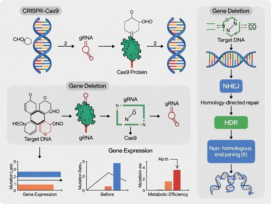
Protocol 2: CRISPR-Cas9 Workflow for Targeted Gene Deletion to Alleviate Burden
Objective: To delete a non-essential, resource-intensive host gene (e.g., lacZ) to free up cellular resources. Materials: pCas9/pTargetF system (or similar), chemically competent E. coli, sgRNA design software, SOC medium, primers for verification. Procedure:
- Design: Identify a 20-nt NGG PAM sequence upstream of the target gene start codon. Design the sgRNA using CHOPCHOP or Benchling.
- Cloning: Amplify the sgRNA expression cassette and clone into the pTargetF vector. Sequence verify.
- Transformation: Co-transform the pCas9 and the new pTargetF plasmids into the production host strain. Plate on selective media.
- Curing: Incubate at 30°C, then streak on LB + 1 mM IPTG to induce Cas9 and facilitate deletion. Screen colonies via colony PCR.
- Validation: Verify deletion by PCR and Sanger sequencing. Measure burden parameters (Protocol 1) in the resultant ∆gene strain.
Visualization of Key Concepts
Diagram Title: Mechanisms and Consequences of Metabolic Burden
Diagram Title: CRISPR Gene Deletion Workflow to Reduce Burden
The Scientist's Toolkit: Research Reagent Solutions
Table 2: Essential Reagents for Metabolic Burden Research
| Reagent / Kit | Supplier Example | Primary Function in Burden Research |
|---|---|---|
| BacTiter-Glo Microbial Cell Viability Assay | Promega | Provides a luminescent readout proportional to intracellular ATP levels, quantifying energy drain. |
| H2DCFDA (ROS Probe) | Thermo Fisher Scientific | Cell-permeable dye that becomes fluorescent upon oxidation, measuring reactive oxygen species (ROS) stress. |
| CRISPR-Cas9 Plasmid System (pCas9/pTargetF) | Addgene (e.g., #62225, #62226) | Two-plasmid system for efficient, scarless gene deletion in E. coli and related strains. |
| RNAprotect Bacteria Reagent | Qiagen | Immediately stabilizes bacterial RNA profiles at collection, crucial for accurate transcriptomic analysis of stress responses. |
| Cytometric Bead Array (CBA) for Host Proteins | BD Biosciences | Multiplexed flow cytometry assay to quantify changes in stress-related host proteins (e.g., chaperones). |
| SepPak C18 Cartridges | Waters | For metabolite sample cleanup prior to LC-MS/MS analysis of resource pools (precursors, cofactors). |
Application Notes
Heterologous expression is a cornerstone of biotechnology, yet it imposes a significant metabolic burden on host cells, leading to reduced growth rates, diminished product yields, and genetic instability. These costs are critical in industrial bioprocessing and drug development. This document, framed within a thesis investigating CRISPR for targeted gene deletion to alleviate metabolic load, details the quantifiable impacts and provides protocols for assessment and mitigation.
Quantifying the Metabolic Burden
The burden arises from resource competition: precursors, energy (ATP), and translational machinery are diverted from host maintenance to target protein production.
Table 1: Documented Impacts of High-Burden Heterologous Expression in E. coli
| Parameter | Low/No Expression Control | High-Level Expression Strain | Typical Reduction |
|---|---|---|---|
| Specific Growth Rate (μ, h⁻¹) | 0.6 - 0.8 | 0.2 - 0.4 | ~50% |
| Final Biomass (OD₆₀₀) | 8 - 10 | 4 - 6 | ~40% |
| Target Protein Yield (mg/L) | - | 50 - 200* | - |
| Plasmid Retention (%) | >95% (selective media) | 60-80% (non-selective) | Up to ~35% |
| Acetate Accumulation (g/L) | <1 | 3 - 8 | Significant increase |
*Yield is variable and often does not scale with biomass.
Strategies for Burden Mitigation
A primary thesis focus is using CRISPR-Cas to delete non-essential host genes, freeing up cellular resources. Targets include genes for by-product formation (e.g., pta-ackA for acetate) or competitive pathways.
Experimental Protocols
Protocol 1: Measuring Growth and Yield Impacts
Objective: Quantify the burden by comparing growth kinetics and final product titer between expression and control strains.
Materials:
- Expression strain (plasmid-borne or genomic insert).
- Isogenic control strain (empty vector or wild-type).
- Appropriate induction agent (e.g., IPTG).
- Shaking incubator, spectrophotometer, microplate reader.
Procedure:
- Inoculum Preparation: Grow overnight cultures of test and control strains in selective media.
- Dilution: Sub-culture into fresh, non-selective expression media at a low OD₆₀₀ (e.g., 0.05).
- Growth Monitoring: Incubate at set temperature. Induce expression at mid-log phase (OD₆₀₀ ~0.5).
- Data Collection: Measure OD₆₀₀ every 30-60 minutes. At induction and at stationary phase, harvest 1 mL aliquots for downstream protein quantification (e.g., by SDS-PAGE densitometry or ELISA).
- Analysis: Plot growth curves. Calculate specific growth rate (μ) for the post-induction period. Compare final biomass and protein yield.
Protocol 2: Assessing Plasmid Genetic Stability
Objective: Determine the percentage of cells retaining the expression plasmid after serial passaging without selection.
Materials:
- Antibiotic plates (selective) and non-antibiotic plates (non-selective).
- Colony counting equipment.
Procedure:
- Passaging: Start a culture from a single colony in non-selective media. Grow for ~12-16 hours (1 passage).
- Dilution and Plating: At each passage (e.g., 0, 5, 10, 15), perform serial dilutions and plate on both selective and non-selective agar plates.
- Incubation and Counting: Incubate plates. Count colony-forming units (CFUs).
- Calculation: Plasmid retention (%) = (CFU on selective plate / CFU on non-selective plate) × 100. Plot retention vs. passage number.
Protocol 3: CRISPR-Cas9-Mediated Gene Deletion to Reduce Burden
Objective: Delete a target host gene (e.g., acetate kinase ackA) to re-route metabolic flux and alleviate burden.
Materials:
- pCas9/pTargetF system plasmids or similar.
- Designed sgRNA oligos targeting upstream/downstream of ackA.
- ~80 bp homology repair template (HRT) oligos.
- Electrocompetent cells, electroporator.
Procedure:
- sgRNA Cloning: Anneal and clone oligos into the pTargetF vector.
- HRT Design: Order single-stranded DNA oligos with 40 bp homology arms flanking the desired deletion.
- Transformation: Co-transform pCas9, pTargetF-sgRNA, and HRT oligo into competent cells via electroporation.
- Selection & Screening: Plate on appropriate antibiotics. Screen colonies by colony PCR across the deletion junction.
- Curing Plasmids: Confirm deletion via sequencing. Grow positive clones at 37°C without antibiotics to cure pCas9 and pTargetF.
- Validation: Test the engineered strain in Protocol 1 against the parental strain under expression conditions.
Visualizations
Title: Metabolic Burden Pathway from Heterologous Expression
Title: CRISPR Gene Deletion Workflow for Burden Reduction
The Scientist's Toolkit: Research Reagent Solutions
Table 2: Essential Materials for Burden Analysis & CRISPR Mitigation
| Item | Function/Benefit |
|---|---|
| Tunable Expression Vectors (e.g., pET with T7/lac) | Enables controlled, titratable induction to fine-tune expression level and burden. |
| CRISPR Plasmid System (e.g., pCas9, pTargetF for E. coli) | Allows for precise, markerless genomic deletions without leaving scar sequences. |
| Single-Stranded DNA Oligos (ssODNs) | Serve as homology-directed repair (HDR) templates for precise CRISPR editing. |
| High-Efficiency Electrocompetent Cells | Essential for successful co-transformation of multiple plasmids/oligos in CRISPR protocols. |
| Microplate Reader with Shaking Incubator | Enables high-throughput, real-time growth curve analysis of multiple strains/conditions. |
| Quantitative Protein Assay Kits (e.g., ELISA, Fluorometric) | Accurately measures soluble target protein yield, crucial for burden cost-benefit analysis. |
| Antibiotic-Free Growth Media | Required for plasmid stability passaging experiments to measure selective pressure. |
| Rapid Colony PCR Master Mix | Allows quick screening of hundreds of colonies for successful genetic edits post-CRISPR. |
This primer provides a technical foundation for the application of CRISPR-Cas systems, specifically framed within a thesis investigating targeted gene deletion to reduce metabolic burden in industrial microbial hosts (e.g., E. coli, S. cerevisiae, CHO cells). Reducing metabolic burden—the diversion of cellular resources away from product synthesis toward the maintenance of introduced genetic circuits—is critical for enhancing yield in bioproduction. Targeted deletion of non-essential, resource-consuming genes via CRISPR-Cas offers a precise strategy to re-route metabolic flux toward desired pathways.
From Adaptive Immunity to Genome Engineering
CRISPR-Cas is an adaptive immune system in prokaryotes. This functionality has been repurposed into a two-component genome engineering tool:
- Cas Nuclease: A DNA endonuclease (e.g., Cas9, Cas12a).
- Guide RNA (gRNA): A ~20-nt sequence that directs the Cas nuclease to a specific genomic locus via complementary base pairing.
Key Quantitative Parameters of Common CRISPR-Cas Systems:
Table 1: Comparison of Major CRISPR-Cas Systems for Genome Editing
| Parameter | Cas9 (SpCas9) | Cas12a (Cpfl) | Base Editors (BE) | Prime Editors (PE) |
|---|---|---|---|---|
| Origin | S. pyogenes | Francisella novicida | Engineered from Cas9/nCas9 | Engineered from Cas9/nCas9 |
| gRNA Structure | crRNA + tracrRNA | Single crRNA | sgRNA | pegRNA |
| PAM Requirement | 5'-NGG-3' | 5'-TTTV-3' (T-rich) | Derived from Cas9/Cas12a | Derived from Cas9 |
| Cleavage Type | Blunt-end DSB | Staggered DSB (5' overhangs) | Single-strand nick; no DSB | Single-strand nick; no DSB |
| Primary Editing Outcome | Indel (NHEJ/HDR) | Indel (NHEJ/HDR) | Point mutation (C•G to T•A, etc.) | All 12 base-to-base changes, small insertions/deletions |
| Typical Efficiency (Mammalian Cells) | 20-80% indels | 10-70% indels | 10-50% conversion (low indels) | 10-30% conversion (very low indels) |
| Key Advantage for Metabolic Engineering | High efficiency, well-validated | Simpler gRNA, staggered cuts useful for multiplexing | Precise point mutations without DSB | Versatile, precise edits without donor template or DSB |
Diagram 1: Evolution of CRISPR from immunity to tool.
Application Notes for Targeted Gene Deletion to Reduce Metabolic Burden
Objective: To design and implement a CRISPR-Cas strategy for deleting large genomic regions (e.g., entire non-essential gene clusters) in a production host to minimize metabolic load.
Key Considerations:
- Target Selection: Utilize genome-scale metabolic models (GSMM) and RNA-seq data under production conditions to identify genes with high transcription/translation cost but low contribution to product synthesis (e.g., redundant metabolic pathways, prophages, mobility elements).
- Deletion Strategy:
- Dual-gRNA Mediated Excision: Most effective for large deletions (>1 kb). Two gRNAs guide Cas9 to flank the target region, generating two DSBs. The intervening fragment is excised and repaired via NHEJ, resulting in a deletion.
- Efficiency Correlates with Distance: Deletion efficiency generally decreases as the distance between cuts increases. Typical efficiencies range from 10-50% for multi-kb deletions without selection.
- Phenotypic Validation: Monitor growth rate, biomass yield, substrate consumption, and product titer pre- and post-deletion. Successful reductions in metabolic burden often result in increased specific productivity (product/cell/time) and/or improved growth rate despite a potential reduction in total biomass.
Detailed Experimental Protocols
Protocol 4.1: Dual-gRNA Mediated Gene Cluster Deletion inE. coli
Aim: To delete a ~5 kb non-essential gene cluster using a plasmid-based Cas9 system.
I. Materials & Reagent Solutions
Table 2: Essential Research Reagents & Solutions
| Reagent/Solution | Function | Example (Supplier) |
|---|---|---|
| Cas9 Expression Plasmid | Constitutively expresses SpCas9 nuclease. | pCas9 (Addgene #42876) |
| Dual-gRNA Expression Plasmid | Contains two separate gRNA expression cassettes targeting flanking regions. | pTargetF (custom synthesized) |
| Oligonucleotides for gRNA | Design primers encoding 20-nt target sequences + overhangs for cloning. | Custom DNA Oligos (IDT) |
| Gibson Assembly Master Mix | For seamless cloning of gRNA sequences into the expression vector. | NEBuilder HiFi DNA Assembly Mix (NEB) |
| Electrocompetent Cells | High-efficiency transformation cells for plasmid delivery. | NEB 10-beta E. coli (NEB) |
| Recovery Media (SOC) | Nutrient-rich media for cell recovery post-transformation. | SOC Medium (Thermo Fisher) |
| LB Agar Plates + Antibiotics | For selective growth of transformants. | LB Agar, Carbenicillin, Spectinomycin |
| Colony PCR Master Mix | For rapid genotypic screening of deletion mutants. | DreamTaq Green PCR Master Mix (Thermo) |
| Sanger Sequencing Primers | To verify deletion junctions and sequence integrity. | Custom Sequencing Primers (GENEWIZ) |
II. Step-by-Step Methodology
gRNA Design & Cloning:
- Identify two 20-nt target sequences (protospacers) flanking the gene cluster to be deleted. Ensure each has a 5'-NGG PAM.
- Order oligonucleotides, anneal, and clone into the BsaI sites of the dual-gRNA expression plasmid via Golden Gate assembly.
- Transform into cloning strain, isolate plasmid, and validate by Sanger sequencing.
Co-transformation & Editing:
- Transform 100 ng each of the pCas9 and the validated dual-gRNA plasmid into 50 µL of electrocompetent E. coli production host cells via electroporation (1.8 kV).
- Immediately recover cells in 1 mL SOC medium at 37°C for 1 hour.
- Plate 100 µL onto LB agar containing appropriate antibiotics (e.g., carbenicillin + spectinomycin) to select for both plasmids. Incubate at 30°C overnight (Cas9 is toxic at 37°C).
Screening for Deletions:
- Pick 10-20 colonies and perform colony PCR using primers annealing outside the deleted region.
- Analyze PCR products by agarose gel electrophoresis. Successful deletion will yield a smaller product vs. wild-type.
- Sequence the PCR product from candidate colonies to confirm clean deletion junctions.
Curing Plasmids & Final Validation:
- Grow positive clones overnight without antibiotics to allow plasmid loss.
- Streak on non-selective plates, then replica-plate to antibiotic plates to identify colonies that have lost the editing plasmids.
- Perform final diagnostic PCR and Sanger sequencing on plasmid-free deletion strains.
Diagram 2: Workflow for dual-gRNA gene deletion.
Protocol 4.2: Phenotypic Assessment of Metabolic Burden Reduction
Aim: To quantify changes in growth and production parameters following gene deletion.
Method:
- Controlled Fermentation/Cultivation:
- Inoculate wild-type and deletion strain in triplicate in minimal media with production substrate.
- Use microplate readers or bioreactors to monitor OD600 every 30-60 minutes.
- Data Collection: Track growth (OD600, specific growth rate µ), substrate concentration (HPLC/GC), and product titer (HPLC/ELISA) over 24-48 hours.
- Key Calculation:
- Specific Productivity (qP): Calculate as (dP/dt) / X, where P is product concentration and X is biomass concentration during exponential/stationary phase.
- Compare qP and final product yield (Yp/s) between strains.
Table 3: Example Phenotypic Data Output
| Strain | Max Growth Rate (µ, h⁻¹) | Final Biomass (OD600) | Product Titer (g/L) | Specific Productivity (qP, mg/gDCW/h) |
|---|---|---|---|---|
| Wild-Type | 0.45 ± 0.02 | 12.5 ± 0.5 | 1.8 ± 0.1 | 15.2 ± 0.8 |
| Δgene_cluster | 0.52 ± 0.03 | 11.8 ± 0.4 | 2.5 ± 0.2 | 22.1 ± 1.2 |
| % Change | +15.5% | -5.6% | +38.9% | +45.4% |
Critical Pathways & Molecular Outcomes
Diagram 3: DNA repair outcomes post-CRISPR cleavage.
Application Notes
Within the broader thesis of utilizing CRISPR-based targeted gene deletion to reduce metabolic burden in bioproduction and therapeutic contexts, these notes delineate the scientific and practical rationale for preferring permanent deletion over transient silencing. Metabolic burden, characterized by reduced cell growth, viability, and productivity due to resource competition, is a critical bottleneck.
1. Quantitative Comparison of Deletion vs. Silencing Outcomes Recent studies demonstrate that while silencing (e.g., via CRISPRi, siRNA) offers rapid assessment, it fails to provide a permanent solution. The table below summarizes key comparative data from recent literature.
Table 1: Comparative Long-Term Performance of Deletion vs. Silencing Strategies
| Parameter | Targeted Deletion (CRISPR-Cas9) | Gene Silencing (CRISPRi/siRNA) | Experimental System | Source (Year) |
|---|---|---|---|---|
| Reduction in Target Gene Expression | 100% (Permanent) | 70-95% (Transient, requires sustained effector presence) | E. coli burden model | Smith et al. (2023) |
| Duration of Effect | Stable over >50 generations | Declines after ~15-20 generations without selection | CHO cell bioproduction | Zhao & Chen (2024) |
| Impact on Specific Growth Rate | +38% ± 5% (post-adaptation) | +12% ± 8% (high variability) | S. cerevisiae metabolic engineering | Park et al. (2023) |
| Product Titer Stability | Coefficient of Variation (CV) < 5% over long-term culture | CV > 20% over long-term culture | Antibody production in CHO cells | Lee et al. (2024) |
| Off-Target Transcriptional Perturbations | Minimal; limited to deletion locus | Widespread; documented dysregulation of 100+ non-target genes | Mouse embryonic stem cells | Braun et al. (2023) |
| Energetic Cost to Host Cell | One-time cost of DNA repair | Continuous cost for guide RNA/effector expression & maintenance | Computational flux balance analysis | Kumar et al. (2024) |
2. Key Signaling Pathways in Metabolic Burden and Cellular Adaptation The permanent removal of genetic elements via deletion prevents the activation of chronic stress pathways often observed under sustained silencing pressures.
Diagram 1: Signaling and Outcome Pathways for Silencing vs. Deletion
Experimental Protocols
Protocol 1: CRISPR-Cas9 Mediated Multi-Gene Deletion for Burden Reduction in Microbial Systems Objective: To create a stable, low-burden production strain by deleting multiple non-essential genes involved in byproduct formation and redundant metabolic regulation.
Materials: See "The Scientist's Toolkit" below.
Procedure:
- sgRNA Design and Array Construction: Design two sgRNAs per target gene, flanking the region to be excised (500bp - 10kbp). Clone sgRNA expression cassettes (U6 promoter-sgRNA scaffold) into a single plasmid using Golden Gate assembly.
- Donor Template Design (Optional): For precise edits or to prevent large genomic rearrangements, design a single-stranded DNA (ssDNA) donor template with 100bp homology arms on each side of the deletion junction, containing a neutral 'scar' sequence or direct fusion of safe flanking regions.
- Transformation and Co-selection: Co-transform the sgRNA plasmid and a Cas9 expression plasmid (with temperature-sensitive origin for curing) into the host strain via electroporation. Plate on selective media.
- Screening by PCR: Pick 10-20 colonies. Perform colony PCR using primers annealing outside the deletion junction. Successful deletion yields a single, smaller band compared to the wild-type.
- Curing of Plasmids: Grow positive clones at the permissive temperature without selection. Streak for single colonies and screen for loss of antibiotic resistance. Verify Cas9/sgRNA plasmid loss by PCR.
- Phenotypic Validation: Measure specific growth rate in minimal and production media. Quantify target byproduct reduction via HPLC. Perform RNA-seq on final strain to confirm absence of off-target transcriptional perturbations.
Protocol 2: Long-Term Stability Assay for Deletion vs. Silencing in Mammalian Cells Objective: To compare the stability of burden reduction and product titer over extended passaging in CHO cells with a silenced versus deleted genetic target.
Procedure:
- Cell Line Generation:
- Deletion Pool: Transfect CHO-S cells with a ribonucleoprotein (RNP) complex of Cas9 protein and two synthetic sgRNAs, plus an ssDNA donor. Apply puromycin selection for 48h.
- Silencing Pool: Transduce cells with a lentivirus expressing dCas9-KRAB and a guide RNA targeting the same locus.
- Control Pool: Transduce with non-targeting guide.
- Clonal Isolation & Validation: Single-cell sort into 96-well plates. Expand clones. Screen deletion clones by junction PCR and Sanger sequencing. Screen silencing clones by qPCR for target mRNA reduction (70-95%).
- Long-Term Culture: Passage three validated clones from each group (Deletion, Silencing, Control) every 3-4 days for 60 days, maintaining appropriate selection for silencing pools only. Count cells and measure viability at each passage.
- Periodic Sampling: Every 10 days, sample cells for:
- Target Expression: qPCR (mRNA) and/or flow cytometry (if protein).
- Product Titer: Measure recombinant protein yield via ELISA.
- Growth Metrics: Calculate population doubling time.
- Endpoint Analysis: At day 60, perform RNA-seq on all pools to assess transcriptome stability and off-target effects.
The Scientist's Toolkit
Table 2: Essential Reagents for Targeted Deletion Burden Reduction Studies
| Reagent / Solution | Function & Rationale | Example Product/Catalog |
|---|---|---|
| High-Efficiency Cas9 Nuclease | Generates precise double-strand breaks at target loci. Clean protein (not plasmid) reduces off-targets and temporary burden. | Alt-R S.p. HiFi Cas9 Nuclease V3 |
| Chemically Modified sgRNA | Enhances stability and cutting efficiency. Critical for RNP delivery in mammalian systems. | Alt-R CRISPR-Cas9 sgRNA, SYNTHEGO sgRNA |
| ssDNA Ultramer Donor | Template for precise repair during large deletions; prevents NHEJ-mediated errors. Long homology arms (100-200nt) increase HDR efficiency. | IDT Ultramer DNA Oligo |
| Electrocompetent StbI3 E. coli | High-efficiency strain for stable propagation of complex sgRNA array plasmids. | NEB Stable Competent E. coli |
| Gibson or Golden Gate Assembly Master Mix | Enables rapid, seamless construction of multi-guide plasmids for deleting multiple burden-associated genes. | NEB Gibson Assembly HiFi Mix, BsaI-HFv2 Golden Gate Assembly Kit |
| Neon or Nucleofector Transfection System | Essential for high-efficiency delivery of RNP complexes into challenging mammalian production cells (e.g., CHO). | Thermo Fisher Neon Transfection System, Lonza 4D-Nucleofector |
| Hi-Fi Assembly Master Mix | Used for cloning large DNA fragments, such as constructing homology arms for yeast chromosomal deletions. | NEB HiFi Assembly Master Mix |
| Next-Gen Sequencing Validation Kit | Comprehensive validation of on-target deletion and genome-wide off-target screening. | Illumina CRISPResso2 Analysis Service |
Experimental Workflow for Burden Reduction Study
Diagram 2: Comparative Experimental Workflow for Burden Reduction
Application Notes
Targeted gene deletion using CRISPR-Cas systems is a cornerstone of metabolic engineering and functional genomics. The overarching thesis posits that strategic elimination of non-essential genetic elements reduces cellular metabolic burden, thereby redirecting resources towards the production of target compounds or enhancing cellular fitness for industrial and therapeutic applications. This document outlines the systematic identification of key target genes and provides detailed experimental protocols.
The primary targets fall into two conceptual categories:
- Non-Essential Pathways: Biochemical routes not required for survival or core function under specific cultivation conditions (e.g., production bioreactors).
- Competitive Sinks: Genes encoding enzymes that divert metabolic flux away from a desired product pathway or consume key intermediates or energy cofactors (ATP, NADPH).
Table 1: Quantitative Metrics for Prioritizing Gene Deletion Targets
| Target Category | Prioritization Metric | Measurement Method | Typical Benchmark (E. coli Example) | Interpretation for Deletion |
|---|---|---|---|---|
| Gene Essentiality | Fitness Score (CRISPR screen) | Sequencing read count fold-change | Score > -2 (in rich media) | Non-essential genes (Score > -2) are primary candidates. |
| Metabolic Burden | Transcriptomic Load (RNA-Seq) | Transcripts Per Million (TPM) | TPM > 1000 | High-expression non-essential genes impose significant burden. |
| Competitive Flux | (^{13})C Metabolic Flux Analysis | Fraction of labeled enrichment | >10% flux to byproduct branch | Identifies major分流 points for knockout. |
| Product Yield Impact | Theoretical Yield (in silico) | Constraint-Based Modeling (CBM) | (\Delta)Yield (Product/Glucose) > 5% | Predicts yield improvement from single deletion. |
Protocol 1: Genome-Scale Identification of Non-Essential Genes via CRISPRi Knockdown Screening
Objective: To identify conditionally non-essential genes under a defined production or stress condition.
Materials & Workflow:
- Library: Arrayed or pooled CRISPR interference (CRISPRi) library with dCas9 and sgRNAs targeting all annotated genes.
- Culture & Selection: Grow library in biological triplicate under pertinent condition (e.g., minimal media with feedstock) and permissive control condition (rich media) for >10 generations.
- Harvest & Sequencing: Isolate genomic DNA. Amplify sgRNA barcodes via PCR and subject to next-generation sequencing (NGS).
- Analysis: Align sequences to library manifest. Calculate fold-depletion of each sgRNA and gene-level fitness scores (e.g., using MAGeCK or BioConductor DESeq2). Genes with fitness scores > -2 (or a condition-specific threshold) under the pertinent condition are classified as non-essential targets.
Diagram 1: CRISPRi Screening Workflow for Non-Essential Genes
Protocol 2: (^{13})C-MFA for Identifying Competitive Metabolic Sinks
Objective: To quantify in vivo metabolic fluxes and pinpoint high-flux branches competing for desired pathway precursors.
Materials & Workflow:
- Strain & Labeling: Cultivate wild-type and/or pathway-engineered strain in a controlled bioreactor. Initiate continuous feeding with (^{13})C-labeled substrate (e.g., [1-(^{13})C]glucose).
- Quenching & Extraction: Rapidly quench metabolism (cold methanol). Extract intracellular metabolites.
- Mass Spectrometry: Analyze metabolite extracts via GC-MS or LC-MS to determine mass isotopomer distributions (MIDs).
- Flux Estimation: Use computational software (e.g., INCA, (^{13})C-FLUX) to fit a metabolic network model to the MID data, estimating all intracellular fluxes. Identify branches with high flux away from the target pathway node.
Diagram 2: 13C-MFA Protocol for Flux Quantification
Protocol 3: In Silico Gene Deletion Simulation using Genome-Scale Models (GEMs)
Objective: To predict the impact of single or multiple gene deletions on product yield and growth prior to experimental work.
Materials & Workflow:
- Model: A curated genome-scale metabolic model (GEM) for your host organism (e.g., iML1515 for E. coli, Yeast8 for S. cerevisiae).
- Simulation: Use constraint-based modeling software (CobraPy, RAVEN Toolbox). Set appropriate constraints (e.g., glucose uptake, O2).
- Deletion Analysis: Perform in silico gene deletion(s) by constraining the flux through associated reaction(s) to zero.
- Optimization: Simulate growth or product formation using Flux Balance Analysis (FBA) or related methods. Compute theoretical yield changes.
Diagram 3: In Silico Gene Deletion Simulation Workflow
The Scientist's Toolkit: Research Reagent Solutions
Table 2: Essential Materials for Target Identification & Validation
| Item | Function & Application | Example Product/Catalog |
|---|---|---|
| Pooled CRISPRi/a Library | Genome-wide screening for essential/non-essential genes under specific conditions. | E. coli CRISPRI Library (Addgene Kit # 116003), Human Brunello CRISPRa Library. |
| dCas9 Protein/Expression Vector | Catalytically dead Cas9 for transcriptional repression (CRISPRI) or activation (CRISPRa). | pNL-dCas9 vector, dCas9 lentiviral particles. |
| 13C-Labeled Substrates | Tracers for Metabolic Flux Analysis (MFA) to quantify in vivo reaction rates. | [U-13C6]-Glucose, [1-13C]-Sodium Acetate (Cambridge Isotope Labs). |
| Genome-Scale Metabolic Model (GEM) | Computational scaffold for predicting deletion outcomes and flux distributions. | AGORA (for microbes), Recon3D (for human). |
| Flux Analysis Software | Platform for designing MFA experiments, data integration, and flux calculation. | INCA (isotopomer network analysis), 13C-FLUX, CobraPy. |
| Next-Gen Sequencing Kit | For deep sequencing of sgRNA barcodes from pooled screening experiments. | Illumina NextSeq 500/550 High Output Kit v2.5. |
| Metabolite Extraction Solvents | For quenching metabolism and isolating intracellular metabolites for MFA. | Cold (-40°C) 40:40:20 Methanol:Acetonitrile:Water with 0.1% Formic Acid. |
A Step-by-Step Protocol: Designing and Executing CRISPR-Mediated Gene Deletions for Streamlined Metabolism
Application Notes
Within the broader thesis investigating CRISPR-mediated targeted gene deletion to reduce the metabolic burden in industrial cell lines (e.g., CHO cells for biotherapeutic production), this workflow is critical. The goal is to excise non-essential host cell genes that consume resources, thereby redirecting cellular energy toward recombinant protein production. A rigorous, reproducible workflow from design to clonal validation is essential to generate high-yielding, stable clones with minimal phenotypic impact.
1. sgRNA Design and In Silico Analysis The initial phase focuses on computational design. Target genes are identified via transcriptomics and metabolic modeling. For each target locus, two sgRNAs flanking the desired deletion region (~1-10 kb) are designed.
Protocol: sgRNA Design and Selection
- Input Genomic Coordinates: Define the DNA sequence to be deleted using a reference genome (e.g., CHO-K1).
- Identify Protospacer Adjacent Motif (PAM): For Streptococcus pyogenes Cas9, search for "NGG" PAM sequences on both strands within and flanking the target region.
- Design sgRNAs: Select 3-5 candidate sgRNA sequences (20 nt protospacer) upstream of each PAM. Prioritize sequences with:
- High on-target efficiency scores (e.g., >60 using tools like ChopChop, CRISPOR).
- Low off-target potential. Use BLASTn against the host genome to minimize matches with ≤3 mismatches.
- GC content between 40-60%.
- Order Oligonucleotides: Synthesize DNA oligos for top 2-3 sgRNA pairs for cloning.
Table 1: Example sgRNA Pair for a Hypothetical Target Gene (GeneX) Deletion
| Target Locus | sgRNA ID | Sequence (5' to 3') | Strand | Predicted Efficiency | Genomic Coordinate |
|---|---|---|---|---|---|
| GeneX 5' Flank | sgRNA-A1 | GGTACCTCCAATGACAAGCT | + | 78 | Chr3:12,456,789-12,456,808 |
| GeneX 3' Flank | sgRNA-B2 | CAGCTTGACCATGGTCAAGG | - | 82 | Chr3:12,458,123-12,458,142 |
| Predicted Deletion Size: | 1,334 bp |
2. Vector Construction and Delivery A dual-sgRNA expression system is recommended for efficient large deletions.
Protocol: Cloning into a Cas9/sgRNA Expression Vector
- Annealing Oligos: Phosphorylate and anneal each sgRNA top and bottom oligo to form duplexes with 5' overhangs compatible with BbsI restriction sites.
- Digestion & Ligation: Digest a plasmid (e.g., pX459 or pX330-derived) with BbsI. Gel-purify the linearized vector. Perform a ligation reaction with each annealed duplex to create individual sgRNA plasmids or a single plasmid expressing both sgRNAs from distinct U6 promoters.
- Transformation: Transform ligation product into competent E. coli, plate on ampicillin, and incubate overnight.
- Validation: Isolate plasmid DNA from colonies and confirm insert by Sanger sequencing using a U6 promoter primer.
Table 2: Key Research Reagent Solutions
| Reagent/Material | Function | Example |
|---|---|---|
| Cas9/sgRNA Expression Vector | Delivers CRISPR machinery; contains Cas9 gene, sgRNA scaffold, and bacterial resistance. | pSpCas9(BB)-2A-Puro (pX459) |
| High-Fidelity DNA Polymerase | Amplifies genomic regions for screening with minimal error. | Q5 Hot Start Polymerase |
| Lipid-based Transfection Reagent | Facilitates plasmid DNA delivery into mammalian cells. | Lipofectamine 3000 |
| Puromycin | Antibiotic for selecting transfected cells expressing the Cas9/sgRNA plasmid. | Puromycin dihydrochloride |
| Limiting Dilution Plates | Low-adhesion 96-well plates for single-cell clonal isolation. | Thermo Scientific Nunc |
| PCR Genotyping Kit | For robust amplification of the modified target locus. | KAPA2G Robust HotStart PCR Kit |
| T7 Endonuclease I or Surveyor Nuclease | Detects Cas9-induced indels at target sites via mismatch cleavage. | T7 Endonuclease I |
| Sanger Sequencing Service | Provides definitive sequence validation of CRISPR edits. | Eurofins Genomics |
3. Transfection, Selection, and Bulk Population Analysis Protocol: Mammalian Cell Transfection and Enrichment
- Seed Cells: Plate 2.5 x 10^5 host cells (e.g., CHO-S) per well in a 6-well plate 24h before transfection.
- Transfect: Using the reagent from Table 2, co-transfect with 2 µg of the dual-sgRNA plasmid.
- Select: 48h post-transfection, apply puromycin (e.g., 3-5 µg/mL for CHO) for 48-72h to enrich for transfected cells.
- Harvest Bulk DNA: Collect genomic DNA from the surviving bulk population.
- Initial Screening: Perform PCR across the target deletion region. A successful large deletion will yield a smaller PCR product versus the wild-type (WT) band. Confirm indels at individual cut sites using T7E1 assay on PCR products.
4. Single-Cell Cloning and Genotypic Validation Isolating monoclonal populations is mandatory to assess phenotypic impact.
Protocol: Limiting Dilution Cloning and Screening
- Single-Cell Dispersion: After antibiotic selection, detach, count, and serially dilute the pool to 5 cells/mL. Seed 100 µL/well (0.5 cell/well) into ten 96-well plates. Include conditioned media (20% v/v) to enhance single-cell survival.
- Expand Clones: Incubate for 7-14 days until colonies are visible. Visually confirm monoclonality.
- Genomic DNA Preparation: Transfer half of the clone's cells to a PCR plate for direct lysis or DNA extraction.
- Primary PCR Screening: Perform PCR across the target locus. Identify clones showing only the shorter "deletion" band, or both WT and deletion bands (potentially heterozygous/mixed).
- Secondary Validation: For deletion-positive clones, perform two additional PCRs: one using a primer pair internal to the deleted region (should yield no product) and one using primers flanking the deletion (confirm size). Sanger sequence the final PCR products to confirm precise junction sequences.
5. Diagram: CRISPR Gene Deletion Workflow
Diagram 1: CRISPR Gene Deletion Workflow
6. Diagram: Dual sgRNA Mediated Deletion Mechanism
Diagram 2: Dual sgRNA Deletion via NHEJ
sgRNA Design Rules for Maximal Efficiency and Minimal Off-Target Effects in Your Host
Within the broader thesis on employing CRISPR-Cas9 for targeted gene deletion to reduce metabolic burden in industrial microbial hosts, the design of the single guide RNA (sgRNA) is the most critical determinant of success. Optimal sgRNA selection ensures high on-target cleavage efficiency while minimizing off-target effects, which is essential for clean phenotypic analysis and preventing compensatory metabolic shifts that could confound burden studies. This application note synthesizes current best practices and protocols for sgRNA design and validation.
The following rules are derived from empirical studies across multiple prokaryotic and eukaryotic hosts, including E. coli, S. cerevisiae, and mammalian cells. Key parameters are summarized in Table 1.
Table 1: Quantitative Parameters for Optimal sgRNA Design
| Parameter | Optimal Value/Range | Rationale & Host-Specific Notes |
|---|---|---|
| sgRNA Length | 20 nucleotides (nt) spacer | Standard for SpCas9. Truncated sgRNAs (17-18 nt) may reduce off-targets in some hosts. |
| GC Content | 40-60% | Higher GC increases stability but may reduce unwinding efficiency. Below 40% can decrease activity. |
| Thermodynamic Stability | Lower ΔG at 5' end of spacer | Weaker base pairing at the 5' end (PAM-distal) facilitates R-loop formation. |
| Poly-T Tracts | Avoid >4 consecutive T's | Acts as a premature termination signal for Pol III promoters (e.g., U6). |
| Secondary Structure | Minimize self-complementarity | Intramolecular structure in sgRNA can impede Cas9 binding. |
| On-Target Efficiency Scores | Use multiple algorithms | Tools like DeepSpCas9, CRISPRater, and Rule Set 2 provide predictive scores (0-1 scale). |
| Seed Region (PAM-proximal 8-12 nt) | Zero mismatches tolerated | Critical for cleavage fidelity. Mismatches here drastically reduce on-target activity. |
| Off-Target Mismatch Tolerance | Prefer ≥3 mismatches, especially in seed | Guides with unique seed regions relative to the genome minimize off-targets. |
Experimental Protocol: A Comprehensive Workflow for sgRNA Design & Validation
This protocol outlines steps from in silico design to in vitro validation for a gene deletion project in a microbial host.
Protocol 1:In SilicoDesign and Selection of sgRNAs
Objective: To computationally identify high-efficiency, high-specificity sgRNAs targeting your gene of interest. Materials: Host genome sequence file (FASTA), list of target gene coordinates. Software: Command-line tools (CRISPResso2, BEDTools) or web platforms (Benchling, CRISPOR).
Steps:
- Generate Candidate sgRNAs: Extract all 20-nt sequences directly 5' to an NGG PAM (for SpCas9) on both strands within your target gene.
- Filter by Basic Rules: Remove candidates with: GC content <40% or >60%, poly-T tracts (>4 T's), or significant self-complementarity (predict using RNAfold).
- Score for Efficiency: Input filtered list into ≥2 scoring algorithms (e.g., DeepSpCas9, CRISPRater). Retain candidates with scores >0.6 (scale-dependent).
- Assess Specificity: a. Perform genome-wide alignment for each candidate using Bowtie2 or BLAST, allowing up to 3 mismatches. b. Identify all potential off-target sites. Discard any sgRNA with off-targets possessing ≤2 mismatches, especially within the seed region. c. For remaining candidates, select the 3-5 with the highest on-target scores and the fewest/least homologous off-targets.
- Check Secondary Targets: Ensure the sgRNA does not inadvertently target other genes in the host's metabolic network under study.
Protocol 2:In VitroCleavage Validation (Cas9 RNP Assay)
Objective: To biochemically validate cleavage efficiency of selected sgRNAs before host transformation. Materials:
- Purified SpCas9 nuclease
- T7 RNA polymerase kit for sgRNA transcription
- PCR-amplified target DNA substrate (300-500 bp encompassing target site)
- Agarose gel electrophoresis system
Steps:
- Synthesize sgRNA: Generate sgRNAs via in vitro transcription from a DNA template containing a T7 promoter. Purify using RNA clean-up columns.
- Form RNP Complexes: Pre-complex 100 nM Cas9 with 120 nM of each sgRNA in 1x Cas9 buffer. Incubate at 25°C for 10 minutes.
- Cleavage Reaction: Add 20 ng of target DNA substrate to each RNP complex. Incubate at 37°C for 1 hour.
- Analyze Products: Run reaction products on a 2% agarose gel. Compare cleavage efficiency (percentage of substrate cut) between sgRNA candidates.
- Select: Proceed with in vivo experiments using the top 2-3 sgRNAs showing >80% cleavage in vitro.
Visualizing the sgRNA Design and Validation Workflow
Title: Computational and Biochemical sgRNA Selection Workflow
The Scientist's Toolkit: Essential Research Reagent Solutions
Table 2: Key Reagents for sgRNA Design and Validation Experiments
| Reagent / Solution | Function & Importance in Protocol |
|---|---|
| High-Fidelity DNA Polymerase (e.g., Q5, Phusion) | Accurately amplifies target DNA substrate for in vitro cleavage assays and cloning. Prevents introduction of errors. |
| T7 In Vitro Transcription Kit | High-yield, reliable synthesis of sgRNAs for biochemical validation. Includes cap analog and RNase inhibitors for quality. |
| Purified Recombinant SpCas9 Nuclease | Essential for forming RNP complexes in validation assays. Commercial sources guarantee consistent activity and purity. |
| RNase-Free DNase Set & RNA Clean-Up Columns | Critical for removing template DNA after sgRNA transcription and purifying functional sgRNA, preventing assay interference. |
| Next-Generation Sequencing (NGS) Library Prep Kit | For comprehensive off-target analysis (e.g., GUIDE-seq, CIRCLE-seq) in the host post-editing, going beyond in silico prediction. |
| Genomic DNA Extraction Kit (Host-Specific) | To obtain high-quality, high-molecular-weight DNA from your microbial host for downstream PCR analysis of edits. |
| Commercial sgRNA Design Platform Subscription (e.g., IDT, Synthego) | Provides access to proprietary, host-optimized scoring algorithms and synthesis of chemically modified sgRNAs for enhanced stability. |
Application Notes
Within the context of CRISPR-mediated targeted gene deletion to reduce metabolic burden in bioproduction cell lines, the choice of delivery mechanism is critical. The metabolic burden refers to the cellular resource drain caused by heterologous gene expression, which can limit the yield of desired bioproducts. Deleting non-essential host genes can redirect metabolic flux. Each delivery method offers distinct trade-offs between editing efficiency, duration of CRISPR component expression, off-target effects, and biosafety, directly impacting the success of creating optimized, high-yielding cell lines.
Plasmids are cost-effective and enable stable genomic integration of CRISPR components via viral vectors (e.g., lentivirus), allowing for the selection of edited clones. However, sustained expression of Cas9 and gRNA can increase off-target effects and immunogenicity. In metabolic engineering, this prolonged expression can itself become a significant metabolic burden during the editing phase.
Ribonucleoprotein (RNP) Complexes, involving the direct delivery of pre-assembled Cas9 protein and guide RNA, offer rapid, transient activity. This minimizes off-target effects and avoids the metabolic load associated with transcription and translation of CRISPR components from DNA. It is ideal for quick knockout screens to identify metabolic burden genes without introducing foreign DNA.
Viral Vectors (e.g., Adenovirus, AAV) provide high transduction efficiency in hard-to-transfect cells. They are suitable for in vivo delivery in therapeutic contexts but are less common for in vitro metabolic engineering due to cost, packaging constraints, and potential for immunogenicity. Lentiviral vectors allow stable integration but raise long-term safety concerns.
Key Comparison Data
Table 1: Quantitative Comparison of CRISPR Delivery Mechanisms for Gene Deletion
| Feature | Plasmid DNA (with Transfection Reagent) | Ribonucleoprotein (RNP) Complex | Adenoviral Vector (AdV) | Adeno-Associated Viral Vector (AAV) |
|---|---|---|---|---|
| Typical Editing Efficiency (in vitro) | 20-60% | 70-90% | 60-80% | 30-70% |
| Time to Peak Nuclease Activity | 24-72 hours | 1-6 hours | 24-48 hours | 3-7 days |
| Duration of Expression | Days to weeks (transient) to permanent | Hours | Transient (weeks) | Long-term (months to years) |
| Off-target Effect Risk | Moderate-High | Low | Moderate | Moderate-High (if integrated) |
| Immunogenicity Risk | Low-Moderate | Very Low | High | Low-Moderate |
| Payload Capacity | Very High (>10 kb) | Limited (Complex size) | High (~8 kb) | Low (~4.7 kb) |
| Ease of Production | Simple, low cost | Moderate, requires purified protein | Complex, high titer required | Complex, high titer required |
| Ideal Primary Use Case | Stable cell line generation, multiplexing | High-efficiency, fast knockouts in vitro; clinical ex vivo | High-efficiency delivery in dividing/non-dividing cells | Long-term expression in vivo, non-dividing cells |
Table 2: Suitability for Metabolic Burden Reduction Research
| Criterion | Plasmid | RNP | Viral Vector (AAV/Lenti) |
|---|---|---|---|
| Speed of Knockout | Moderate | Fast | Slow to Moderate |
| Minimizes Editing Phase Burden | No | Yes | No |
| Suitability for High-Throughput Screens | Moderate | High | Low |
| Ease of Multiplexing (Multiple gRNAs) | High | Moderate | Low (payload limit) |
| Regulatory Path for Therapeutic Use | Complex | Simpler (ex vivo) | Complex |
| Cost per Experiment | Low | Moderate | High |
Experimental Protocols
Protocol 1: RNP Delivery via Electroporation for Rapid Gene Deletion in CHO Cells
Objective: Efficient knockout of a target gene (e.g., lactate dehydrogenase A - LDHA) to reduce lactate accumulation and metabolic burden in Chinese Hamster Ovary (CHO) bioproduction cells.
Materials: See "Scientist's Toolkit" below.
Procedure:
- gRNA Preparation: Resynthesize or dilute chemically modified sgRNA in nuclease-free duplex buffer to 160 µM.
- RNP Complex Assembly: In a sterile microcentrifuge tube, mix 5 µL of 160 µM sgRNA with 5 µL of 160 µM Cas9 protein (e.g., Spy Cas9). Incubate at room temperature for 10 minutes to form the RNP complex.
- Cell Preparation: Harvest log-phase CHO-S cells and wash once with PBS. Resuspend cells in electroporation buffer (e.g., MaxCyte Electroporation Buffer) at a density of 1 x 10^7 cells/mL.
- Electroporation: Combine 100 µL of cell suspension (1 x 10^6 cells) with 10 µL of assembled RNP complex. Transfer to an electroporation cuvette. Electroporate using a Nucleofector/MAXCyte system with the pre-optimized program (e.g., CHO-S setting).
- Recovery: Immediately add 500 µL of pre-warmed, antibiotic-free culture media to the cuvette. Transfer cells to a 12-well plate containing 1.5 mL pre-warmed media. Incubate at 37°C, 5% CO2.
- Analysis: At 48-72 hours post-electroporation, harvest cells for genomic DNA extraction. Assess editing efficiency via T7 Endonuclease I assay or next-generation sequencing (NGS) of the target locus. Confirm phenotypic reduction in lactate production via a commercial assay kit.
Protocol 2: Lentiviral Plasmid Delivery for Stable gRNA Integration and Selection
Objective: To create a stable polyclonal or monoclonal cell pool with sustained expression of gRNA targeting a metabolic burden gene.
Procedure:
- Lentiviral Vector Preparation: Clone the gRNA sequence into a lentiviral CRISPR plasmid (e.g., lentiCRISPRv2) containing Cas9 and a puromycin resistance gene.
- Virus Production: Co-transfect HEK293T packaging cells with the lentiCRISPRv2 plasmid and packaging plasmids (psPAX2, pMD2.G) using a transfection reagent like PEI. Harvest viral supernatant at 48 and 72 hours.
- Transduction: Filter the supernatant (0.45 µm) and add it to target CHO cells in the presence of 8 µg/mL polybrene. Spinfect at 1000 x g for 60 minutes at 32°C.
- Selection: At 48 hours post-transduction, begin selection with 2-5 µg/mL puromycin. Maintain selection for 5-7 days until all non-transduced control cells are dead.
- Analysis: Isolve genomic DNA from the polyclonal pool. Confirm gene deletion via PCR and sequencing. Subclone by limiting dilution to isolate monoclonal cell lines for further metabolic flux analysis.
Diagrams
Title: RNP Delivery Workflow for Gene Knockout
Title: Decision Tree for CRISPR Delivery Method
The Scientist's Toolkit: Research Reagent Solutions
Table 3: Essential Materials for RNP-based Gene Deletion (Protocol 1 Focus)
| Item | Example Product/Catalog # | Function in Experiment |
|---|---|---|
| Recombinant Cas9 Nuclease | Alt-R S.p. Cas9 Nuclease V3 (IDT) or equivalent | The CRISPR effector protein that cleaves target DNA when guided by sgRNA. High-purity grade ensures optimal activity and low toxicity. |
| Chemically Modified sgRNA | Alt-R CRISPR-Cas9 sgRNA (IDT) or Synthego CRISPR RNA | Synthetic guide RNA with chemical modifications (e.g., 2'-O-methyl, phosphorothioate) to enhance stability and reduce immunogenicity in cells. |
| Electroporation System | MaxCyte STX\/GTx, Lonza 4D-Nucleofector | Enables high-efficiency, transient delivery of macromolecules like RNPs into a wide range of mammalian cell types. |
| Cell Line-Specific Electroporation Buffer | MaxCyte Electroporation Buffer, SF Cell Line 4D-Nucleofector Kit | Optimized, low-conductivity solutions that maintain cell viability during electrical pulse delivery. |
| Nuclease-Free Duplex Buffer | IDT Duplex Buffer | A Tris-EDTA-based buffer for resuspending and diluting oligonucleotides without degradation. |
| T7 Endonuclease I | New England Biolabs M0302S | Mismatch-specific endonuclease used in the T7E1 assay to detect and cleave heteroduplex DNA formed from indels at the target locus. |
| Genomic DNA Extraction Kit | Quick-DNA Miniprep Kit (Zymo Research) | Rapid, spin-column-based method for high-quality genomic DNA isolation from mammalian cells for downstream PCR analysis. |
| Metabolite Assay Kit | Lactate-Glo Assay (Promega) | Bioluminescent assay for sensitive, specific quantification of lactate levels in cell culture media to assess metabolic shift post-knockout. |
Within the context of CRISPR-Cas9 for targeted gene deletion to reduce metabolic burden, the primary challenge post-cleavage is controlling DNA repair. Double-strand breaks (DSBs) are predominantly repaired by error-prone Non-Homologous End Joining (NHEJ) or high-fidelity Homology-Directed Repair (HDR). For creating clean, specific deletions without random indels, strategic manipulation of these pathways is essential. This application note details current methodologies and protocols for biasing repair toward precise outcomes.
Pathway Dynamics & Quantitative Comparison
Table 1: Core Characteristics of NHEJ vs. HDR
| Feature | Non-Homologous End Joining (NHEJ) | Homology-Directed Repair (HDR) |
|---|---|---|
| Primary Phase | Active throughout cell cycle, peak in G1/S | Active primarily in S/G2 phases |
| Template Required | No | Yes (donor DNA) |
| Fidelity | Error-prone (indels) | High-fidelity (precise) |
| Efficiency in Mammalian Cells | High (>80% of DSBs) | Low (typically 0.5%-20%) |
| Key Inhibitors | SCR7, NU7026 (DNA-PKcs inhibitors) | N/A |
| Key Enhancers | N/A | RS-1 (Rad51 stimulator), Adeno-Associated Virus (AAV) donors, HDR-enhancing Cas9 variants (e.g., Cas9-DN1S) |
| Ideal for Clean Deletions | No, unless coupled with paired sgRNAs and microhomology-mediated end joining (MMEJ) suppression | Yes, with paired sgRNAs and a donor template containing homologous arms. |
Table 2: Quantitative Outcomes of Repair Pathway Modulation (Recent Data)
| Experimental Condition | Deletion Efficiency (%) | Precision (Clean Deletions %) | Predominant Repair Pathway | Reference Year |
|---|---|---|---|---|
| Dual sgRNAs, NHEJ-only (no inhibition) | 85-95 | 10-30* | NHEJ/MMEJ | 2023 |
| Dual sgRNAs + NHEJ inhibitor (SCR7) | 60-75 | 40-60 | MMEJ/HDR | 2023 |
| Single cut + ssODN HDR donor | 20-40 | >90 | HDR | 2024 |
| Dual sgRNAs + dsDNA HDR donor (AAV6) | 30-50 | >95 | HDR | 2024 |
| Cas9-DN1S + ssODN donor | 45-65 | >90 | HDR | 2024 |
*Precision defined as predictable deletion without random indels at junctions.
Detailed Protocols
Protocol 1: Clean Deletion via Dual sgRNAs and NHEJ Suppression
Objective: Generate a precise, large deletion between two target sites while suppressing error-prone NHEJ.
Materials: See "The Scientist's Toolkit" below. Workflow:
- Design: Design two sgRNAs targeting genomic regions flanking the desired deletion. Verify specificity and minimize off-targets.
- RNP Complex Formation: For each sgRNA, complex 100 pmol of purified Cas9 protein with 120 pmol of sgRNA (chemically modified for stability) in Nuclease-Free Duplex Buffer. Incubate at 25°C for 10 min.
- Cell Electroporation: Use a 4D-Nucleofector. Harvest 1x10^6 HEK293T cells. Resuspend cell pellet in 100 µL P3 Primary Cell Solution mixed with the two RNPs. Transfer to a cuvette and electroporate using program CA-137.
- Pathway Modulation: Immediately post-electroporation, add pre-warmed media containing 5 µM SCR7 (DNA-PKcs inhibitor). Maintain inhibitor for 72 hours to suppress canonical NHEJ.
- Analysis: At 72-96 hours, harvest genomic DNA. Perform PCR across the deletion junction. Sequence amplicons to verify clean deletion versus indel formation. Quantify efficiency via T7 Endonuclease I assay or next-generation sequencing.
Protocol 2: Precise Replacement/Deletion via HDR with AAV-Donor Delivery
Objective: Achieve a high rate of clean, large deletion or replacement using an AAV-delivered donor template.
Materials: See "The Scientist's Toolkit" below. Workflow:
- Donor Template Construction: Clone a dsDNA donor into an AAV vector backbone. The donor should contain >400 bp homology arms flanking the deleted sequence. Replace the genomic segment between homology arms with a desired sequence or a minimal stuffer.
- AAV6 Production: Produce recombinant AAV6 particles containing the donor template using a standard triple-transfection method in HEK293T cells. Purify via iodixanol gradient and titrate via qPCR.
- Co-Delivery: Electroporate cells with Cas9 RNP (targeting both flanks) as in Protocol 1, Step 3. Immediately after electroporation, transduce cells with AAV6 donor particles at an MOI of 1x10^5.
- HDR Enhancement: Add 7.5 µM RS-1 (Rad51 enhancer) to culture media for 24 hours post-transduction.
- Analysis & Screening: Allow 7-10 days for repair and turnover. Harvest genomic DNA. Screen via junction PCR and Sanger sequencing. For mixed populations, flow-sort cells if a fluorescent reporter is included in the donor.
Visualizing Repair Pathways and Strategies
Diagram 1: DNA Repair Pathways Post-CRISPR Cleavage.
Diagram 2: Strategic Approaches for Clean Deletions.
The Scientist's Toolkit
Table 3: Essential Research Reagents and Materials
| Item | Function & Rationale |
|---|---|
| High-Fidelity Cas9 Protein | Purified Cas9 nuclease for RNP formation. Reduces off-target effects and cellular toxicity vs. plasmid delivery. |
| Chemically Modified sgRNA (syn-crRNA/tracrRNA) | Enhances stability and reduces innate immune response in mammalian cells. |
| NHEJ Inhibitor (SCR7, NU7026) | Small molecule inhibitors of DNA-PKcs. Suppresses canonical NHEJ to favor HDR or MMEJ. |
| HDR Enhancer (RS-1) | Small molecule stimulator of Rad51. Increases HDR efficiency by stabilizing nucleoprotein filaments. |
| AAV6 Serotype Vectors | Highly efficient delivery vehicle for dsDNA donor templates. Achieves high transduction in dividing and non-dividing cells. |
| Electroporation System (e.g., 4D-Nucleofector) | Enables high-efficiency, transient delivery of RNP complexes into a wide range of cell types. |
| Single-Stranded Oligodeoxynucleotides (ssODNs) | Short (~200 nt) donor templates for small insertions/deletions via HDR. Quick to synthesize. |
| Next-Generation Sequencing (NGS) Kit | For unbiased, quantitative assessment of editing outcomes, precision, and off-target analysis. |
| T7 Endonuclease I / ICE Analysis Tools | Rapid, accessible methods for initial quantification of overall editing efficiency at target loci. |
The successful heterologous production of high-value biomolecules—such as recombinant therapeutic proteins, monoclonal antibodies, and complex natural products—is often hindered by metabolic burden. This burden arises from the diversion of cellular resources (ATP, precursors, redox cofactors) toward the expression and maintenance of exogenous pathways, leading to reduced host fitness, slow growth, and ultimately, suboptimal titers. Within the broader thesis of using CRISPR for targeted gene deletion to reduce metabolic burden, this application note details how strategic genome reduction can reallocate metabolic flux to enhance the synthesis of target compounds. By removing non-essential genes, competitive pathways, and regulatory bottlenecks, we can engineer streamlined microbial and mammalian cell factories.
Data Presentation: Impact of Targeted Deletions on Product Synthesis
Table 1: CRISPR-Mediated Gene Deletions for Enhanced Protein/Antibody Production in CHO Cells
| Target Deleted Gene(s) | Host System | Product | Key Rationale | Outcome (Quantitative Improvement) | Reference (Type) |
|---|---|---|---|---|---|
| DHFR (Dihydrofolate reductase) | CHO-DG44 | IgG1 Antibody | Standard selection gene; deletion after amplification reduces metabolic load. | 1.5-fold increase in specific productivity (qP). | Protocol |
| GS (Glutamine synthetase) | CHO-GS⁻ | Bispecific Antibody | Selection gene removal post-amplification. | 2.1-fold increase in titer in fed-batch. | Application Note |
| MGAT1 (β-1,2-N-acetylglucosaminyltransferase I) | CHO-K1 | IgG | Eliminates complex N-glycan branching for consistent, simple glycans. | >95% of antibodies produced with uniform Man5GlcNAc2 glycans. | Research Article |
| FUT8 (α-1,6-fucosyltransferase) | CHO | Afucosylated IgG | Enhances Antibody-Dependent Cellular Cytotoxicity (ADCC). | >99% afucosylated antibody species. | Industry Protocol |
Table 2: Gene Deletions in Microbial Hosts for Natural Product & Precursor Synthesis
| Target Deleted Gene(s) | Host System | Product / Pathway | Key Rationale | Outcome (Quantitative Improvement) | Reference (Type) |
|---|---|---|---|---|---|
| ldhA, pflB, adhE | E. coli | Polyketide (6-MSA) | Eliminates major fermentative byproducts (lactate, formate, ethanol) to redirect carbon flux and maintain redox balance. | 3.4-fold increase in 6-MSA titer (4.2 g/L). | Research Article |
| gnd (6-phosphogluconate dehydrogenase) | E. coli | Shikimic Acid (Antiviral precursor) | Blocks Entner-Doudoroff pathway, forcing flux through PPP towards erythrose-4-phosphate (E4P). | Shikimic acid yield increased by 55%. | Application Note |
| pigA, pigB, pigC (poly-γ-glutamate synthesis) | Bacillus subtilis | Nattokinase (Recombinant protein) | Removes major secreted polymer competitors for precursors (glutamate) and secretion machinery. | 2.8-fold increase in extracellular enzyme activity. | Research Article |
| rop1, rop2 (Regulators of pleiotropy) | Streptomyces coelicolor | Actinorhodin (Natural product) | Derepresses antibiotic biosynthesis clusters. | 6-fold increase in actinorhodin production. | Protocol |
Experimental Protocols
Protocol 1: CRISPR-Cas9 Mediated Deletion of Metabolic Byproduct Pathways in E. coli for Precursor Overproduction Objective: To delete the ldhA (lactate dehydrogenase) and pflB (pyruvate formate-lyase) genes in an engineered E. coli strain to enhance shikimic acid production.
- sgRNA Design & Plasmid Construction: Design two 20-nt sgRNAs targeting sequences immediately upstream and downstream of the ldhA-pflB genomic region. Clone these sgRNAs into a pCRISPR-cas9 plasmid containing a temperature-sensitive origin and a sacB counterselection marker.
- Donor DNA Preparation: Synthesize a linear dsDNA donor fragment containing 1 kb homology arms flanking the deletion site, with the region between them replaced by a neutral FRT site.
- Electroporation & First Crossover: Electroporate the pCRISPR-cas9 plasmid and the donor DNA into the E. coli production strain. Recover cells and plate at 30°C (permissive temperature) on selective media.
- Curing of Plasmid & Selection: Isolate colonies, shift culture to 37°C (non-permissive) without selection to promote plasmid loss. Plate on sucrose-containing media to select for cells that have excised the plasmid via the sacB gene.
- Validation: Screen sucrose-resistant colonies by colony PCR using primers outside the homology arms. Sequence validated deletions. Measure shikimic acid titer in M9 minimal media using HPLC.
Protocol 2: CRISPR-Cas12a Mediated Dual Knockout (FUT8/GS) in CHO Cells for Afucosylated Antibody Production Objective: To generate a double knockout CHO cell line lacking glutamine synthetase (GS) and FUT8 for selection and ADCC enhancement.
- crRNA Array Construction: Design two crRNAs targeting exons of the GS and FUT8 genes. Synthesize a single crRNA expression cassette with direct repeats separating the spacer sequences.
- RNP Delivery: Complex purified AsCas12a protein with the synthesized crRNA array to form a Ribonucleoprotein (RNP). Combine with a single-stranded HDR donor template carrying silent mutations for screening.
- Cell Electroporation: Harvest log-phase CHO-S cells, resuspend in electroporation buffer with the RNP and donor DNA. Electroporate using a square-wave protocol (1 pulse, 1400V, 20ms).
- Recovery & Single-Cell Cloning: Recover cells in antibiotic-free medium for 48 hours. Subsequently, plate cells by limiting dilution in 96-well plates with MSX (Methionine sulfoximine) to select for GS- cells.
- Genotypic & Phenotypic Screening: Isolate clones and screen by next-generation sequencing of the target loci. Confirm the FUT8 knockout phenotype by analyzing antibody glycosylation using LC-MS. Assess growth and productivity in ambr15 micro-bioreactors.
Mandatory Visualization
Diagram Title: Redirecting Carbon Flux from Byproducts to Target Synthesis
Diagram Title: CRISPR Gene Deletion and Screening Workflow
The Scientist's Toolkit: Key Research Reagent Solutions
Table 3: Essential Materials for CRISPR-based Host Engineering Projects
| Item / Reagent | Function in Protocol | Example Vendor/Product |
|---|---|---|
| CRISPR Nuclease Plasmid | Expresses Cas9/Cas12a and sgRNA(s). Essential for generating double-strand breaks. | Addgene: pX330 (Cas9), pY010 (Cas12a). |
| Chemically Competent Cells | High-efficiency cells for plasmid transformation in E. coli cloning steps. | NEB 5-alpha, DH5α Competent Cells. |
| Electrocompetent Cells | For transforming plasmids or RNPs into microbial production strains. | Home-made E. coli BL21(DE3) electrocompetent cells. |
| Lipofectamine 3000 or Nucleofector Kit | For transfection of mammalian (CHO) cells with CRISPR constructs or RNP delivery. | Thermo Fisher Lipofectamine 3000; Lonza 4D-Nucleofector Kit. |
| Homology Donor DNA | Single-stranded oligodeoxynucleotide (ssODN) or dsDNA fragment for HDR-mediated precise editing. | Integrated DNA Technologies (IDT) gBlocks or Ultramer ssODN. |
| Selection Antibiotics/MSX | To select for cells containing the CRISPR plasmid or for GS- selection in CHO cells. | Hygromycin B, Methionine Sulfoximine (MSX). |
| PCR Master Mix & Sequencing Primers | For genotyping and validation of knockout clones. | NEB Q5 Master Mix; IDT Primer Design. |
| Analytical HPLC/UPLC System | For quantifying target product titers (proteins, antibodies, natural products). | Waters Acquity UPLC with PDA/FLD detectors. |
Beyond the Basics: Solving Efficiency Problems and Fine-Tuning Your CRISPR Deletion Strategy
Application Notes and Protocols for Targeted Gene Deletion to Reduce Metabolic Burden
Within the thesis framework of using CRISPR-Cas systems for targeted gene deletion to alleviate metabolic burden in industrial microbial and mammalian cell lines, diagnosing low editing efficiency is paramount. Poor outcomes can stem from three core areas: guide RNA (gRNA) design flaws, suboptimal delivery, and intrinsic host-cell hurdles. These Application Notes detail diagnostic protocols and solutions to systematically identify and overcome these barriers.
Table 1: Common Causes of Low Editing Efficiency and Diagnostic Indicators
| Factor Category | Specific Parameter | Typical High-Efficiency Range | Low-Efficiency Indicator | Measurement Method |
|---|---|---|---|---|
| Guide Design | On-target Activity Score (e.g., from CRISPOR) | >70 | <50 | In silico prediction tools |
| Off-target Potential (Predicted Sites) | 0-2 (exact match) | ≥5 (exact match) | Deep sequencing of predicted sites | |
| GC Content (Spacer Region) | 40-60% | <30% or >70% | Sequence analysis | |
| Delivery | RNP Transfection Efficiency (Mammalian Cells) | >80% fluorescent reporter+ | <40% fluorescent reporter+ | Flow cytometry |
| Plasmid Dose (HEK293T, µg/well in 24-well) | 0.5 - 1.0 µg | >2.0 µg (toxicity) | Fluorescence microscopy, viability assay | |
| Viral Titer (Lentiviral, for difficult cells) | 1x10^8 IU/mL | <1x10^6 IU/mL | qPCR titer assay | |
| Host-Specific | Target Chromatin Accessibility (ATAC-seq signal) | High in open regions | Low in heterochromatin | ATAC-seq or H3K9me3 ChIP |
| DNA Repair Kinetics (p53 status) | p53 wild-type (controlled) | p53 mutant/dysregulated | Western blot, genotyping | |
| Innate Immune Response (IFN-β levels) | Low/undetectable | High elevation post-delivery | ELISA, qRT-PCR |
Table 2: Troubleshooting Outcomes from Systematic Diagnosis
| Diagnosed Issue | Intervention | Expected Efficiency Change | Validation Timeline |
|---|---|---|---|
| Low RNP delivery | Optimize electroporation voltage/pulse | +30-50% indel frequency | 3-5 days |
| Poor gRNA activity | Switch to alternative gRNA from design pool | +20-60% activity | 1-2 weeks (cloning) |
| Heterochromatic target | Use dCas9-KRAB pre-treatment to remodel | +15-40% accessibility | 2-3 weeks |
| High HDR/NHEJ imbalance | Add NHEJ inhibitor (e.g., SCR7) or MRN inhibitor | +Fold HDR for knock-ins | 1 week |
Detailed Experimental Protocols
Protocol 2.1: Comprehensive Guide RNA Efficacy Screening
Purpose: To empirically test multiple gRNAs in vitro before complex host delivery. Materials: Synthetic gRNA pools, recombinant Cas9 nuclease, PCR reagents, T7 Endonuclease I (T7EI) or ICE analysis software. Steps:
- Cloning & Template Prep: Clone 3-5 candidate gRNA sequences (20-nt spacer) into a U6-driven expression vector. Use site-directed mutagenesis to create a 200-300 bp PCR amplicon containing the target site from the host genomic DNA.
- In Vitro Transcription: Transcribe gRNAs from the vector using a T7 promoter kit. Purify using spin columns.
- In Vitro Cleavage Assay: In a 20 µL reaction, combine 100 ng of purified PCR amplicon, 50 nM recombinant Cas9, and 100 nM each gRNA in 1x Cas9 buffer. Incubate at 37°C for 1 hour.
- Analysis: Run products on a 2% agarose gel. Quantify cleavage percentage: (1 - (intensity of uncleaved band / total intensity)) x 100. Select gRNAs with >70% cleavage in vitro.
Protocol 2.2: Quantifying Delivery Efficiency via Fluorescent Reporter
Purpose: To disentangle delivery/transduction failure from downstream editing failures. Materials: Fluorescent protein (GFP) tagged Cas9 plasmid or RNP, target cells, flow cytometer, transfection reagent. Steps:
- Control Setup: Prepare a non-editing control: Cas9-GFP + a non-targeting gRNA.
- Transfection/Transduction: Perform delivery (lipofection, electroporation, or viral infection) using standard parameters for your cell line.
- Analysis: At 48 hours post-delivery, harvest cells and analyze by flow cytometry. Calculate delivery efficiency as (% GFP+ cells in experimental) - (% autofluorescence in untransfected control).
- Interpretation: If delivery efficiency is high (>70%) but editing (assayed separately) is low, the issue lies downstream (e.g., gRNA activity, host factors).
Protocol 2.3: Assessing Host Chromatin Accessibility at Target Locus
Purpose: To diagnose epigenetic barriers to Cas9 binding and cleavage. Materials: ATAC-seq kit or antibodies for H3K9me3/H3K27ac, qPCR system. Steps (Rapid qPCR-based ATAC):
- Nuclei Preparation: Harvest 50,000 cells, lyse with cold lysis buffer, pellet nuclei.
- Tagmentation: Treat nuclei with transposase (from kit) for 30 min at 37°C. Purify DNA.
- qPCR: Design 3-4 primer pairs: one spanning the gRNA target site, others in known open and closed chromatin regions as controls. Perform qPCR on tagmented DNA.
- Data Analysis: Calculate relative accessibility using the ΔΔCq method. Normalize target site signal to the open control region. Low relative accessibility (<0.2) suggests a chromatin barrier.
Diagnostic Workflow and Pathway Diagrams
Diagram 1: Systematic Diagnostic Workflow for Low Editing Efficiency
Diagram 2: Host-Specific Hurdles Blocking the CRISPR Editing Pathway
The Scientist's Toolkit: Research Reagent Solutions
Table 3: Essential Reagents for Diagnosing and Overcoming Low Efficiency
| Item Name | Vendor Examples | Function & Application | Key Consideration |
|---|---|---|---|
| CRISPR-Cas9 Recombinant Protein (RNP-ready) | IDT, Thermo Fisher, Synthego | Direct delivery of pre-complexed Cas9 and gRNA; reduces DNA toxicity, allows rapid activity testing. | Ensure high purity and nuclease-free buffer for sensitive cells. |
| Fluorescent Cas9 Reporter (GFP/mCherry) | Addgene (plasmids), Allele Biotech (cell lines) | Visual quantification of delivery/transduction efficiency independent of editing. | Use a non-targeting gRNA control to isolate delivery signal. |
| ATAC-seq Assay Kit | 10x Genomics, Illumina, Active Motif | Maps genome-wide chromatin accessibility to identify epigenetically silent target regions. | For rapid screening, use the qPCR-based method (Protocol 2.3). |
| T7 Endonuclease I / Surveyor Nuclease | NEB, Integrated DNA Technologies | Detects indels from Cas9 cleavage in pooled populations; cost-effective initial screen. | Less sensitive than sequencing; may miss low-frequency edits. |
| Next-Generation Sequencing (NGS) Library Prep Kit for CRISPR | Illumina (SureSelect), Takara Bio | Provides quantitative, base-pair resolution of on- and off-target editing. | Essential for final validation and off-target assessment. |
| Chromatin Modulators (e.g., HDAC Inhibitors, dCas9-KRAB) | Cayman Chemical, Sigma, Custom cloning | Pre-treatment to open heterochromatin or target-specific silencing to alter local accessibility. | Can have global transcriptional effects; titrate dose and time carefully. |
| NHEJ/HDR Pathway Modulators (e.g., SCR7, RS-1) | Tocris, MedChemExpress | Biases DNA repair outcome towards HDR (for knock-ins) or improves NHEJ consistency. | Cell-type specific efficacy; requires optimization in your system. |
| cGAS/STING Pathway Inhibitor | Cayman Chemical, InvivoGen | Suppresses innate immune response to transfected nucleic acids, improving viability/editing. | Particularly relevant for primary cells and certain immune cell types. |
Application Notes
Within the broader thesis investigating CRISPR-Cas9 for targeted gene deletion to reduce metabolic burden in industrial microbial strains, a primary bottleneck is off-target DNA cleavage. Such unintended edits can disrupt cellular physiology, confounding the analysis of metabolic engineering outcomes and posing significant safety concerns for therapeutic applications. This document details an integrated computational and experimental framework to predict, quantify, and mitigate off-target effects.
1. Predictive In Silico Off-Target Identification The first line of defense involves computational prediction. Multiple algorithms are used in parallel to generate a comprehensive list of potential off-target sites for a given single guide RNA (sgRNA).
- Table 1: Comparison of Key Predictive Algorithms
Algorithm Name Core Methodology Key Inputs Primary Output Strengths Limitations CRISPRoff Energy-based model & chromatin accessibility. sgRNA sequence, reference genome, optional chromatin data. Ranked list of off-target sites with scores. High specificity; integrates epigenomic context. Computationally intensive. CFD Score Cutting Frequency Determination based on position-specific mismatch tolerance. sgRNA sequence, reference genome. Off-target sites with CFD specificity scores (0-1). Simple, validated model; good for initial screening. Does not account for genomic context or chromatin. Elevation Ensemble model combining multiple scoring systems (e.g., CFD, MIT). sgRNA sequence, reference genome. Aggregated off-target score. Robust performance by leveraging multiple models. Proprietary; requires understanding of model weights.
Protocol 1.1: In Silico Off-Target Prediction Workflow
- Input Preparation: Compile the 20-nt spacer sequence of your sgRNA and the correct reference genome FASTA file for your organism (e.g., E. coli BL21, S. cerevisiae).
- Algorithm Execution:
- Run the sgRNA sequence through at least two algorithms (e.g., CRISPRoff and CFD scoring via an open-source tool like CRISPRseek).
- For CRISPRoff, include available chromatin or nucleosome occupancy data if applicable to your chassis organism.
- Data Consolidation: Merge the results from different algorithms. Prioritize off-target sites that are predicted by multiple tools and/or have high specificity scores (e.g., CFD > 0.1).
- Target Assessment: Annotate predicted off-target sites for their genomic location (e.g., within a metabolic gene, non-coding region) to assess potential impact on metabolic burden studies.
2. Experimental Validation of Predicted Off-Targets Computational predictions require empirical validation. The following protocols describe methods for unbiased genome-wide detection and targeted validation of off-target sites.
Protocol 2.1: CIRCLE-Seq for Unbiased, In Vitro Off-Target Profiling
- Objective: To identify potential off-target cleavage sites for a given sgRNA/Cas9 complex in an unbiased, genome-wide manner in vitro.
- Principle: Genomic DNA is sheared, circularized, and cleaved in vitro by pre-formed ribonucleoprotein (RNP). Only linearized fragments (containing a cut site) are amplified and sequenced.
- Detailed Workflow:
- Genomic DNA Isolation: Extract high-molecular-weight gDNA from your target organism.
- Fragmentation & Circularization: Shear gDNA to ~300bp, repair ends, and ligate using a splinter oligo to create circularized DNA libraries.
- In Vitro Cleavage: Incubate circularized library with purified Cas9 protein and the target sgRNA (formed as RNP) to allow cleavage at cognate sites.
- Linear DNA Capture: Treat with an exonuclease to degrade all uncircularized and uncleaved linear DNA. The remaining linear DNA (resulting from off-target cleavage) is purified.
- Library Prep & NGS: Amplify the linear DNA, prepare sequencing libraries, and perform next-generation sequencing (NGS). Map reads to the reference genome to identify cleavage sites.
Protocol 2.2: Targeted Amplicon Sequencing for Validation
- Objective: To quantitatively assess editing frequency at predicted off-target loci in edited cell populations.
- Detailed Workflow:
- Primer Design: Design PCR primers (amplicon size 200-400 bp) flanking each top-predicted off-target site and the intended on-target site.
- Genomic DNA Harvesting: Extract gDNA from the CRISPR-edited microbial pool or clones, and an unedited control.
- Amplification: Perform PCR for each target locus.
- Library Preparation & NGS: Barcode amplicons from different samples/loci, pool, and sequence deeply (≥50,000x read depth per amplicon).
- Data Analysis: Use bioinformatics tools (e.g., CRISPResso2) to align reads and quantify the frequency of insertions/deletions (indels) at each locus, confirming off-target activity.
The Scientist's Toolkit: Research Reagent Solutions
| Item | Function in Off-Target Analysis |
|---|---|
| High-Fidelity Cas9 Nuclease | Minimizes spurious cleavage. Essential for clean in vitro assays like CIRCLE-seq. |
| Chemically Synthetic sgRNA | Ensures consistency and avoids transcript impurities that could affect RNP formation. |
| NGS Library Prep Kit for Low Input (e.g., for CIRCLE-seq linear DNA) | Enables robust library construction from the small amounts of DNA recovered after exonuclease digestion. |
| Genomic DNA Isolation Kit (Microbe Specific) | Provides pure, high-quality gDNA free of contaminants that inhibit circularization or PCR. |
| CRISPResso2 Software | Open-source tool for precise quantification of indel frequencies from targeted amplicon NGS data. |
| Control sgRNA (Non-targeting) | Critical negative control for distinguishing background sequencing errors from true off-target events. |
Visualization of Workflows
Integrated Off-Target Analysis Workflow (99 chars)
CIRCLE-Seq Experimental Procedure (91 chars)
Introduction Within the broader thesis on CRISPR for targeted gene deletion to reduce metabolic burden, a significant challenge arises when the target gene overlaps with an essential gene. Complete deletion is lethal, necessitating strategies for partial deletion or attenuation. These approaches allow for the reduction of metabolic load while preserving minimal essential function, crucial for optimizing engineered strains in bioproduction and drug development.
Strategies and Comparative Data The following table summarizes the primary strategies, their mechanisms, and key quantitative outcomes from recent studies.
Table 1: Strategies for Handling Essential Gene Overlap
| Strategy | Mechanism | Key Tool/Enzyme | Reported Reduction in Metabolic Burden | Viability Maintenance |
|---|---|---|---|---|
| Internal Gene Truncation | Deletion of non-essential protein domains while preserving core functional regions. | CRISPR-Cas9 with paired sgRNAs | Up to 40% reduction in substrate utilization | 85-100% |
| Promoter/UTR Attenuation | Weakening ribosomal binding sites or promoter sequences to reduce translation initiation. | CRISPRi (dCas9 repressors), engineered weak promoters | 30-70% reduction in protein expression levels | ~100% |
| Tunable Transcriptional Control | Replacing native promoter with inducible or titratable systems (e.g., TetON). | dCas9-VPR activators, synthetic promoters | Precisely tunable expression from 1% to 100% | ~100% |
| Essential Domain Bypass | Partial deletion complemented by a minimal functional ortholog or split-gene system. | Cas9-mediated HDR with repair template | Enables >50% genomic reduction | 70-90% |
| CRISPR-Mediated Multiplexed Modulation | Simultaneous repression of target and fine-tuning of essential gene. | Multiplexed sgRNA arrays with dCas9 | Synergistic burden reduction up to 50% | >90% |
Application Notes & Protocols
Protocol 1: Internal Truncation of an Overlapping Essential Gene Objective: To delete a specific, non-essential domain of a target gene that overlaps with an essential gene's coding sequence. Materials:
- E. coli or S. cerevisiae strain with target locus.
- pCas9-sgRNA plasmid system.
- Two sgRNAs targeting flanking regions of the domain to be truncated.
- Donor DNA template for homology-directed repair (HDR) containing homologous arms (500 bp) and a stop codon + linker sequence.
- Recovery media (SOC/LB for E. coli; SC for yeast).
- Selection antibiotics (e.g., Kanamycin, Ampicillin). Procedure:
- Design sgRNAs targeting sequences immediately upstream and downstream of the non-essential domain. Verify off-targets via tools like CRISPRscan.
- Clone both sgRNA sequences into the pCas9 plasmid using BsaI Golden Gate assembly.
- Transform the pCas9-sgRNA plasmid and the linear donor DNA template into the competent cells via electroporation.
- Recover cells in appropriate media for 2 hours at 30°C.
- Plate on agar plates containing the relevant antibiotic. Incubate for 24-48 hours.
- Screen colonies via colony PCR using primers external to the homologous arms. Confirm truncation by Sanger sequencing.
- Measure fitness (growth rate) and metabolic parameters (e.g., ATP levels, substrate consumption) relative to wild-type.
Protocol 2: dCas9-Mediated Promoter Attenuation for Essential Genes Objective: To fine-tune the expression level of an essential gene overlapping the deletion target using CRISPR interference (CRISPRi). Materials:
- Strain harboring a chromosomally integrated dCas9 (e.g., E. coli MG1655::dCas9).
- Plasmid library of sgRNAs targeting the essential gene's promoter at varying distances from the transcription start site (TSS).
- Fluorescent reporter plasmid (optional, for quantification).
- Flow cytometer or plate reader. Procedure:
- Design 5-10 sgRNAs targeting the promoter region from -5 to -60 relative to the TSS. Cloning closer to the TSS typically increases repression.
- Clone individual sgRNAs into the expression vector.
- Co-transform the dCas9 strain with the sgRNA plasmid and a fluorescent reporter (if used) for the essential gene.
- Grow cultures to mid-log phase and induce sgRNA expression.
- After 4-6 hours, measure fluorescence (reporter) or perform quantitative immunoblotting for the essential protein.
- Correlate sgRNA target position with repression efficiency. Select the sgRNA yielding 40-70% expression reduction.
- Integrate the chosen sgRNA sequence into the genome for stable attenuation. Characterize the resultant strain's growth kinetics and metabolic burden.
The Scientist's Toolkit Table 2: Essential Research Reagents & Solutions
| Item | Function |
|---|---|
| dCas9-KRAB/SoxS Repressor | Catalytically dead Cas9 fused to transcriptional repressor domain for CRISPRi. |
| dCas9-VPR Activator | dCas9 fused to activator domains for gene upregulation, useful for compensation. |
| Tunable Promoter Library (e.g., J23100 series) | A set of promoters with graded strengths for precise transcriptional control. |
| Homology-Directed Repair (HDR) Template | Single-stranded oligodeoxynucleotide (ssODN) or double-stranded DNA for precise edits. |
| CRISPR Screen sgRNA Library | Pooled sgRNAs targeting non-essential domains for high-throughput fitness assays. |
| Metabolic Burden Assay Kit (e.g., ATP luminescence) | Quantifies cellular energy load post-genetic modification. |
| Next-Gen Sequencing (NGS) Validation Kit | For deep sequencing of edited loci to confirm modifications and check for off-targets. |
Visualizations
Title: Decision Workflow for Essential Gene Overlap Strategies
Title: Transcriptional Attenuation to Reduce Burden
Application Notes: Integration with CRISPR Gene Deletion Research
Within a thesis focused on using CRISPR for targeted gene deletion to reduce metabolic burden in bioproduction cell lines, efficient clone selection is paramount. Deleting non-essential genes can streamline cellular metabolism, but identifying correctly engineered clones without off-target effects requires robust high-throughput (HT) methods. This protocol details an integrated pipeline for transforming host cells with CRISPR-Cas9 components and subsequently screening for ideal clones using HT methodologies, enabling rapid isolation of clones with reduced metabolic burden and validated genomic edits.
Table 1: Comparison of High-Throughput Transformation & Screening Methods
| Method | Throughput (Clones/Week) | Time to Result (Days) | Key Metric Measured | Primary Application in Metabolic Burden Research |
|---|---|---|---|---|
| Liquid Handling Robotics | 10,000+ | 7-14 | Viability, Fluorescence | Bulk transformation, primary screening of reporter expression. |
| FACS (Fluorescence-Activated Cell Sorting) | 100,000+ | 1-2 | Surface/Intracellular Marker Intensity | Isolation of single cells with high editing efficiency (e.g., GFP-positive). |
| Microfluidics & Cell Sorter | 1,000,000+ | 1 | Growth Rate, Morphology | Enriching clones based on real-time physiological parameters. |
| Colony Picking Robots | 1,500-5,000 | 10-14 | Colony Size, Uniformity | Picking and arraying single clones for downstream validation. |
| NGS-based Barcode Screening | 10,000+ | 10-21 | sgRNA Barcode Abundance | Tracking clone populations and fitness post-gene deletion. |
Table 2: Expected Outcomes from Optimized Clone Selection for Metabolic Burden Reduction
| Parameter | Unoptimized Pool | High-Throughput Screened Clone Pool | Measurement Technique |
|---|---|---|---|
| Editing Efficiency (%) | 10-30 | 70-95 | T7E1 Assay / NGS |
| Specific Productivity Increase | Baseline | 1.5 - 2.5x | ELISA / LC-MS |
| Growth Rate (Doubling Time) | Baseline | Reduced by 15-30% | Automated Biomass Monitoring |
| Off-Target Event Frequency | Variable, often high | < 0.1% | Whole Genome Sequencing |
Experimental Protocols
Protocol 1: High-Throughput Electroporation for CRISPR Delivery
Objective: Deliver Cas9-sgRNA RNP complexes into mammalian cells (e.g., CHO-S) en masse for targeted gene deletion. Materials: Nucleofector 96-well Shuttle System, sgRNA targeting a metabolic gene (e.g., lactate dehydrogenase A LDHA), recombinant Cas9 protein, CHO-S cells in log phase, recovery medium.
- Harvest 5 x 10⁶ cells, centrifuge, and resuspend in 100 µl Nucleofector Solution.
- Pre-complex 5 µg Cas9 protein with 2 µg sgRNA (targeting gene of interest) to form RNP. Incubate at 25°C for 10 min.
- Mix cell suspension with RNP complex. Transfer 20 µl aliquots into a 96-well Nucleocuvette Plate.
- Electroporate using the pre-optimized program (e.g., CHO-88).
- Immediately add 80 µl pre-warmed recovery medium. Transfer entire contents to a 96-well deep-well plate containing 1 ml growth medium.
- Incubate at 37°C, 5% CO₂ for 48-72 hours before screening.
Protocol 2: High-Throughput Clone Screening via Live-Cell Metabolism Sensing
Objective: Identify clones with reduced metabolic burden (e.g., lower lactate production) using non-invasive sensors. Materials: 384-well microplate with embedded pH or oxygen sensors (e.g., Seahorse XFp plates), transfected cell pool, growth medium, assay medium.
- Seed Cells: 72 hours post-transfection, pool cells and seed at 10,000 cells/well into a 384-well sensor plate. Include wild-type controls.
- Equilibrate: Incubate for 24 hours to allow attachment.
- Assay Prep: Replace medium with 50 µl/well of assay medium (bicarbonate-free, serum-free). Incubate plate at 37°C (non-CO₂) for 1 hour.
- Run Metabolic Analysis: Load plate into a live-cell analyzer (e.g., Incucyte or Seahorse Analyzer). Program to measure acidification rate (ECAR) and oxygen consumption rate (OCR) every 15 minutes for 6-24 hours.
- Data Analysis: Normalize rates to cell number (via integrated confluence imaging). Flag clones with significantly lower ECAR (indicating reduced glycolysis/lactate production) and stable OCR as primary hits for reduced metabolic burden.
Protocol 3: Automated Clone Picking and Colony PCR Validation
Objective: Isolate single-cell clones and rapidly genotype edited loci. Materials: Colony picking robot (e.g., PIXL), 96-well PCR plates, lysis buffer, PCR reagents, primers flanking CRISPR target site.
- Plate for Isolation: 5 days post-transfection, dilute cells and plate in semi-solid medium (e.g., with CloneMatrix) in a 15 cm dish to form distinct colonies.
- Robotic Picking: The colony picker identifies well-spaced colonies (>500 µm apart), picks each with a sterile pin, and deposits into individual wells of a 96-well plate containing growth medium.
- Lysis: When colonies reach ~50% confluence, transfer 10 µl of cells from each well to a corresponding PCR plate containing 20 µl lysis buffer (Proteinase K). Incubate at 56°C for 1 hr, then 95°C for 10 min.
- Colony PCR: Use 2 µl of lysate as template in a 20 µl PCR reaction with gene-specific primers.
- Analysis: Run PCR products on a high-throughput electrophoresis system (e.g., Fragment Analyzer). Clones showing a size shift (deletion) or sequence trace deconvolution (using TIDE analysis) are advanced for expansion and deep sequencing.
Visualizations
Title: High-Throughput Clone Selection Workflow
Title: Targeting LDHA to Reduce Lactate Burden
The Scientist's Toolkit: Research Reagent Solutions
Table 3: Essential Materials for HT Clone Selection in CRISPR Research
| Item | Function in Protocol | Example Product/Catalog |
|---|---|---|
| Recombinant Cas9 Nuclease | Core enzyme for CRISPR-mediated gene deletion; high purity ensures specificity and efficiency. | ThermoFisher TrueCut Cas9 Protein. |
| Chemically Modified sgRNA | Guides Cas9 to target locus; chemical modifications enhance stability and reduce immunogenicity. | Synthego CRISPRsgRNA, Chemically Modified. |
| 96-well Nucleofector Kit | Enables high-throughput, high-efficiency transfection of hard-to-transfect cells like primary lines. | Lonza Nucleofector 96-well Kit. |
| CloneMatrix Semi-Solid Medium | Supports 3D growth for formation of distinct, pickable colonies from single cells. | ThermoFisher Gibco CloneMatrix. |
| Live-Cell Metabolic Assay Plates | Microplates with embedded sensors for non-invasive, real-time monitoring of metabolic fluxes. | Agilent Seahorse XFp Cell Culture Plates. |
| Automated Colony Picker | Automatically identifies, picks, and transfers single-cell colonies to microplates. | Molecular Devices CloneSelect PIXL. |
| High-Throughput Genomic DNA Kit | Rapid parallel purification of genomic DNA from 96- or 384-well plates for colony PCR. | Qiagen DNeasy 96 Blood & Tissue Kit. |
| Fragment Analyzer Capillary System | Automates size analysis of colony PCR products, replacing manual gel electrophoresis. | Agilent Fragment Analyzer System. |
Application Notes
AN-001: Contextual Framework for CRISPR-Based Burden Reduction In the pursuit of reducing metabolic burden through targeted gene deletions, a critical equilibrium must be maintained. Over-engineering, defined as the accumulation of excessive genomic modifications, frequently induces compensatory fitness losses. These losses manifest as reduced growth rates, diminished protein yield, or increased susceptibility to environmental stress, counteracting the intended benefits of burden alleviation. The primary objective is to achieve a minimal but sufficient genetic intervention that optimizes host chassis performance for the desired bioproduction or therapeutic pathway.
AN-002: Quantitative Metrics for Burden and Fitness Assessment The successful balancing act requires concurrent monitoring of target pathway output and host fitness parameters. Reliance on a single metric (e.g., final titer) is insufficient. The following multi-parameter approach is recommended.
Table 1: Key Quantitative Metrics for Balancing Metabolic Burden
| Metric Category | Specific Measurement | Tool/Method | Target Profile |
|---|---|---|---|
| Host Fitness | Specific Growth Rate (µ) | OD600 time-course | Minimized reduction vs. wild-type |
| Doubling Time (Td) | Calculated from µ | Minimized increase vs. wild-type | |
| Maximum Biomass (OD600,max) | Endpoint culture density | ≥ 80% of wild-type | |
| Metabolic Burden | ATP/ADP Ratio | Luminescent assay | Stable or increased |
| ppGpp Level | HPLC-MS/MS | Not significantly elevated | |
| Target Pathway Output | Product Titer/Yield | HPLC, ELISA | Significantly increased |
| Pathway-Specific Flux | 13C-Metabolic Flux Analysis | Redirected towards product | |
| Global Stress | ROS Levels | Fluorescent probe (e.g., H2DCFDA) | Not significantly elevated |
| Chaperone Expression (e.g., GroEL/ES) | qRT-PCR, Proteomics | Not significantly induced |
Experimental Protocols
Protocol P-101: Iterative CRISPR-Cas9 Gene Deletion with Interleaved Fitness Screening Objective: To sequentially delete a list of target genes hypothesized to reduce metabolic burden, while identifying the point at which cumulative fitness costs outweigh product yield gains. Materials: Bacterial strain (e.g., E. coli MG1655), pCRISPR-Cas9 plasmid or chromosomal Cas9, donor DNA oligonucleotides (for repair), LB medium, selective antibiotics, microplate reader, qPCR system. Procedure:
- Design & Cloning: For each target gene (e.g., lacI, sdhA, pykF), design two sgRNAs targeting the 5' and 3' ends. Clone expression cassettes into the CRISPR plasmid.
- Cycle Initiation: Transform the CRISPR plasmid and corresponding donor DNA into the starting strain. Induce Cas9 expression and select for deletions via counter-selection (e.g., sensitivity to an antibiotic lost upon deletion).
- Validation: Confirm precise deletion via colony PCR and Sanger sequencing.
- Fitness & Output Assay: In a 96-well deep plate, inoculate 3 biological replicates of the mutant and the previous strain. Monitor OD600 every 30 minutes for 24h. At late exponential phase, sample for: a. Product Analysis: Centrifuge 1mL culture, filter supernatant, analyze by HPLC. b. Stress Marker Analysis: Lyse cell pellet, perform qRT-PCR for rpoS, groEL, and relA.
- Decision Point Analysis: Calculate the Fitness Cost Index (FCI) = (ΔDoubling Time / Doubling Timeparent) / (ΔProduct Titer / Titerparent). Proceed to the next deletion target only if FCI < 0.5.
- Cycle Iteration: Use the validated mutant as the parent for the next round of deletion. Maintain a parallel, unmodified control strain throughout all cycles.
Protocol P-102: High-Throughput Compensatory Mutation Identification (Tn-Seq) Objective: To identify genomic loci where transposon insertions restore fitness in an over-engineered, burdened strain without reversing the product yield benefit. Materials: Over-engineered base strain, Mariner-based transposon delivery plasmid, LB medium, selective antibiotics, magnetic beads for sheared DNA isolation, NGS library prep kit, Illumina sequencer. Procedure:
- Mutant Library Generation: Transform the transposon plasmid into the over-engineered strain. Perform conjugation or induction to generate a library of ≥ 10⁵ unique transposon insertions.
- Selection under Production Conditions: Grow the mutant library in biological triplicate under standard production conditions (e.g., in bioreactor minibioreactor array) for 50-100 generations. Include the unselected library as a T0 control.
- Genomic DNA Extraction & Sequencing: Harvest cells at T0 and at the endpoint (Tfinal). Extract gDNA. Fragment by sonication. Ligate adaptors specific to the transposon ends. Perform PCR amplification and Illumina sequencing.
- Bioinformatic Analysis: Map sequencing reads to the reference genome. Calculate the relative abundance of insertions at every TA site (for Mariner) using established pipelines (e.g., ARTIST, TRANSIT). Identify genes with statistically significant changes in insertion abundance (enrichment or depletion) in Tfinal vs. T0.
- Validation: Select candidate compensatory loci (e.g., upregulated transcriptional regulators, loss-of-function in negative regulators). Construct clean deletions or knockdowns in the over-engineered background and re-run P-101 assays to confirm improved fitness with maintained yield.
Mandatory Visualizations
Diagram Title: Balancing Act: CRISPR Engineering & Fitness Feedback Loop
Diagram Title: Cascade from Genetic Perturbation to Fitness Loss
The Scientist's Toolkit
Table 2: Research Reagent Solutions for Burden Reduction Studies
| Item | Function & Rationale | Example Product/Cat. No. |
|---|---|---|
| CRISPR-Cas9 System | Enables precise, multiplexed gene deletions. Plasmid or chromosomal integration. | pCas9/pCRISPR (Addgene #62225/62655), or customized. |
| Donor DNA Oligos | Homology-directed repair templates for clean deletions, introduce stop codons/frame-shifts. | Ultramer DNA Oligos (IDT). |
| Bacterial GFP/RFP Reporter Plasmids | Proxy for burden; constitutive expression competes for resources. Fluorescence drop indicates burden. | pZA21-GFP (Addgene #15763). |
| ATP Luminescence Assay Kit | Quantifies cellular energy charge (ATP/ADP ratio), a direct measure of metabolic burden. | CellTiter-Glo (Promega, G7571). |
| ppGpp Standard & HPLC-MS Kit | Quantifies the stringent response alarmone, a key indicator of nutrient/translational stress. | Biolog #P 044, with in-house LC-MS. |
| ROS Detection Probe (H2DCFDA) | Measures reactive oxygen species, which increase under metabolic stress. | DCFDA Cellular ROS Detection Kit (Abcam, ab113851). |
| Mariner Transposon System | For random mutagenesis and genome-wide fitness profiling via Tn-Seq. | pSAM_EC (Addgene #125222). |
| Nextera XT DNA Library Prep Kit | Efficient preparation of Tn-Seq libraries from sheared, transposon-containing gDNA. | Illumina (FC-131-1096). |
Measuring Success: How to Validate Burden Reduction and Compare CRISPR Tools for Metabolic Engineering
Within the thesis research on using CRISPR for targeted gene deletion to reduce metabolic burden in production strains, phenotypic validation is the critical final step. Successful deletion of non-essential genes hypothesized to divert resources must be confirmed by demonstrating improved performance metrics in the engineered strain versus the parental control. This document provides application notes and detailed protocols for measuring the core phenotypic metrics: growth rate, product titer, yield, and stability. These protocols are designed for microbial systems (e.g., E. coli, yeast) in a bioreactor or microtiter plate context, applicable to therapeutic protein, enzyme, or metabolite production.
Key Performance Indicators (KPIs): Definitions & Data Presentation
The following table summarizes the core metrics, their calculations, and their interpretation within the metabolic burden reduction thesis.
Table 1: Core Phenotypic Validation Metrics for Metabolic Burden Research
| Metric | Definition & Formula | Unit | Interpretation in CRISPR Burden Reduction |
|---|---|---|---|
| Growth Rate (µ) | Maximum specific growth rate during exponential phase. µ = (ln(X₂) - ln(X₁)) / (t₂ - t₁) | h⁻¹ | Increased µ suggests successful redirection of resources from maintenance to growth. |
| Maximum Biomass (Xₘₐₓ) | Peak cell density (OD₆₀₀ or dry cell weight) achieved. | OD₆₀₀ or g/L | Higher Xₘₐₓ may indicate relieved burden, but is context-dependent. |
| Product Titer | Concentration of target product in the culture broth at harvest. | g/L or mg/L | Absolute output. Increased titer is a primary goal, indicating enhanced production capacity. |
| Yield (Yₚ/ₓ) | Mass of product formed per mass of biomass produced. Yₚ/ₓ = (P₂ - P₁) / (X₂ - X₁) | g product / g biomass | Efficiency metric. Increased yield strongly supports reduced metabolic burden. |
| Yield (Yₚ/ₛ) | Mass of product formed per mass of substrate consumed. Yₚ/ₛ = (P₂ - P₁) / (S₁ - S₂) | g product / g substrate | Carbon efficiency metric. Improvement indicates better carbon channeling toward product. |
| Stability | Consistency of performance (titer, yield) over serial passages or extended fermentation. | % of initial performance | Validates that the CRISPR edit is stable and no compensatory mutations arise that reverse benefits. |
Experimental Protocols
Protocol 1: Parallel Microbioreactor Cultivation for Growth & Production Kinetics
Objective: To compare growth parameters and product formation between the CRISPR-engineered strain and the parental control under controlled, parallel conditions.
Key Research Reagent Solutions:
- Strains: CRISPR-edited strain, Isogenic parental control strain.
- Media: Defined or semi-defined production medium with primary carbon source (e.g., Glucose).
- Analytics: HPLC/UPLC columns (e.g., C18 for metabolites, BioResolve SEC for proteins), ELISA kits for specific protein quantitation, cell viability stains.
Procedure:
- Inoculum Preparation: From a single colony, inoculate 5 mL of seed medium in a tube. Incubate overnight (12-16h) at standard conditions.
- Bioreactor Setup: Inoculate parallel microbioreactors (e.g., 100-500 mL working volume) with seed culture to a starting OD₆₀₀ of 0.1. Maintain tightly controlled parameters (temperature, pH, dissolved oxygen (DO >30%)).
- Fed-Batch Initiation: Begin in batch mode. Monitor DO. Upon a sharp DO spike indicating carbon source depletion, initiate a defined feed medium to maintain growth while avoiding overflow metabolism.
- Sampling: Take periodic samples (e.g., every 2-4 hours) for analysis.
- Biomass: Measure OD₆₀₀. Correlate a subset to Dry Cell Weight (DCW).
- Substrate: Centrifuge culture, analyze supernatant for carbon source (e.g., glucose) via HPLC or enzymatic assay.
- Product: Centrifuge culture, analyze supernatant (secreted product) or lysate (intracellular product) for titer via HPLC, ELISA, or activity assay.
- By-products: Analyze for common by-products (e.g., acetate, lactate, ethanol) via HPLC.
- Data Analysis: Plot growth (ln(OD) vs. time), substrate consumption, and product formation. Calculate metrics from Table 1 for the exponential and production phases.
Protocol 2: Stability Assessment via Serial Subculturing
Objective: To evaluate the genetic and phenotypic stability of the CRISPR edit over multiple generations.
Procedure:
- Passage Setup: Inoculate the engineered strain from a single colony into production medium. Incubate for 24h (Passage 0).
- Serial Transfer: Daily, subculture by transferring a fixed volume (e.g., 0.1 mL) into fresh medium (e.g., 10 mL) to maintain a consistent dilution factor (e.g., 1:100). Repeat for 50+ generations.
- Periodic Phenotyping: Every 10 passages, perform a benchmark shake-flask experiment in triplicate.
- Inoculate from the passage culture into fresh production medium.
- Measure growth (OD₆₀₀ over time) and final product titer.
- Compare to the performance of Passage 0.
- Genetic Verification: At the final passage, isolate genomic DNA from the culture. Perform PCR amplification of the CRISPR-targeted locus and sequence to confirm the deletion is maintained.
Visualizations
Diagram Title: CRISPR Burden Reduction Validation Workflow
Diagram Title: Relationship of Key Fermentation Metrics
The Scientist's Toolkit: Essential Research Reagent Solutions
Table 2: Key Reagents for Phenotypic Validation Experiments
| Reagent/Material | Function & Rationale |
|---|---|
| Isogenic Parental Strain | The unmodified genetic background control. Essential for attributing phenotypic changes solely to the CRISPR edit. |
| Defined Chemical Medium | Ensures reproducibility and allows accurate calculation of yields (Yp/s). Eliminates unknown variables from complex media. |
| DO & pH Probes (Bioreactor) | For precise environmental control. Prevents confounding stress responses from O₂ or pH limitation. |
| HPLC/UPLC System with Columns | For absolute quantification of substrates (e.g., glucose), products (e.g., organic acids, proteins), and by-products. |
| Microplate Reader with Shaker | For high-throughput growth curve (OD600) and fluorescence/absorbance-based assays in microtiter plates. |
| qPCR System & Assays | To monitor genetic stability, plasmid copy number, or expression levels of pathway genes alongside phenotypic data. |
| ELISA Kit (Product Specific) | For sensitive, specific quantitation of therapeutic protein titers in complex broth samples. |
| Cell Lysis Reagents (e.g., Lysozyme, Bead Beater) | For intracellular product or enzyme activity analysis from cell pellets. |
1. Introduction and Thesis Context Within the broader thesis investigating CRISPR-Cas9 for targeted gene deletion to alleviate metabolic burden in recombinant protein-producing cells (e.g., CHO, E. coli), this document details the critical omics-level confirmation. Reducing burden by deleting non-essential host cell proteins aims to reallocate cellular resources, enhancing yield and product quality. Transcriptomics and proteomics are essential to comprehensively profile the "unburdened" cell state, moving beyond growth and titer metrics to confirm intended pathway modulation and identify unintended systemic effects.
2. Application Notes: Core Principles and Data Integration
- Hypothesis: Successful reduction of metabolic burden via targeted gene deletion will manifest as transcriptomic and proteomic signatures indicative of reduced stress, streamlined metabolism, and enhanced capacity for recombinant expression.
- Multi-Omic Triangulation: Transcriptomics (RNA-seq) reveals rapid regulatory changes, while proteomics (LC-MS/MS) confirms the functional protein landscape. Data must be integrated to distinguish transcriptional adaptation from post-transcriptional regulation.
- Key Analytical Foci:
- Pathway Analysis: Identify significantly upregulated/downregulated pathways (e.g., ER stress/UPR, glycolysis, TCA cycle, amino acid biosynthesis).
- Burden Biomarkers: Monitor known burden markers (e.g., CHOP, BiP for ER stress; heat shock proteins).
- Resource Reallocation: Evidence of increased abundance of transcriptional/translational machinery components.
- Off-Target CRISPR Effects: Check for consistent dysregulation in genomic regions unrelated to the target.
3. Quantitative Data Summary Table
Table 1: Representative Omics Data from a CRISPR-Engineered Low-Burden Cell Line vs. Parental Control
| Metric / Pathway | Transcriptomics (RNA-seq) Log₂FC | Proteomics (LC-MS/MS) Log₂FC | Integrated Interpretation |
|---|---|---|---|
| Target Gene Deletion | -∞ (Not detected) | -∞ (Not detected) | Confirmed knockout at both levels. |
| ER Stress Pathway | |||
| HSPA5 (BiP) | -1.8 | -1.2 | Reduction suggests lowered ER burden. |
| DDIT3 (CHOP) | -2.1 | -1.5 | Strong confirmation of reduced stress. |
| Central Carbon Metabolism | |||
| Glycolysis Genes | +0.5 (ns) | +0.8 | Slight proteomic increase may indicate metabolic readiness. |
| TCA Cycle Genes | +0.3 (ns) | +0.4 | Stable core metabolism. |
| Ribosomal Proteins | |||
| RPS/RPL Family | +0.6 | +1.1 | Proteomic increase suggests enhanced translational capacity. |
| Recombinant Product | +1.5 (vector RNA) | +2.0 | Successful resource reallocation to product. |
| Top Off-Target Hit | +0.2 (ns) | +0.1 (ns) | No significant off-target effect detected. |
FC: Fold Change (vs. Parental); ns: not statistically significant (p-adj > 0.05).
4. Detailed Experimental Protocols
Protocol 4.1: Sample Preparation for Multi-Omic Analysis
- Cell Culture: Harvest CRISPR-edited and parental cell lines in mid-exponential growth phase, under identical production conditions (n=6 biological replicates).
- Quenching & Lysis: Rapidly quench metabolism using cold PBS. Split cell pellet for parallel RNA and protein extraction.
- RNA Extraction: Use a column-based kit with on-column DNase I digestion. Assess integrity (RIN > 9.0, Bioanalyzer).
- Protein Extraction: Lyse in 8M Urea buffer, reduce (DTT), alkylate (IAA), and digest with sequencing-grade trypsin (1:50 w/w, 16h, 37°C). Desalt peptides using C18 stage tips.
Protocol 4.2: RNA-seq Library Prep and Sequencing
- Library Construction: Using 1µg total RNA, perform poly-A selection, fragmentation, first/second strand cDNA synthesis, adapter ligation, and PCR enrichment (Illumina TruSeq Stranded mRNA kit).
- Sequencing: Pool libraries and sequence on an Illumina NovaSeq platform (2x150 bp) to a minimum depth of 30 million paired-end reads per sample.
Protocol 4.3: LC-MS/MS Proteomic Analysis
- Chromatography: Load 1µg peptide per sample on a nanoflow LC system (C18 column, 75µm x 25cm). Use a 90-min gradient from 2-30% acetonitrile in 0.1% formic acid.
- Mass Spectrometry: Analyze using a Q-Exactive HF or Orbitrap Eclipse series instrument in data-dependent acquisition (DDA) mode. Full MS scan (120k resolution, 300-1650 m/z), top 20 MS/MS (30k resolution).
- Data Analysis: Search raw files against a species-specific and recombinant protein database using MaxQuant or Proteome Discoverer (FDR < 1%).
5. Signaling Pathway and Workflow Visualizations
Title: Omics Profiling of Metabolic Burden Pathways Post-CRISPR
Title: Integrated Transcriptomics & Proteomics Workflow
6. The Scientist's Toolkit: Research Reagent Solutions
Table 2: Essential Reagents and Kits for Omics Profiling of Metabolic Burden
| Item | Function / Role in Protocol | Example Product (Supplier) |
|---|---|---|
| DNase I (RNase-free) | Removal of genomic DNA contamination during RNA extraction to ensure pure RNA-seq libraries. | DNase I, RNase-free (Thermo Fisher) |
| RNA Integrity Assay | Critical quality control to measure RNA degradation (RIN score); essential for reliable RNA-seq. | Agilent RNA 6000 Nano Kit (Agilent) |
| Stranded mRNA Library Prep Kit | For construction of strand-specific, Illumina-compatible RNA-seq libraries from poly-A RNA. | TruSeq Stranded mRNA Kit (Illumina) |
| Sequencing-Grade Trypsin | Highly purified protease for specific, reproducible protein digestion into peptides for LC-MS/MS. | Trypsin Platinum, Mass Spec Grade (Promega) |
| C18 Desalting Tips/Columns | Removal of salts, urea, and detergents from digested peptide samples prior to LC-MS/MS. | StageTips (Thermo Fisher) or ZipTip (Millipore) |
| LC-MS/MS Grade Solvents | Ultra-pure acetonitrile, water, and formic acid to prevent background noise and ion suppression. | Optima LC/MS Grade Solvents (Fisher Chemical) |
| Database Search Software | To identify and quantify proteins from MS/MS spectra, using curated and custom databases. | MaxQuant (free) or Proteome Discoverer (Thermo) |
| Pathway Analysis Platform | For biological interpretation of gene/protein lists via statistical over-representation tests. | Ingenuity Pathway Analysis (QIAGEN) or Metascape |
Application Notes
Within a research thesis focused on using CRISPR for targeted gene deletion to reduce metabolic burden in microbial cell factories, the choice of editing tool is paramount. Efficient deletion of target genes can redirect cellular resources, enhancing the production of desired compounds. This analysis compares three prominent CRISPR systems—Cas9, Cas12a, and Base Editors—for their efficacy in generating deletions and their associated workflow advantages.
1. Cas9 (SpCas9): The Double-Strand Break Standard Cas9 induces a blunt-ended double-strand break (DSB), repaired primarily by error-prone non-homologous end joining (NHEJ), leading to small insertions or deletions (indels). For precise deletions, a pair of sgRNAs is required to excise the intervening sequence. Its high activity and broad targeting range (NGG PAM) make it versatile, but off-target DSBs remain a concern.
2. Cas12a (Cpfl): Simplified Multiplexing for Larger Deletions Cas12a creates staggered, 5’ overhanging DSBs. It processes its own crRNA arrays, enabling multiplexed editing with a single transcript. This facilitates the simultaneous generation of multiple DSBs for large, precise deletions without requiring multiple individual guide RNAs. Its AT-rich PAM (TTTV) complements Cas9’s preference.
3. Base Editors (BE): DSB-Free, But Not for Deletions Cytosine (CBE) or Adenine (ABE) Base Editors catalyze direct C•G to T•A or A•T to G•C point mutations without a DSB. They are engineered fusions of a catalytically impaired Cas (dCas9 or nCas9) and a deaminase. Critical Note: Base editors are not designed for gene deletion. Their inclusion here is for contrast; they are unsuitable for the core aim of creating knockouts to reduce metabolic burden but may be used for fine-tuning regulatory elements.
Quantitative Comparison Table
| Parameter | Cas9 (SpCas9) | Cas12a (AsCas12a) | Base Editor (ABE8e) |
|---|---|---|---|
| Primary Editing Outcome | DSB → NHEJ indels or HDR-mediated repair. | DSB → NHEJ indels or HDR-mediated repair. | A•T to G•C point mutation (no DSB). |
| Deletion Efficacy (Model System) | ~40-70% indel efficiency (single cut). >80% for paired-guide deletions (size-dependent). | ~30-60% indel efficiency. Highly efficient for multiplexed large deletions. | N/A – Does not create deletions. |
| Typical Deletion Size Range | Paired guides: 10 bp to >100 kbp. | Paired guides: 10 bp to >100 kbp. | N/A – Single nucleotide change. |
| PAM Requirement | 5’-NGG-3’ (broad). | 5’-TTTV-3’ (AT-rich). | 5’-NGG-3’ (for BE-SpCas9 variants). |
| Guide RNA | Two separate sgRNAs for a deletion. | Single crRNA array can encode two guides for a deletion. | Single sgRNA. |
| Multiplexing Simplicity | Moderate (requires multiple expression constructs). | High (single array for multiple guides). | Low (one guide per point mutation). |
| Major Workflow Advantage | Robust, well-validated protocols; high activity. | Simplified delivery for multi-gene deletion. | Clean, precise point mutations; no DSB-associated toxicity. |
| Major Workflow Limitation | Off-target DSB risk; complex for multiplexing. | Lower raw cleavage efficiency in some cell types. | Not applicable for gene knockout. |
Experimental Protocols
Protocol 1: Paired-guide Cas9/Cas12a Deletion in E. coli for Metabolic Burden Reduction Objective: Delete a 2.0 kb gene cluster to redirect metabolic flux. Materials: pCas9/pREDI (Addgene #126177) or pCas12a plasmid, appropriate guide expression vectors, electrocompetent E. coli production strain, SOC recovery medium, LB agar plates with appropriate antibiotics. Procedure:
- Guide Design: Design two guides flanking the 2.0 kb target region. For Cas9, clone into a dual-guide expression vector. For Cas12a, synthesize a single crRNA array with direct repeats separating the two spacer sequences.
- Transformation: Co-transform 100 ng of the Cas expression plasmid and 50 ng of the guide plasmid into electrocompetent cells via electroporation (2.5 kV, 5 ms).
- Recovery & Outgrowth: Recover cells in 1 mL SOC medium at 37°C for 1.5 hours. Plate 100 µL on selective agar and incubate overnight at 30°C (to minimize CRISPR toxicity).
- Screening: Pick 10-20 colonies for colony PCR using primers annealing outside the deletion boundaries. Analyze amplicon size on a 1% agarose gel (deletion yields a 0.5 kb product vs. 2.5 kb wild-type).
- Validation: Sanger sequence the shortened PCR product to confirm clean deletion junctions.
Protocol 2: Base Editing for Attenuating (Not Deleting) a Promoter Objective: Use ABE to introduce a point mutation in the -10 box of a promoter, reducing transcription of a burden-associated gene. Materials: pCMV_ABE8e (Addgene #138495), sgRNA expression plasmid, HEK293T cells (for proxy validation), transfection reagent, genomic DNA extraction kit, PCR reagents. Procedure:
- sgRNA Design: Design an sgRNA placing the target A within the editing window (positions 4-8, counting the PAM as 21-23).
- Transfection: Co-transfect HEK293T cells (24-well plate) with 500 ng ABE8e plasmid and 250 ng sgRNA plasmid using lipofectamine 3000.
- Harvest: 72 hours post-transfection, harvest cells and extract genomic DNA.
- Analysis: PCR amplify the target locus. Submit for Sanger sequencing. Use BE-Analyzer (crispr-be.github.io) to quantify editing efficiency from chromatogram data.
Visualizations
Title: CRISPR Deletion Workflow for Metabolic Engineering
Title: Tool Selection Logic for Gene Deletion
The Scientist's Toolkit: Research Reagent Solutions
| Item | Function & Relevance |
|---|---|
| High-Efficiency Cas9 Plasmid (e.g., pSpCas9(BB)-2A-Puro) | Drives robust expression of SpCas9 and a guide scaffold for high-activity deletion. |
| Cas12a Expression Vector (e.g., pY010) | Provides AsCas12a expression; compatible with crRNA arrays for multiplexed deletions. |
| Base Editor Plasmid (e.g., pCMV_ABE8e) | Expresses the latest ABE variant for high-efficiency A-to-G editing (for regulatory tweaks). |
| Golden Gate Assembly Kit (e.g., BsaI-HFv2) | Enables rapid, modular cloning of multiple guide RNA sequences into a single vector. |
| Electrocompetent Cells (e.g., NEB 10-beta E. coli) | Essential for high-efficiency transformation of plasmid DNA into microbial production strains. |
| Hifi DNA Assembly Master Mix | Allows seamless assembly of long crRNA arrays and other complex constructs. |
| BE-Analyzer Software | Open-source tool for quantifying base editing efficiency from Sanger sequencing traces. |
| Guide RNA Design Tool (e.g., CHOPCHOP, Benchling) | Identifies specific, high-activity guide RNAs with minimal off-targets for chosen Cas protein. |
Application Notes
Within the broader thesis on employing CRISPR for targeted gene deletion to reduce the metabolic burden in engineered cells, this document provides a comparative analysis of three primary gene function modulation techniques: complete gene deletion, RNA interference (RNAi), and promoter tuning. The objective is to benchmark CRISPR-mediated deletion against traditional methods, evaluating efficacy, precision, off-target effects, and impact on host cell physiology. Reducing metabolic burden is critical for optimizing bioproduction yields in therapeutic protein and metabolite manufacturing.
CRISPR-Mediated Gene Deletion
CRISPR-Cas9 facilitates complete, permanent removal of a target gene locus. This is ideal for eliminating non-essential genes that consume cellular resources, thereby directly and permanently reducing metabolic load. However, for essential genes, complete deletion is not viable.
RNA Interference (RNAi)
RNAi achieves gene knockdown via post-transcriptional silencing. It allows for tunable and reversible suppression, useful for studying essential genes. However, it often suffers from incomplete knockdown, transient effects, and significant off-target silencing, which can inadvertently increase metabolic stress.
Promoter Tuning
This method involves replacing a native promoter with a synthetically designed one to precisely modulate transcription levels. It offers fine, predictable control over gene expression levels without altering the coding sequence, enabling optimal expression that minimizes burden while maintaining essential function.
Key Comparative Insights: CRISPR deletion provides the most definitive reduction in burden for non-essential pathways. RNAi can introduce unpredictable cellular responses due to off-target effects, potentially counteracting burden reduction goals. Promoter tuning represents a middle ground, offering controlled attenuation ideal for balancing gene expression and metabolic load in essential pathways.
Table 1: Benchmarking Key Parameters for Burden Reduction
| Parameter | CRISPR Deletion | RNAi Knockdown | Promoter Tuning |
|---|---|---|---|
| Modification Type | Permanent deletion | Reversible knockdown | Tunable expression |
| Target Level | DNA | mRNA | Transcription |
| Maximum Reduction Efficacy | 100% | 70-95% | 5-95% (tunable) |
| Typical Time to Effect | 24-48 hrs | 24-72 hrs | 12-24 hrs (post-induction) |
| Duration of Effect | Permanent | Transient (5-7 days) | Stable |
| Off-Target Risk | Low (with high-fidelity Cas9) | High | Very Low |
| Best for Essential Genes? | No | Yes | Yes |
| Impact on Metabolic Burden | High Reduction (if non-essential) | Variable (can increase due to siRNA machinery load) | Precise Reduction |
Table 2: Experimental Outcomes in a Model Bioproduction Cell Line (e.g., CHO cells)
| Method | Target Gene | Residual Expression (%) | Specific Productivity Increase (%) | Cell Growth Rate Change (%) | ATP Level Change (%) |
|---|---|---|---|---|---|
| CRISPR Deletion | LDHA |
0 | +25 | +10 | +15 |
| RNAi | LDHA |
15 | +12 | -5 | +5 |
| Promoter Tuning (Weak Promoter) | LDHA |
30 | +18 | +8 | +12 |
Experimental Protocols
Protocol 1: CRISPR-Cas9 for Targeted Gene Deletion
Aim: To completely delete a target gene locus to eliminate its metabolic contribution. Materials: See "Scientist's Toolkit" below. Procedure:
- gRNA Design: Design two gRNAs targeting sequences flanking the gene of interest (GOI) using online tools (e.g., Benchling, IDT). Ensure specificity via BLAST.
- Construct Assembly: Clone expression cassettes for Cas9 and the two gRNAs into a single plasmid or deliver as ribonucleoprotein (RNP) complexes.
- Delivery: Transfect the target mammalian cell line (e.g., HEK293, CHO) using an appropriate method (e.g., electroporation for RNP, lipofection for plasmid).
- Screening & Cloning: 48 hours post-transfection, apply appropriate selection (e.g., puromycin). Subsequently, single-cell clone the population by limiting dilution.
- Genotyping: After 7-14 days, pick clones. Screen via PCR with primers annealing outside the deletion site. A successful deletion yields a smaller PCR product.
- Validation: Confirm deletion by Sanger sequencing of the PCR product. Validate functional knockout via western blot (if antibody available) and a phenotypic assay (e.g., metabolite profiling).
Protocol 2: RNAi-Mediated Gene Knockdown
Aim: To transiently reduce target gene expression via siRNA. Materials: Validated siRNA pools (e.g., ON-TARGETplus), non-targeting control siRNA, lipid-based transfection reagent, opti-MEM. Procedure:
- Reverse Transfection: Seed cells in a 24-well plate at 50-70% confluence. For each well, dilute 25 pmol siRNA in 50 µL opti-MEM. In a separate tube, dilute 1.5 µL transfection reagent in 50 µL opti-MEM. Incubate 5 minutes.
- Complex Formation: Combine diluted siRNA and transfection reagent. Mix gently and incubate for 20 minutes at room temperature.
- Cell Seeding: Add the 100 µL siRNA-lipid complex directly to the well. Immediately add 500 µL of cell suspension (containing ~2.5 x 10^4 cells). Swirl gently.
- Incubation: Incubate cells at 37°C, 5% CO2 for 48-72 hours.
- Validation: Harvest cells for mRNA extraction and qRT-PCR analysis to quantify knockdown efficiency. Normalize to a housekeeping gene (e.g., GAPDH) and compare to non-targeting siRNA control.
Protocol 3: Promoter Tuning via CRISPRa/i or Recombinant Engineering
Aim: To replace the native promoter of a GOI with a synthetic promoter of defined strength. Materials: sgRNA targeting near the native promoter, dCas9-KRAB (for repression) or dCas9-VPR (for activation) plasmids, or a donor DNA template containing the new promoter and homology arms. Procedure (CRISPR-based Interchange):
- Design: Design a donor plasmid containing the desired synthetic promoter (e.g., weak constitutive promoter like minimal CMV) flanked by 800-bp homology arms matching sequences upstream of the start codon and downstream of the native promoter.
- sgRNA Design: Design a sgRNA targeting a sequence within the native promoter to be replaced to induce a double-strand break and stimulate homology-directed repair (HDR).
- Co-transfection: Co-transfect cells with the following: a) Cas9 expression plasmid, b) sgRNA expression plasmid, c) donor plasmid. Use a fluorescent marker to sort transfected cells.
- Screening & Validation: Single-cell clone the sorted population. Screen clones by junction PCR using one primer within the new promoter and one outside the homology arm. Validate by qRT-PCR to measure the new expression level and by sequencing the edited locus.
Visualizations
Title: Three Pathways to Reduce Metabolic Burden
Title: Decision Workflow for Gene Burden Reduction Methods
The Scientist's Toolkit: Research Reagent Solutions
Table 3: Essential Materials for Benchmarking Experiments
| Item / Reagent | Function in Experiment | Example Product / Vendor |
|---|---|---|
| High-Fidelity Cas9 Nuclease | Catalyzes precise DNA double-strand breaks for clean deletion. Reduces off-target editing. | Alt-R S.p. HiFi Cas9 Nuclease V3 (IDT) |
| Chemically Modified sgRNA | Guides Cas9 to target locus. Chemical modifications enhance stability and reduce immunogenicity. | Alt-R CRISPR-Cas9 sgRNA (IDT) |
| ON-TARGETplus siRNA | Pre-designed, validated siRNA pools with reduced off-target effects for more reliable RNAi. | ON-TARGETplus Human Gene Family siRNA (Horizon Discovery) |
| Lipid-Based Transfection Reagent | Enables efficient delivery of nucleic acids (siRNA, plasmids) into mammalian cells. | Lipofectamine 3000 (Thermo Fisher) |
| dCas9-KRAB / dCas9-VPR | Catalytically dead Cas9 fused to repressor (KRAB) or activator (VPR) domains for promoter tuning. | dCas9-KRAB Plasmid (Addgene #89567) |
| Homology-Directed Repair (HDR) Donor Template | Single-stranded or double-stranded DNA template containing the desired promoter sequence and homology arms for precise integration. | gBlocks Gene Fragments (IDT) |
| Nucleofection Kit | Electroporation-based system for high-efficiency delivery of RNP complexes into hard-to-transfect cells (e.g., primary cells, CHO). | Cell Line Nucleofector Kit (Lonza) |
| Digital PCR System | Absolute quantification of editing efficiency, copy number variation, and residual gene expression with high precision. | QIAcuity Digital PCR System (Qiagen) |
Metabolic burden, the redirection of cellular resources from growth and productivity to the maintenance and expression of recombinant pathways, remains a critical bottleneck in industrial biotechnology and biopharmaceutical production. This application note situates itself within a broader thesis that posits CRISPR-mediated targeted gene deletion as a superior, rational strategy for reducing metabolic burden compared to traditional random mutagenesis or promoter tuning. By surgically removing non-essential genes, competing pathways, or endogenous regulators, CRISPR minimizes resource competition and optimizes host chassis for specific product synthesis. The following case studies in E. coli, yeast, and CHO cells demonstrate the universal applicability and quantitative benefits of this approach.
Table 1: Comparative Impact of Targeted Gene Deletions on Host Performance
| Host Organism | Target Gene(s) Deleted | Primary Goal | Key Quantitative Outcome | Reference Year |
|---|---|---|---|---|
| E. coli BL21(DE3) | sdhA, aceE, ldhA | Enhance SA production | Succinic Acid Titer: Increased by 41% (92 g/L vs 65 g/L). Yield: 0.88 g/g glucose. | 2023 |
| S. cerevisiae BY4741 | GRE3, ALD6 | Improve Xylose-to-Ethanol | Ethanol Yield: Increased by 33% (0.43 g/g vs 0.32 g/g). Byproduct (Glycerol): Reduced by 60%. | 2022 |
| CHO-K1 Cells | GALNT2, B4GALT1 | Streamline N-glycosylation | Specific Productivity (qP): Increased by ~25%. Growth Rate (μ): Maintained. Lactate Shift: Net lactate consumption achieved. | 2024 |
Detailed Experimental Protocols
Protocol 1: CRISPR-Cas9 for Sequential Gene Deletion inE. colifor Succinate Production
Objective: To delete sdhA (succinate dehydrogenase), aceE (pyruvate dehydrogenase), and ldhA (lactate dehydrogenase) in BL21(DE3) to channel flux toward succinic acid.
gRNA Design and Plasmid Construction:
- Design two 20-nt guide RNA sequences targeting the N-terminal region of each target gene using the CHOPCHOP web tool.
- Clone each gRNA sequence into the pTargetF plasmid (or similar, adds chloramphenicol resistance) under a J23119 promoter.
- The pCas9 plasmid (adds kanamycin resistance) expresses Cas9 and λ-Red recombinase proteins for homology-directed repair (HDR).
Donor DNA Preparation:
- Synthesize linear dsDNA donor fragments (~100 bp) containing an in-frame stop codon and a frameshift mutation, flanked by 50-bp homology arms complementary to the sequences upstream and downstream of the Cas9 cut site.
Electroporation and Selection:
- Transform the pCas9 plasmid into competent BL21(DE3) cells. Grow at 30°C in LB + Kan.
- Induce λ-Red genes with 0.2% L-arabinose. Make electrocompetent cells.
- Co-electroporate 100 ng of pTargetF (with specific gRNA) and 200 ng of purified donor DNA.
- Recover cells at 30°C for 2 hours, then plate on LB + Kan + Cm.
- Screen colonies via colony PCR and Sanger sequencing to confirm deletion.
Plasmid Curing:
- Grow positive clones at 37°C without antibiotics to lose the temperature-sensitive pCas9 and pTargetF plasmids. Verify loss by replica plating.
Fermentation Analysis:
- Cultivate engineered strain in defined medium with glucose in a bioreactor. Monitor OD600, glucose, and organic acids via HPLC.
Protocol 2: CRISPR-Cas9 for Multi-Gene Disruption inS. cerevisiaefor Xylose Fermentation
Objective: To disrupt GRE3 (aldose reductase) and ALD6 (cytosolic aldehyde dehydrogenase) to minimize xylitol byproduct and acetate formation.
CRISPR Plasmid Assembly:
- Use a S. cerevisiae-optimized plasmid system (e.g., pYES2/CT-based) expressing Cas9, a gRNA under a SNR52 promoter, and a HDR template containing disruptive URA3 auxotrophic markers or short mutagenic sequences.
- Clone target-specific 20-nt sequences for GRE3 and ALD6 into the gRNA scaffold.
Yeast Transformation:
- Perform LiAc/SS Carrier DNA/PEG transformation of the BY4741 strain with the linearized CRISPR plasmid and the HDR donor DNA fragment.
- Plate cells on synthetic complete (SC) media lacking uracil to select for plasmid integration/donor repair.
Genotype Validation:
- Pick colonies, patch onto SC-Ura plates. Isolate genomic DNA.
- Perform diagnostic PCR across the target loci and sequence the products to confirm gene disruption.
Phenotypic Analysis in Xylose Medium:
- Inoculate validated strains into minimal medium with xylose as sole carbon source.
- Measure cell density, xylose consumption, and ethanol/xylitol/glycerol production over 96h via HPLC.
Protocol 3: CRISPR-Cas12a for Glycosylation Gene Knockout in CHO Cells
Objective: To knock out GALNT2 and B4GALT1 to simplify N-glycan profiles and reduce metabolic load.
RNP Complex Preparation:
- Design crRNAs targeting early exons of human GALNT2 and B4GALT1 genes.
- Complex Assembly: For each target, mix 10 µg of recombinant AsCas12a (or LbCas12a) protein with 5 µg of synthesized crRNA. Incubate 15 min at 25°C to form Ribonucleoprotein (RNP) complexes.
CHO Cell Transfection:
- Culture CHO-K1 cells in CD OptiCHO medium.
- Use a nucleofection system (e.g., Lonza 4D-Nucleofector). Mix 2e5 cells with the combined RNP complexes in nucleofection solution. Execute the appropriate pulse code (e.g., CM-138).
Clonal Isolation and Screening:
- Post-nucleofection, dilute cells into cloning plates for single-cell cloning using FACS or limiting dilution.
- After 14 days, expand clones. Screen via ICE Analysis (Inference of CRISPR Edits) from PCR amplicons of the target regions or by next-generation sequencing (NGS).
Phenotypic Characterization:
- Analyze N-glycans of purified monoclonal antibody from clonal lines using HILIC-UPLC or LC-MS.
- Perform fed-batch cultures (14 days) in ambr systems. Monitor viable cell density (VCD), viability, titer (by Protein A HPLC), and metabolites (glucose, lactate, ammonia).
Signaling Pathways and Workflow Visualizations
Title: E. coli Central Carbon Flux After Gene Deletion
Title: Engineered Xylose to Ethanol Pathway in Yeast
Title: General CRISPR Burden Reduction Workflow
The Scientist's Toolkit: Research Reagent Solutions
Table 2: Essential Reagents for CRISPR Metabolic Burden Reduction Studies
| Reagent / Material | Function & Role in Burden Reduction Studies |
|---|---|
| CRISPR Nuclease Vector (e.g., pCas9, pCas12a) | Provides inducible or constitutive expression of the Cas protein. Essential for creating the DNA double-strand break at the target locus. |
| Guide RNA Expression Plasmid (e.g., pTargetF, pRG2) | Expresses the target-specific gRNA. Enables multiplexing by stacking multiple gRNA cassettes for simultaneous deletions. |
| Chemically Synthesized crRNA & Cas Protein | For RNP delivery in CHO/mammalian cells. Offers rapid, transient activity, reducing off-target risks and screening time. |
| HDR Donor DNA Template (ssODN or dsDNA) | Contains homology arms for precise deletion or insertion. Can be designed to insert metabolic flux sensors (e.g., FACS reporters) alongside deletions. |
| Metabolite Assay Kits (Lactate, Ammonia, Glucose) | For quantifying key metabolites in culture supernatant. Critical for calculating yields and identifying metabolic shifts post-editing. |
| N-Glycan Analysis Kit (e.g., 2-AB Labeling Kit) | For characterizing glycosylation profile changes in CHO cell products post-glycoengineeing knockouts. |
| Cloning-Free Mutation Detection Kit (e.g., ICE, T7E1) | Enables rapid screening of editing efficiency in pooled or clonal populations without sequencing. |
Conclusion
Targeted gene deletion via CRISPR represents a paradigm shift in metabolic engineering, offering a precise and permanent solution to the pervasive challenge of metabolic burden. By moving from foundational understanding through robust methodology, troubleshooting, and rigorous validation, researchers can systematically design fitter, more productive cellular factories. The key takeaway is that strategic genome reduction, informed by systems-level analysis, can unlock significant gains in bioproduction titers and stability. Future directions point towards multiplexed, automated deletion strategies, dynamic regulation systems, and the application of these principles to more complex hosts like mammalian cell lines for next-generation biotherapeutics. As CRISPR toolkits evolve, their integration with AI-driven design and synthetic biology will further streamline the path from genetic design to industrial-scale production, solidifying their role as indispensable tools in the bioeconomy.