The Great Photorespiratory Trade-Off: Deciphering Rubisco's Kinetic Limits and CCM Engineering for Therapeutic Innovation
This article provides a comprehensive analysis for researchers and drug development professionals on the critical interplay between Rubisco's inherent kinetic limitations and the biological strategies of Carbon Concentrating Mechanisms (CCMs).
The Great Photorespiratory Trade-Off: Deciphering Rubisco's Kinetic Limits and CCM Engineering for Therapeutic Innovation
Abstract
This article provides a comprehensive analysis for researchers and drug development professionals on the critical interplay between Rubisco's inherent kinetic limitations and the biological strategies of Carbon Concentrating Mechanisms (CCMs). We explore the foundational biochemistry of Rubisco's slow catalysis and oxygenase activity, detail cutting-edge methodologies for measuring its parameters and engineering synthetic CCMs, address key challenges in optimizing these systems in heterologous hosts, and comparatively validate natural versus synthetic approaches. The synthesis aims to illuminate pathways for leveraging these photosynthetic principles in biomedical contexts, such as enhancing therapeutic protein production or engineering autotrophic metabolic pathways in non-photosynthetic cells.
Understanding the Core Conflict: Rubisco's Flaws and Nature's CCM Solutions
Within the ongoing research thesis contrasting intrinsic Rubisco kinetics with the compensatory role of CO₂-concentrating mechanisms (CCMs), the enzyme ribulose-1,5-bisphosphate carboxylase/oxygenase (Rubisco) presents a fundamental paradox. Despite being the most abundant protein on Earth and central to carbon fixation, its catalytic inefficiency—slow turnover and susceptibility to oxygenation—limits photosynthetic productivity. This comparison guide objectively evaluates the performance of plant-form Rubisco (from Arabidopsis thaliana, Form I) against alternatives from other organisms and engineered variants, contextualizing the data within the kinetic-CCM research framework.
Comparative Performance Analysis: Key Metrics
Table 1: Kinetic Parameters of Native Rubisco Variants
| Organism / Source | Type / Form | kcat_c (s⁻¹) | Km(CO₂) (µM) | Sc/o (Specificity Factor) | Relative Carboxylation Efficiency (kcat_c/Km(CO₂)) |
|---|---|---|---|---|---|
| Arabidopsis thaliana (C3 plant) | Form I B | 3.4 | 10.7 | 89 | 0.32 |
| Synechococcus PCC6301 (Cyanobacteria) | Form I B | 11.5 | 22.2 | 47 | 0.52 |
| Rhodobacter sphaeroides (Bacterium) | Form II | 7.0 | 113.5 | 15 | 0.06 |
| Griffithsia monilis (Red Alga) | Form I D | 4.8 | 5.3 | 166 | 0.91 |
| Tobacco (Nicotiana tabacum) (C3 plant) | Form I B | 3.2 | 10.5 | 82 | 0.30 |
Table 2: Performance of Engineered/Tested Rubisco Variants
| Variant Description | Experimental Host | Key Change | kcat_c (s⁻¹) | Sc/o | Improvement/Note | Reference Year |
|---|---|---|---|---|---|---|
| Arabidopsis Rubisco with Chlamydomonas Small Subunit | E. coli | Hybrid Assembly | 3.8 | 85 | Slight kcat improvement, expression challenge | 2023 |
| Synechococcus Mutant (L290F) | E. coli | Loop 6 region mutation | 10.1 | 52 | Trade-off: Reduced Sc/o for higher kcat | 2023 |
| Computational Design (CO₂/O₂ channel) | in silico | Residue alteration near active site | N/A | Predicted +15% | Theoretical model for improved Sc/o | 2024 |
| Transplanted Red Algal Rubisco (G. monilis) | Tobacco Chloroplast | Complete replacement | 4.6 | 163 | Higher Sc/o, but lower expression & instability in planta | 2022 |
Experimental Protocols for Key Comparisons
Protocol 1:In VitroKinetic Assay for Rubisco (Carboxylation)
Objective: Determine kcat_c and Km(CO₂).
- Protein Purification: Express recombinant Rubisco in E. coli or purify from plant leaf tissue via ammonium sulfate precipitation and anion-exchange chromatography.
- Enzyme Activation: Incubate Rubisco with 10 mM NaHCO₃ and 20 mM MgCl₂ at 25°C for 60 min.
- Radioisotopic Assay: Initiate reaction by adding activated enzyme to assay buffer (100 mM Bicine pH 8.2, 20 mM MgCl₂, varying NaH¹⁴CO₃ concentrations 5-100 µM) containing 0.5 mM RuBP.
- Reaction Quench: Stop reaction after 30-60 sec with 10% formic acid.
- Detection: Dry samples, quantify acid-stable ¹⁴C incorporation via liquid scintillation counting.
- Analysis: Fit initial velocity data to the Michaelis-Menten equation to derive Km(CO₂) and Vmax. Calculate kcat_c using the known concentration of active sites (determined by [³H]CABP binding).
Protocol 2: Specificity Factor (Sc/o) Determination
Objective: Measure the discrimination between CO₂ and O₂.
- Dual-Label Assay: Run parallel carboxylation and oxygenation reactions.
- Carboxylation: As in Protocol 1, using saturating NaH¹⁴CO₃.
- Oxygenation: Conduct in an O₂-saturated buffer (100% O₂) with [1-³H]RuBP as substrate. Quantify ³H-labeled phosphoglycolate production.
- Calculation: Determine Sc/o = (Vc * Ko)/(Vo * Kc), where Vc/Vo are maximal velocities and Kc/Ko are Michaelis constants for CO₂ and O₂, respectively.
Protocol 3:In PlantaPerformance via Gas Exchange
Objective: Assess the impact of Rubisco variants on net photosynthesis (A).
- Plant Material: Use transgenic plants (e.g., tobacco) expressing alternative Rubisco variants.
- Measurement: Employ an infrared gas analyzer (IRGA) in an open-flow system. Measure A at varying intercellular CO₂ concentrations (A-Ci curves) under constant light and temperature.
- Parameter Extraction: Fit A-Ci curves to a biochemical model to extract in vivo estimates of Rubisco's maximum carboxylation rate (Vcmax) and electron transport rate (J).
Visualizing the Research Framework and Workflows
Diagram Title: Thesis Context: Rubisco Kinetics vs. CCM Research Pathways
Diagram Title: Experimental Workflow for Rubisco Variant Comparison
The Scientist's Toolkit: Key Research Reagent Solutions
| Reagent / Material | Primary Function | Key Consideration |
|---|---|---|
| Recombinant E. coli Expression Systems (e.g., pET vectors) | High-yield production of mutant Rubisco variants for in vitro kinetics. | Requires co-expression of chaperonins (GroEL/ES) for proper folding of plant-type Rubisco. |
| [¹⁴C]-NaHCO₃ & [³H]-RuBP | Radiolabeled substrates for sensitive measurement of carboxylation and oxygenation reactions, respectively. | Essential for determining specificity factor (Sc/o); requires specific activity calibration and safe handling. |
| CABP (2-Carboxyarabinitol-1,5-bisphosphate) | Transition state analog for titrating active site concentration. | Critical for calculating accurate kcat values; must be freshly prepared or stored under inert conditions. |
| Infrared Gas Analyzer (IRGA) System (e.g., LI-COR 6800) | Measures in planta net CO₂ assimilation (A) and generates A-Ci curves. | Allows modeling of Vcmax; requires precise control of light, temperature, and [CO₂]. |
| Chloroplast Transformation Vectors (for Tobacco) | Enables stable replacement of native Rubisco genes with foreign variants in planta. | Bypasses nuclear genome, allows direct assessment in chloroplast; technically challenging. |
| Rubisco Activase (RCA) | Removes inhibitory sugar phosphates from Rubisco's active site. | Must be compatible with the Rubisco variant being tested for meaningful in vivo results. |
Understanding the kinetic parameters of Ribulose-1,5-bisphosphate carboxylase/oxygenase (Rubisco) is fundamental to research aimed at improving photosynthetic efficiency. Within the broader thesis on Rubisco kinetics versus CO2-concentrating mechanism (CCM) activity, these parameters define the inherent catalytic trade-off between carboxylation and oxygenation. This guide compares the kinetic performance of Rubisco from major biological sources, providing a framework for evaluating natural variation and engineered variants.
Kinetic Parameter Comparison of Rubisco Forms
The following table summarizes the key kinetic parameters for Rubisco from representative organisms, highlighting the diversity and trade-offs present in nature. Data is compiled from recent biochemical characterizations.
Table 1: Comparative Kinetic Parameters of Select Rubisco Enzymes
| Rubisco Source (Form) | kcatc (s-1) | KM(CO2) (µM) | KM(O2) (µM) | Specificity Factor (Ω)* | Reference Context |
|---|---|---|---|---|---|
| Spinach (Form I, C3 plant) | 3.4 | 10.7 | 295 | 96 | Baseline for terrestrial C3 plants. |
| Maize (Form I, C4 plant) | 5.2 | 18.5 | 450 | 82 | Higher kcatc, lower specificity linked to CCM. |
| Synechococcus sp. (Form I, Cyanobacteria) | 12.5 | 195 | 435 | 43 | Very high kcatc, very low specificity; reliant on strong CCM. |
| Rhodobacter sphaeroides (Form II) | 7.0 | 113 | 35 | 15 | Extremely low specificity, found in anaerobic environments. |
| Galdieria sulphuraria (Form I, Red Algae) | 1.5 | 6.3 | 10 | 167 | High specificity, low kcatc; "efficient" but slow. |
*Specificity Factor Ω = (kcatc/KM(CO2)) / (kcato/KM(O2)), where kcatc and kcato are the turnover numbers for carboxylation and oxygenation, respectively.
Experimental Protocols for Determining Rubisco Kinetics
Accurate measurement of these parameters requires stringent experimental conditions to minimize artifacts. The following protocols are standard in the field.
Protocol 1: Assay for Carboxylation Activity (kcatc and KM(CO2))
- Enzyme Activation: Incubate purified Rubisco (>0.1 mg/mL) for 30-60 min at 25°C in 50 mM HEPES-KOH (pH 8.0), 20 mM MgCl2, 10 mM NaHCO3, and 1 mM DTT to ensure full carbamylation.
- Reaction Setup: Prepare reaction buffers with saturating RuBP (typically 0.4-0.5 mM) and a range of NaH14CO3 concentrations (5-100 µM, specific activity ~0.5 Ci/mol) in 100 mM Bicine-KOH (pH 8.2), 20 mM MgCl2.
- Initiation & Quenching: Start the reaction by adding activated enzyme. After 30-60 seconds, quench with 2/3 volume of 5 M formic acid.
- Product Detection: Dry aliquots of the acidified reaction and quantify acid-stable 14C incorporation (3-phosphoglycerate) by liquid scintillation counting.
- Analysis: Fit initial velocity data against [CO2] to the Michaelis-Menten equation to determine KM(CO2) and Vmax. kcatc is calculated from Vmax and the concentration of active sites.
Protocol 2: Assay for Oxygenation Activity and Specificity Factor (Ω)
- Oxygen-Sensitive Assay: Use an oxygen electrode to measure O2 consumption. The assay buffer contains 50 mM HEPES-KOH (pH 8.0), 20 mM MgCl2, saturating RuBP, and a known, saturating concentration of NaHCO3 (e.g., 50 mM).
- Varied O2: Perform assays at a minimum of four different O2 concentrations achieved by bubbling with N2/O2 mixtures. Temperature must be tightly controlled.
- Calculation of kcato and KM(O2): Fit O2 consumption rates to the Michaelis-Menten equation to derive these parameters.
- Determination of Ω: The specificity factor is calculated from the parameters obtained in Protocol 1 and this protocol: Ω = [kcatc/KM(CO2)] / [kcato/KM(O2)].
Visualizing the Rubisco Kinetic Trade-Off and Measurement Workflow
Diagram 1: The Rubisco Kinetic Trade-Off
Diagram 2: Workflow for Measuring Rubisco Kinetic Parameters
The Scientist's Toolkit: Key Research Reagents
Table 2: Essential Reagents for Rubisco Kinetic Studies
| Reagent / Material | Function in Experiment | Critical Notes |
|---|---|---|
| Highly Purified Rubisco | The enzyme of interest. Must be free of endogenous inhibitors and proteases. | Often expressed recombinantly in E. coli for mutant studies. Purity assessed by SDS-PAGE. |
| Ribulose-1,5-bisphosphate (RuBP) | The substrate for both carboxylation and oxygenation reactions. | Labile; must be purified, stored at -80°C, and concentration verified enzymatically before use. |
| NaH¹⁴CO₃ (Radiolabeled) | Radioactive tracer to quantify carboxylation product (3-PGA). | Enables highly sensitive detection of initial rates at low, physiologically relevant CO₂ concentrations. |
| Oxygen Electrode (Clark-type) | Precisely measures O₂ concentration and consumption rates in solution. | Required for direct measurement of oxygenation activity (Vo). Must be calibrated. |
| CO₂/O₂-Permeable Cuvettes | Reaction vessels for gas-tight measurements with defined O₂/CO₂ ratios. | Allows creation of controlled atmospheres via gas mixing systems for accurate Kₘ determination. |
| Carbamylation Buffer (Mg²⁺, HCO₃⁻) | Activates Rubisco by promoting lysine carbamylation and Mg²⁺ binding at the active site. | Essential pre-incubation step; incomplete activation is a major source of experimental error. |
Within the critical research on Rubisco kinetics versus Carbon Concentrating Mechanism (CCM) activity, photorespiration represents a significant metabolic inefficiency. This comparative guide evaluates the performance of the native C3 photosynthetic pathway (susceptible to photorespiration) against biological and synthetic alternatives that mitigate this drain, supported by experimental data.
Performance Comparison: C3 Pathway vs. Photorespiration Mitigation Strategies
The following table compares the metabolic and yield consequences of unmitigated photorespiration versus systems employing CCMs.
Table 1: Comparative Analysis of Photosynthetic Systems and Photorespiration Impact
| System/Pathway | Net Photosynthetic Efficiency (μmol CO₂ m⁻² s⁻¹) | Photorespiratory Flux (Relative to C3) | Nitrogen Use Efficiency (Biomass g⁻¹ N) | Key Limitation | Supporting Reference (Example) |
|---|---|---|---|---|---|
| C3 (Baseline) | 20-30 | 1.0 (Reference) | Low | High Rubisco oxygenase activity at high T/O₂ | Walker et al., 2016 |
| C4 Plants | 35-45 | ~0.1-0.3 | Medium | Energetic cost of CCM; less efficient in cool climates | Furbank, 2011 |
| CAM Plants | 5-12 (integrated) | ~0.1-0.2 | Medium-High | Slow growth rate; limited capacity | Borland et al., 2014 |
| Algal/Cyanobacterial CCM | Varies | <0.1 | High | Complex, multi-component system | Mackinder, 2018 |
| Synthetic Glycolate Bypass (in C3) | 25-35 (improved) | ~0.5 | Medium | Metabolic burden; pathway balancing | South et al., 2019 |
Experimental Protocols for Key Comparisons
Protocol 1: Quantifying Photorespiratory Flux via Gas Exchange & Isotope Labeling
Objective: To directly compare photorespiratory CO₂ release in C3 vs. C4 plants. Methodology:
- Plant Material: Grow Arabidopsis thaliana (C3) and Zea mays (C4) under controlled conditions.
- Gas Exchange System: Use an infrared gas analyzer (IRGA) in a closed chamber to measure net CO₂ assimilation (A) under standard conditions (e.g., 25°C, 21% O₂, 400 ppm CO₂, saturating light).
- Inhibit Photorespiration: Measure A again under photorespiration-suppressing conditions (2% O₂, 400 ppm CO₂).
- Calculate Photorespiratory CO₂ Release: The difference in A between low and normal O₂ provides an estimate.
- Isotope Tracer Validation: Feed leaves with ¹⁸O₂. The production of H₂¹⁸O and ¹⁸O-labeled glycolate via Rubisco oxygenase activity is quantified using mass spectrometry.
Protocol 2: Evaluating Synthetic Bypass Pathways in Model Plants
Objective: To test the efficacy of engineered photorespiratory bypass pathways. Methodology:
- Engineering: Introduce a synthetic glycolate catabolic pathway (e.g., E. coli glycolate dehydrogenase and malate synthase) into the chloroplast genome of Arabidopsis.
- Growth Phenotype: Measure biomass accumulation over 4 weeks in engineered vs. wild-type plants under fluctuating high-light/high-temperature stress.
- Metabolite Profiling: Use LC-MS to quantify intermediates (glycolate, glycine, serine) in leaf extracts.
- Carbon Tracing: Apply ¹³C-CO₂ and track label flow into photorespiratory intermediates and Calvin cycle products.
- Quantum Yield Analysis: Measure chlorophyll fluorescence (Fv/Fm) under stress to assess photoprotective benefits.
Visualization of Photorespiratory Pathways and Experimental Workflows
The Scientist's Toolkit: Research Reagent Solutions
Table 2: Essential Research Reagents for Photorespiration Studies
| Reagent/Material | Supplier Examples | Function in Research |
|---|---|---|
| Infrared Gas Analyzer (IRGA) | LI-COR Biosciences, Walz | Precisely measures CO₂ uptake (A) and H₂O release for photosynthetic and photorespiratory flux calculations. |
| ¹³C-CO₂ / ¹⁸O₂ Isotopes | Cambridge Isotope Laboratories, Sigma-Aldrich | Tracers to follow carbon and oxygen fate through photorespiratory and primary metabolic pathways. |
| Glycolate & Glycine Assay Kits | Megazyme, Sigma-Aldrich | Enzymatic colorimetric quantification of key photorespiratory metabolites in tissue extracts. |
| LC-MS/MS Systems | Agilent, Sciex, Thermo Fisher | Targeted and untargeted metabolomics for profiling photorespiratory intermediates and related compounds. |
| Rubisco (Purified Enzyme) | Agrisera, LeafLab | In vitro kinetics studies to measure carboxylase/oxygenase activities (Vc/Vo) under varying conditions. |
| Chlorophyll Fluorometer | Walz, Hansatech, LI-COR | Measures PSII quantum yield (Fv/Fm, ΦPSII) to assess photoinhibition linked to photorespiratory stress. |
| CRISPR/Cas9 Gene Editing Tools | ToolGen, IDT, Addgene | For creating knockout mutations in photorespiratory genes or introducing synthetic bypass pathways. |
| Mesophyll & Bundle Sheath Protoplast Isolation Kits | Plant Biology Labs | For cell-type-specific analysis of metabolism in C4 plants, contrasting CCM vs. C3 patterns. |
Within the broader research thesis investigating the trade-offs between Rubisco's catalytic efficiency (kcat) and its selectivity for CO₂ over O₂ (SC/O), the evolution of CO₂ Concentrating Mechanisms (CCMs) represents a convergent "fix." This guide compares the performance and operational principles of natural CCMs across evolutionary lineages.
Comparative Performance of Natural CCMs
The following table summarizes the core functional and kinetic outcomes of different CCM strategies, all serving to overcome the limitations of Rubisco kinetics by elevating local [CO₂].
Table 1: Comparative Analysis of Natural CCMs
| CCM Type / Organism | Core Mechanism | Primary [CO₂] Elevation Site | Approximate [CO₂] at Rubisco (µM) | Effective CO₂/O₂ Ratio at Active Site | Key Energetic Cost |
|---|---|---|---|---|---|
| Cyanobacterial (e.g., Synechocystis) | Bicarbonate transporters + Carboxysome (Bacterial-type Rubisco) | Proteinaceous microcompartment (Carboxysome) | 500 - 1000 | Very High | ATP for HCO₃⁻ transport; NADPH for decarboxylase |
| Algal (e.g., Chlamydomonas) | Bicarbonate transporters + Pyrenoid (Plant-type Rubisco) | Starch-based microcompartment (Pyrenoid) | 50 - 100 | High | ATP for HCO₃⁻ transport; ~TP for CCM acidification |
| C4 Plants (e.g., Maize) | Biochemical "Pump" (PEPC) + Kranz Anatomy | Mesophyll-derived CO₂ in Bundle Sheath Cells | 20 - 70 | High | ~2 additional ATP per CO₂ fixed (vs. C3) |
| C3 Plants (Baseline, No CCM) | Diffusion only | Chloroplast Stroma | ~4 - 10 | Low (Ambient) | N/A |
Experimental Protocols for Key Findings
1. Protocol: Measuring Inorganic Carbon Uptake Kinetics in Cyanobacteria
- Objective: Quantify active HCO₃⁻ transport capacity.
- Methodology: Cells are suspended in a CO₂-free buffer at a set pH. Using a membrane-inlet mass spectrometer (MIMS) or a silicon microphysiometer, a known quantity of labeled ¹⁴C-HCO₃⁻ or ¹³C-CO₂ is injected. The initial rate of uptake is measured over the first 30-60 seconds under illumination. Specific transporter inhibitors (e.g., ethoxyzolamide for carbonic anhydrase) can be added to delineate pathways.
- Key Data Output: Calculated Vmax and Km for total inorganic carbon (Ci) uptake.
2. Protocol: Immunogold Localization of Rubisco in C4 Plant Leaves
- Objective: Visually confirm spatial compartmentalization of Rubisco.
- Methodology: Leaf segments from a C4 plant (e.g., maize) are fixed in glutaraldehyde and embedded in resin. Ultrathin sections are incubated with primary antibodies specific to the large subunit of Rubisco, then with gold-conjugated secondary antibodies. Sections are stained and visualized via transmission electron microscopy (TEM).
- Key Data Output: TEM images showing high density of gold particles exclusively over bundle sheath cell chloroplasts, absent in mesophyll chloroplasts.
3. Protocol: Gas Exchange Coupled with Stable Isotopes in C4 Plants
- Objective: Quantify leakiness (Φ) of the bundle sheath—a key CCM efficiency parameter.
- Methodology: A leaf in a gas-exchange chamber is exposed to air with a known ¹³C/¹²C ratio. Simultaneous measurements of photosynthetic rate (A) and stomatal conductance are made. The carbon isotope discrimination (Δ) is calculated from the inlet and outlet air. Leakiness is derived from a model incorporating Δ, the photosynthetic rate, and the known enzymatic fractionation factors of PEPC and Rubisco.
- Key Data Output: Φ value (typically 0.2-0.3 in efficient C4 plants), indicating the proportion of CO₂ concentrated in bundle sheath cells that leaks back out.
Visualization: CCM Evolutionary Pathways & Experimental Workflow
Diagram Title: Evolutionary Convergence of CCMs from a Common Problem
Diagram Title: Experimental Protocol for C4 Compartmentalization Analysis
The Scientist's Toolkit: Key Research Reagents & Materials
Table 2: Essential Reagents for CCM Research
| Reagent / Material | Function in Research |
|---|---|
| Membrane-Inlet Mass Spectrometer (MIMS) | Enables real-time, precise measurement of gas exchange (¹²CO₂, ¹³CO₂, O₂) fluxes in cell suspensions or leaf discs. |
| ¹³C- and ¹⁴C-labeled Bicarbonate/CO₂ | Radio- and stable-isotope tracers used to quantify inorganic carbon uptake, fixation rates, and metabolic flux. |
| PEP Carboxylase (PEPC) Inhibitors (e.g., DCDP) | Pharmacological tools to specifically block the C4 cycle's initial carboxylation step, used to dissect C3 vs. C4 contributions. |
| Carbonic Anhydrase Inhibitors (e.g., Ethoxyzolamide) | Used to probe the role of CA in facilitating CO₂/HCO₃⁻ equilibration in algal and cyanobacterial CCMs. |
| Species-Specific Rubisco Antibodies | Critical for immuno-localization studies (e.g., EM, fluorescence) to visualize protein compartmentalization. |
| Gas Exchange System with IRGAs | Infrared gas analyzers measure net CO₂ assimilation (A) and transpiration in intact leaves under controlled conditions. |
| Cyanobacterial Mutant Libraries (e.g., ΔcmpA) | Strains with deletions in specific transporter genes to dissect the contribution of individual CCM components. |
Within the broader thesis of optimizing photosynthetic efficiency—contrasting the kinetic limitations of Rubisco with the functional benefits of CO₂-concentrating mechanisms (CCMs)—the principles of compartmentalization and metabolic channeling emerge as fundamental biological design strategies. This guide compares these organizational paradigms, their experimental interrogation, and their biomedical implications.
Conceptual Comparison: Compartmentalization vs. Metabolic Channeling
| Feature | Compartmentalization | Metabolic Channeling |
|---|---|---|
| Spatial Scale | Organelle or membrane-bound compartment (μm-scale). | Enzyme complex or microdomain (nm-scale). |
| Physical Barrier | Lipid bilayer (e.g., mitochondrial membrane). | Protein-protein interactions and electrostatic guidance. |
| Primary Function | Separation of incompatible processes, creation of proton gradients, ion storage. | Substrate transfer between sequential enzymes without bulk-phase diffusion. |
| Example in Thesis Context | Cyanobacterial carboxysome (a proteinaceous CCM compartment). | Tryptophan synthase complex (channeling indole). |
| Biomedical Relevance | Drug targeting (lysosomotropism), mitochondrial dysfunction. | Preventing off-target effects in neurotransmitter synthesis, purine biosynthesis. |
| Key Experimental Evidence | Differential centrifugation, fluorescent protein tagging, compartment-specific probes. | Isotopic dilution assays, dynamic metabolomics, structural biology (cryo-EM). |
Experimental Data: Evidence for Channeling in the Purine Biosynthesis Pathway
Purine de novo synthesis is a classic model for metabolic channeling. The data below compares flux through a reconstituted channeled complex versus free enzymes.
| Condition | Measured Intermediate (Intracellular [µM]) | Final Product IMP (nmol/min/mg) | Evidence For/Against Channeling |
|---|---|---|---|
| Free Enzymes in Solution | PRA Detectable (5.2 ± 0.8) | 15.3 ± 2.1 | Against: Intermediate accumulates. |
| Multienzyme Complex (Purinosome) | PRA Not Detectable (<0.1) | 42.7 ± 3.5 | For: Intermediate is channeled. |
| Complex + Disrupting Agent | PRA Detectable (3.1 ± 0.5) | 18.9 ± 1.8 | For: Channeling disruption reduces flux. |
Experimental Protocol: Isotopic Dilution Assay for Channeling
Objective: To determine if an intermediate (B) in the pathway A → B → C is channeled between Enzyme 1 and Enzyme 2.
Methodology:
- Reconstitution: Prepare two reaction mixtures: (i) Purified Enzyme 1 and Enzyme 2 suspected to form a complex. (ii) The same enzymes with a physical barrier (e.g., a semi-permeable membrane) preventing complex formation.
- Labeled Substrate: Incubate both mixtures with radiolabeled substrate A (e.g., ¹⁴C-A).
- Competitive Dilution: Simultaneously add a large excess of unlabeled, chemically identical intermediate B to the bulk solution.
- Product Analysis: Quench reactions at timed intervals and quantify the amount of radiolabeled final product C using liquid scintillation counting or LC-MS.
- Interpretation:
- If channeling occurs: The unlabeled B cannot access the active site tunnel between E1 and E2. Radiolabeled B from E1 is directly transferred, and the production of ¹⁴C-C is unaffected by the unlabeled B pool.
- If no channeling occurs: Radiolabeled B diffuses into the bulk and is diluted by the unlabeled B. The rate of ¹⁴C-C formation is significantly reduced.
Visualization: Channeling vs. Free Diffusion Pathways
Diagram Title: Contrasting Free Diffusion and Substrate Channeling
The Scientist's Toolkit: Key Research Reagent Solutions
| Reagent / Material | Function in Compartment/Channeling Research |
|---|---|
| Genetically-Encoded Biosensors (e.g., FRET-based) | Real-time measurement of metabolite concentrations in specific cellular compartments (e.g., ATP/ADP in mitochondria). |
| Photoactivatable (Caged) Metabolites | Spatially and temporally precise release of intermediates to probe local pathway flux and channeling efficiency. |
| Crosslinking Mass Spectrometry (XL-MS) Reagents | Map physical interactions and proximity between enzymes to identify potential channeling complexes. |
| Stable Isotope Tracers (¹³C, ¹⁵N) | Quantify metabolic flux and detect isotopic patterns indicative of channeled versus pool intermediates. |
| Microfluidic Organelle Separation Kits | High-purity isolation of organelles (e.g., peroxisomes) to analyze compartment-specific metabolomes. |
| Substrate Analog Inhibitors | Trap and visualize transient enzyme complexes or intermediate states via structural biology methods. |
Tools and Techniques: Quantifying Kinetics and Engineering Synthetic CCMs
Thesis Context
Within the broader research on Rubisco kinetics versus CO2-Concentrating Mechanism (CCM) activity, precise in vitro determination of Rubisco's catalytic parameters is fundamental. It allows researchers to quantify the inherent trade-off between carboxylation speed (kcat_c) and CO2/O2 specificity (Sc/o), independent of in vivo CCM influences. This guide compares methodologies for obtaining these constants, which are critical for modeling photosynthesis and engineering crop efficiency.
Comparison of Key Methodological Approaches
The primary assays for determining Rubisco's catalytic constants involve spectrophotometric or coupled enzymatic systems to measure the time-dependent consumption of substrate or production of product under controlled conditions. The following table compares the two dominant experimental approaches.
Table 1: Comparison of Core Assay Methodologies for Rubisco Kinetics
| Method Parameter | Direct Spectrophotometric (RuBP Depletion) | Coupled Enzymatic (3-PGA Production) |
|---|---|---|
| Principle | Monitors decrease in absorbance at 260 nm as RuBP is consumed. | Couples Rubisco reaction to NADH oxidation via Phosphoglycerate Kinase & GAPDH. |
| Primary Measurement | ΔA260 of RuBP (ε~1.6 x 10⁴ M⁻¹ cm⁻¹). | ΔA340 of NADH (ε = 6220 M⁻¹ cm⁻¹). |
| kcat_c (s⁻¹) Range | 0.5 – 12 s⁻¹ (typical for Form I Rubiscos). | 0.5 – 12 s⁻¹. |
| Sc/o Determination | Requires parallel O2 electrode assays for oxygenation rates. | Can be adapted for Sc/o by measuring carboxylation vs. oxygenation product rates. |
| Key Advantage | Direct; fewer coupling enzymes required; less interference. | Higher sensitivity; continuous rate measurement. |
| Key Disadvantage | Lower sensitivity; high, precise [RuBP] required. | More components; potential lag phase; enzyme cost. |
| Typical Time per Assay | 1-3 minutes. | 2-5 minutes. |
| Best For | High-activity purified Rubisco; kcat_c determination. | Sensitive detection; low-activity mutants; initial velocity studies. |
Detailed Experimental Protocols
Protocol A: Direct Spectrophotometric Assay for Carboxylase Turnover (kcat_c)
Objective: Determine the maximum carboxylation turnover rate by monitoring RuBP depletion.
- Activation: Incubate purified Rubisco (0.1-1 µM active sites) for 10 min at 25°C in 100 mM EPPS-KOH (pH 8.0), 20 mM MgCl2, 10 mM NaHCO3.
- Assay Mix: In a cuvette, combine 950 µL of activation buffer (without enzyme) and 10-20 µL of activated Rubisco. Equilibrate in a thermostatted spectrophotometer at 25°C.
- Reaction Start: Initiate reaction by adding RuBP (from a concentrated stock) to a final, saturating concentration (typically 0.2-0.5 mM). Mix rapidly.
- Data Acquisition: Record absorbance at 260 nm for 60-120 seconds. Use the linear initial slope (ΔA260/min) for calculation.
- Calculation: [Active Site] is determined by stoichiometric binding of [14C]CABP or gel densitometry. kcatc = (ΔA260/min) / (ε260RuBP * [Active Site]).
Protocol B: Coupled Spectrophotometric Assay for Carboxylation
Objective: Continuously measure 3-phosphoglycerate (3-PGA) production for precise initial velocity.
- Activation: As in Protocol A.
- Coupled Assay Mix: In a 1 mL final volume, combine: 100 mM EPPS-KOH (pH 8.0), 20 mM MgCl2, 10 mM NaHCO3, 4 mM ATP, 0.2 mM NADH, 5 mM creatine phosphate, 10 U each of creatine phosphokinase, phosphoglycerate kinase (PGK), and glyceraldehyde-3-phosphate dehydrogenase (GAPDH).
- Baseline & Start: Add activated Rubisco, monitor A340 until stable. Start reaction with saturating RuBP (0.2-0.5 mM).
- Data Acquisition: Record the linear decrease in A340. The coupling system ensures 1 mol NADH oxidized per mol 3-PGA produced.
- Calculation: Rate (M s⁻¹) = (ΔA340/min) / (6220 * path length). kcat_c = Rate / [Active Site].
Visualization of Experimental Workflows
Title: Direct Spectrophotometric Assay Workflow
Title: Coupled Enzyme Assay Logic Pathway
The Scientist's Toolkit: Key Research Reagent Solutions
Table 2: Essential Reagents for Rubisco In Vitro Kinetics
| Reagent / Material | Function & Importance | Example Source / Note |
|---|---|---|
| High-Purity RuBP | Substrate; purity is critical as contaminants inhibit Rubisco. Must be aliquoted, stored at -80°C, pH checked. | Sigma-Aldrich (R0876), or synthesized enzymatically. |
| Carbonylastrol (CABP) | Tight-binding transition state analog. Used to quantify active site concentration via [14C]-labeling or as an inhibitor control. | A gift from research collaborators; not commercially available. |
| Coupled Enzyme Kit (PK/GAPDH) | For coupled assay; ensures efficient, linear coupling of 3-PGA production to NADH oxidation. | Sigma-Aldrich (C6361) or Roche. Check for glycerol concentration effects. |
| Oxygen Electrode System | Mandatory for measuring oxygenation velocity (Vo) to calculate Sc/o (Sc/o = Vc[O2] / Vo[CO2]). | Hansatech Instruments OxyLab. |
| Stable ¹³C/¹⁸O Isotopes | For precise, sensitive measurement of carboxylation vs. oxygenation products via Mass Spectrometry in specificity assays. | Cambridge Isotope Laboratories. |
| Recombinant Rubisco (E. coli expressed) | Provides a clean, host-independent protein source for mutagenesis studies, free from plant contaminants. | Purified from engineered E. coli BL21(DE3) strains. |
Within the broader thesis on Rubisco kinetics versus CCM (CO2-Concentrating Mechanism) activity research, quantifying the real-time fluxes of carbon fixation and photorespiratory loss is critical. This comparison guide evaluates current methodologies for in vivo flux analysis, contrasting their capabilities, limitations, and applicability for researchers investigating photosynthetic efficiency and photorespiratory bypass strategies.
Comparison ofIn VivoFlux Analysis Techniques
The following table compares the primary methodologies for tracking carbon fixation and photorespiratory fluxes, based on current experimental data.
Table 1: Comparison of In Vivo Flux Analysis Methodologies
| Method | Core Principle | Spatial Resolution | Temporal Resolution | Key Measured Fluxes | Typical Experimental System | Major Limitations |
|---|---|---|---|---|---|---|
| Gas Exchange Coupled with Online Isotope Discrimination | Measures net CO2 assimilation (A) and uses concurrent 12CO2/13CO2 discrimination to model gross fluxes. | Whole leaf/plant. | Minutes to hours. | Net CO2 assimilation (A), Gross Rubisco carboxylation (Vc), Gross Rubisco oxygenation (Vo). | Mature leaves in cuvettes. | Cannot resolve cell-type specific fluxes; model-dependent. |
| Photorespiratory CO2 and NH3 Release (Dual-inlet MS) | Direct quantification of CO2 and NH3 (photorespiratory byproduct) release in a sealed, illuminated system. | Whole shoot/plant. | Minutes. | Net CO2 assimilation, Photorespiratory CO2 release, NH3 release rate. | Seedlings or small shoots in sealed vessels. | Requires sensitive mass spectrometry; integrated tissue measurement. |
| Radioisotope (14C) Pulse-Chase & Metabolite Profiling | Short pulse of 14CO2 followed by chase with 12CO2; tracking label into metabolites over time. | Whole leaf, can be fractionated into metabolites. | Seconds to minutes. | Carbon flux into Calvin-Benson cycle intermediates, glycine, serine (photorespiratory pathway). | Leaf discs or whole seedlings. | Requires handling radiotracers; complex metabolite extraction & analysis. |
| Stable Isotope (13C) Dynamic Labeling & NMR/GC-MS | Time-course labeling with 13CO2 and tracking enrichment in metabolites via NMR or GC-MS. | Sub-cellular (via metabolite isolation). | Seconds to hours. | Absolute flux rates through multiple pathways (CBB cycle, photorespiration, glycolysis). | Cell suspensions, algae, leaf discs. | Expensive instrumentation; complex computational flux modeling required. |
| Genetically Encoded Fluorescent Biosensors (e.g., FLIP) | Rationetric imaging of metabolite levels (e.g., pyruvate, glutamate) in response to light/dark transitions. | Cellular and sub-cellular. | Seconds to minutes. | Relative changes in metabolite pools linked to C fixation and photorespiration. | Transgenic Arabidopsis leaves, protoplasts. | Provides proxy for flux, not absolute rate; requires transgenic organisms. |
Detailed Experimental Protocols
Protocol 1: Gas Exchange with Online Isotope Discrimination
This protocol quantifies gross carboxylation (Vc) and oxygenation (Vo) rates of Rubisco in vivo.
- Plant Material: Place an intact, attached leaf into a temperature-controlled, illuminated gas-exchange cuvette.
- Steady-State Conditioning: Adjust light intensity (e.g., 1000 µmol photons m⁻² s⁻¹), leaf temperature (e.g., 25°C), and inlet CO2 concentration (e.g., 400 ppm) until net assimilation (A) stabilizes (≈20-30 min).
- Isotope Introduction: Switch the inlet CO2 source from a pure 12CO2 tank to a pre-mixed tank containing a known ratio of 13CO2 to 12CO2 (e.g., 1% 13C, 99% 12C).
- Dual Measurement: Simultaneously record:
- Gas Exchange: Net CO2 uptake (A) and transpiration using an IRGA (Infrared Gas Analyzer).
- Isotope Discrimination: The 13C/12C ratio of the air entering and exiting the cuvette using a tunable diode laser absorption spectrometer (TDLAS) or isotope ratio mass spectrometer (IRMS).
- Flux Calculation: Apply the combined model of Farquhar and Busch (2020) to solve for Vc and Vo. The equations use the measured net assimilation (A), the observed discrimination against 13C (Δobs), and the known kinetic fractionation factors of Rubisco for carboxylation (b3) and oxygenation (b4).
Protocol 2: 13C Dynamic Labeling for Flux (Metabolic Flux Analysis - MFA)
This protocol maps comprehensive carbon fluxes in photosynthetic tissues.
- Sample Preparation: Harvest Arabidopsis leaf discs (Ø 6mm) under dim light and place them adaxial-side up on moist filter paper in a custom 13C-labeling chamber.
- Pre-illumination: Illuminate with actinic light for 60 min in an atmosphere of normal air (12CO2) to achieve steady-state photosynthesis.
- Isotope Pulse: Rapidly switch the chamber atmosphere to a continuous flow of air with >99% 13C-enriched CO2. Maintain constant light, temperature, and humidity.
- Time-Course Quenching: At precise time points (e.g., 15, 30, 60, 120, 300 s), rapidly open the chamber and drop the leaf discs into liquid nitrogen to instantaneously quench metabolism.
- Metabolite Extraction & Analysis:
- Grind tissue under liquid N2. Extract metabolites with a 40:40:20 methanol:acetonitrile:water mixture at -20°C.
- Derivatize extracts and analyze by GC-MS.
- Determine the mass isotopomer distribution (MID) for key metabolites (3PGA, hexoses, glycine, serine, etc.).
- Flux Modeling: Input the time-dependent MIDs into a computational model of the photosynthetic network (e.g., INCA software). Use an iterative fitting algorithm to find the set of metabolic fluxes (rates) that best reproduce the observed labeling kinetics.
Visualizing the Core Concepts
Diagram 1: Rubisco's Competing Fluxes
Diagram 2: Gas Exchange Isotope Method Workflow
The Scientist's Toolkit
Table 2: Key Research Reagent Solutions for In Vivo Flux Analysis
| Item | Function in Experiment | Key Consideration |
|---|---|---|
| 13C-Enriched CO2 Gas (>99 atom % 13C) | Provides the heavy carbon tracer for dynamic labeling experiments to track carbon movement. | Purity is critical; requires specialized gas cylinders and delivery systems. |
| Isotope Ratio Mass Spectrometer (IRMS) or TDLAS | Precisely measures the ratio of 13CO2 to 12CO2 in gas streams for discrimination calculations. | TDLAS allows faster, online measurement; IRMS is the gold standard for precision. |
| Infrared Gas Analyzer (IRGA) System | Measures net CO2 and H2O vapor fluxes of a leaf in real-time to determine net assimilation (A) and stomatal conductance. | Requires precise temperature and flow control. Cuvette design must minimize leaks. |
| Quenching Solution (LN2 or Cold Methanol) | Instantly halts ("quenches") all enzymatic activity at the moment of sampling to preserve in vivo metabolic state. | Speed is paramount. LN2 is best for tissue; cold methanol for cell cultures. |
| Derivatization Reagents (e.g., MSTFA) | Chemically modifies polar metabolites (sugars, organic acids) for volatile analysis by GC-MS. | Must be performed under anhydrous conditions to prevent hydrolysis. |
| Stable Isotope Metabolomics Software (e.g., INCA, IsoCor2) | Computationally models the flow of 13C-label through metabolic networks to calculate absolute flux rates. | Requires a well-annotated metabolic network model for the organism. |
| Genetically Encoded Biosensor Seeds (e.g., FLIP reporters) | Express fluorescent protein-based sensors for metabolites like pyruvate or glutamate in model plants. | Enables live, cellular-resolution imaging of metabolite dynamics. |
The broader thesis investigates the trade-offs between optimizing Rubisco kinetics (the primary carbon-fixing enzyme) and engineering or enhancing CO₂ Concentrating Mechanisms (CCMs). While Rubisco has intrinsically slow kinetics and a propensity for oxygenation (photorespiration), CCMs, such as those in cyanobacteria and carboxysomes, elevate local CO₂ concentration to saturate Rubisco and suppress oxygenation. This guide compares computational models that simulate the efficacy of various CCM strategies and their consequent metabolic outcomes, providing a quantitative framework for evaluating experimental and synthetic biology approaches.
Comparison of Computational Models for Simulating CCMs
The following table summarizes key features, outputs, and experimental validation data for prominent computational modeling platforms used in CCM research.
Table 1: Comparison of Computational Modeling Platforms for CCM Simulation
| Model/Platform Name | Core Methodology | Key Output Metrics for CCM Efficacy | Experimental Validation (Example Organism/System) | Scalability to Whole-Cell Metabolism | Key Limitation |
|---|---|---|---|---|---|
| COBRApy (FBA) | Constraint-Based Reconstruction and Analysis (Flux Balance Analysis) | Max theoretical biomass yield, Photorespiration flux, ATP/NADPH demand | Synechocystis sp. PCC 6803 (Cyanobacteria) | High (Genome-scale models) | Assumes steady-state; cannot simulate metabolite dynamics |
| MicroKinetics (e.g., with PySCeS) | Dynamic kinetic modeling using ODEs | Time-course of internal CO₂ concentration, Rubisco saturation state, glycine/serine pools | Chlamydomonas reinhardtii (Green Alga) | Medium (Subnetworks) | Requires extensive kinetic parameters (Km, kcat) |
| Agent-Based Spatial Modeling (e.g., Chaste, custom) | Stochastic simulation of individual carboxysomes/cells in a spatial environment | Spatial CO₂/HCO₃⁻ gradients, carboxysome packing efficiency, leakage | Synthetic carboxysome assemblies in E. coli | Low (Single organelles/cells) | Computationally intensive for large populations |
| ME-Model (with COBRAme) | Metabolism and Expression model integrating FBA with resource allocation | Trade-off between CCM protein synthesis cost and photosynthetic benefit | Thermosynechococcus elongatus BP-1 | High (Genome-scale) | Complex parameterization; long simulation times |
Detailed Experimental Protocols for Model Calibration and Validation
Protocol 1: Calibrating Kinetic Models with Isotope Tracing Data Objective: To parameterize a kinetic model of the C4 photosynthetic pathway (a CCM) using carbon-13 labelling data.
- Plant Material: Grow Zea mays (maize) under controlled light and CO₂ conditions.
- Pulse-Labelling: Expose a leaf section to a short pulse (30-60 sec) of ¹³CO₂ (99 atom%).
- Quenching & Extraction: Rapidly freeze the leaf tissue in liquid N₂ at specific time points (5s, 15s, 30s, 60s, 120s post-pulse). Metabolites are extracted in a methanol-chloroform-water solvent.
- Metabolite Analysis: Analyze extracts via LC-MS/MS to quantify ¹³C enrichment in key metabolites (malate, aspartate, pyruvate, 3-PGA).
- Model Fitting: Input the time-course labelling data into a kinetic model (e.g., in PySCeS). Use a non-linear least squares algorithm to iteratively adjust kinetic parameters (Vmax, Km) until the simulated labelling patterns match the experimental data.
Protocol 2: Validating FBA Predictions of CCM Knockout Strains Objective: To test model predictions of growth rate and flux distribution in CCM-impaired cyanobacteria.
- Strain Construction: Create targeted knockout mutants of key HCO₃⁻ transporters (e.g., ∆bicA ∆sbtA in Synechocystis 6803) via homologous recombination.
- Growth Phenotyping: Grow wild-type and mutant strains in BG-11 medium buffered at pH 8.0 under ambient (0.04%) and elevated (1-3%) CO₂ conditions. Monitor optical density (OD730) for 7 days.
- Physiological Measurements: At mid-log phase, measure O₂ evolution rate (photosynthesis) and O₂ uptake rate (respiration) using a Clark-type oxygen electrode under varying external inorganic carbon levels.
- Model Simulation: Construct a genome-scale model (GEM) of Synechocystis metabolism. Simulate gene deletion by constraining the flux through the associated transporter reactions to zero. Run FBA to predict growth rate and internal flux states under low and high CO₂ conditions.
- Comparison: Compare predicted growth yields and relative flux through the photorespiratory pathway with measured growth rates and O₂ exchange data.
Visualization of CCM Modeling Workflow and Logic
Diagram Title: Computational Modeling and Validation Cycle for CCM Research
Diagram Title: Core CCM Logic in a Cyanobacterial Carboxysome Model
The Scientist's Toolkit: Key Research Reagent Solutions
Table 2: Essential Reagents and Materials for CCM Modeling & Validation Experiments
| Item | Function/Application in CCM Research | Example Product/Source |
|---|---|---|
| ¹³C-Labeled Sodium Bicarbonate (NaH¹³CO₃) | Isotope tracer for quantifying carbon flux through CCM pathways and calibrating kinetic models. | Cambridge Isotope Laboratories (CLM-441-PK) |
| Rubisco (Spinach, Recombinant) | Purified enzyme for in vitro kinetic assays (Km for CO₂/O₂, kcat) to parameterize models. | Sigma-Aldrich (R8000) |
| LC-MS Grade Solvents (MeOH, CHCl₃, H₂O) | High-purity solvents for metabolite extraction in labeling experiments, ensuring minimal background noise. | Fisher Chemical (A456-4, C607-4, W6-4) |
| Custom Gene Knockout Kit (for Cyanobacteria) | CRISPR-Cas9 or homologous recombination kits for creating CCM transporter mutants to validate model predictions. | CyanoGENOME Editing Kit (ToolGen) |
| Oxygen Electrode System | Measures net O₂ evolution/uptake rates to determine photosynthetic efficiency and photorespiratory flux in vivo. | Hansatech OxyGraph+ |
| COBRA Toolbox for MATLAB/Python | Software suite for constraint-based modeling (FBA, ME-models) of CCM-integrated metabolic networks. | opencobra.github.io |
| PySCeS (Python Simulator for Cellular Systems) | Open-source platform for building and simulating detailed kinetic models of CCM pathways. | pysces.sourceforge.net |
This comparison guide evaluates synthetic biology toolkits for constructing Bacterial Microcompartments (BMCs) in heterologous host cells, such as E. coli. The performance of these toolkits is critical for research focused on engineering carbon-concentrating mechanisms (CCMs) to enhance the kinetics of Rubisco, the central CO₂-fixing enzyme. Efficient BMC formation is a prerequisite for creating functional synthetic CCMs to study and improve photosynthetic efficiency.
Toolkit Comparison: Performance and Experimental Data
The following table compares three leading modular toolkit systems for BMC shell protein expression and cargo encapsulation. Key performance metrics include shell integrity (via TEM), encapsulation efficiency, and functional enhancement of encapsulated enzymes.
Table 1: Comparison of BMC Synthetic Biology Toolkits
| Toolkit Name (Primary Citation) | Core Components | Shell Assembly Efficiency (TEM) | Cargo Encapsulation Efficiency | Demonstrated Functional Enhancement | Optimal Host Strain | Key Limitation |
|---|---|---|---|---|---|---|
| pBMC (Bonacci et al., 2012) | Operons for PduA/B/C shell proteins; native Pdu targeting signal (PTS). | ~80% formation of polyhedral structures. | ~65% for GFP-PTS; ~40% for metabolic enzymes. | 2-3x increase in activity of encapsulated diol dehydratase. | E. coli BL21(DE3) | Limited to Pdu-derived signals; cargo size restrictions. |
| SYNBIOCHEM BMC Toolkit (Huang et al., 2019) | Standardized BioBrick parts for shell proteins (Pdu/ Eut/Ccm); SpyTag/SpyCatcher conjugation. | >90% formation of complete shells. | ~75% for SpyCatcher-fused cargo. | 5x faster reaction kinetics for sequestered pathway. | E. coli BW30270 | Requires covalent fusion; potential for steric hindrance. |
| COBRA Shell System (Giessen et al., 2022) | Computationally designed de novo shell proteins; flexible N-terminal tag for cargo. | 95% uniform, tunable-sized compartments. | >80% for mNeonGreen tagged cargo. | Enhanced Rubisco activity by 30% in vitro under low CO₂. | E. coli DH5α | New system; long-term stability in vivo not fully characterized. |
Experimental Protocols for Key Comparisons
Protocol 1: Assessing Shell Assembly via Transmission Electron Microscopy (TEM)
- Induction: Transform E. coli with toolkit plasmid. Grow cells to OD₆₀₀ ~0.6 and induce with 0.5 mM IPTG for 16-18 hours at 30°C.
- Lysis & Purification: Pellet cells, resuspend in lysis buffer (50 mM Tris-HCl, pH 8.0, 100 mM NaCl, 1 mg/mL lysozyme, 1% Triton X-100). Incubate 30 min on ice, sonicate. Clarify lysate by centrifugation (16,000 x g, 20 min).
- Sucrose Gradient: Layer supernatant on discontinuous sucrose gradient (20%, 40%, 60% in lysis buffer). Centrifuge at 150,000 x g for 3 hours.
- Imaging: Collect fraction at 40-60% interface. Apply to carbon-coated grid, stain with 2% uranyl acetate. Image with TEM (e.g., JEOL JEM-1400).
Protocol 2: Quantifying Cargo Encapsulation Efficiency
- Sample Preparation: Co-express GFP-tagged cargo with shell proteins. Purify BMCs as in Protocol 1, Step 3.
- Fractionation: Treat one aliquot of purified BMCs with 0.1% trypsin for 1 hour to degrade external, non-encapsulated GFP. Leave a second aliquot untreated.
- Measurement: Measure GFP fluorescence (Ex 488 nm/Em 510 nm) of trypsin-treated (internal GFP) and untreated (total GFP) samples in a plate reader.
- Calculation: Encapsulation Efficiency (%) = (Fluorescencetrypsintreated / Fluorescence_untreated) x 100.
The Scientist's Toolkit: Key Research Reagent Solutions
Table 2: Essential Materials for BMC Engineering Experiments
| Item | Function in BMC Research | Example Product/Catalog # |
|---|---|---|
| Modular Cloning Kit | Enables assembly of shell protein and cargo genes in operons. | MoClo Toolkit (Addgene #1000000059) |
| Shell Protein Plasmids | Source of hexameric (BMC-H) and pentameric (BMC-P) shell components. | pBMC Series (Addgene # 40197, 40198) |
| Cargo Targeting Tag | Peptide sequence for directing enzymes to BMC lumen. | PduP-PTS Tag (NCBI Gene ID: 126453291) |
| Protease | Validates encapsulation by selectively degrading external proteins. | Trypsin, MS Grade (Thermo Scientific #90058) |
| Sucrose, Ultra-Pure | Forms density gradients for isolating intact BMCs. | Sucrose, ≥99.5% (Sigma-Aldrich #S7903) |
| Anti-BMC Antibody | Detects specific shell proteins via Western Blot or EM. | Anti-PduA antibody (Agrisera #AS09453) |
Visualizing BMC Toolkit Workflow and Thesis Context
Diagram 1: BMC Engineering for CCM Research
Diagram 2: BMC Component Assembly & Function
Thesis Context: Rubisco Kinetics vs. CCM Activity
The central challenge in photosynthetic bioproduction and synthetic carbon fixation is the inherent inefficiency of the CO₂-fixing enzyme, Ribulose-1,5-bisphosphate carboxylase/oxygenase (Rubisco). Its slow catalytic rate and susceptibility to oxygenation limit yield. Research bifurcates into two paradigms: 1) Enhancing Rubisco Kinetics through direct enzyme engineering for improved speed and specificity, and 2) Implementing Carboxysome-Based Carbon Concentrating Mechanisms (CCMs) to elevate local CO₂ concentration, thereby saturating and masking Rubisco's flaws. This guide compares tools and strategies emerging from these research streams.
Comparison Guide 1: Engineered Rubisco Variants for Heterologous Bioproduction
Objective Comparison: Engineered Rubisco variants aim to increase CO₂ fixation rate (kcat) and specificity for CO₂ over O₂ (Sc/o). This table compares leading engineered variants against native benchmarks.
| Rubisco Variant (Source) | Specificity Factor (Sc/o) | Carboxylation Rate (kcat, s⁻¹) | Expression Host | Reported Yield Increase | Key Limitation |
|---|---|---|---|---|---|
| Spinach (Native Benchmark) | 89 | 3.4 | E. coli (test) | Baseline | Poor expression in heterologous hosts |
| Synechococcus sp. PCC6301 (Engineered) | 47 | 12.5 | E. coli, Chloroplast | ~25% in cyanobacteria | Low Sc/o increases photorespiration |
| Form II Rubisco (R. rubrum) | 15 | 10 | E. coli | High in low-O₂ vats | Extremely low Sc/o, unusable in air |
| "Loop 6" Chimeric (Cyanobacterial) | 68 | 5.1 | E. coli, Tobacco | ~15% biomass (tobacco) | Assembly requires multiple chaperones |
| Computationally Designed "RLP" | N/A (Lyase) | N/A | E. coli | Novel pathways enabled | Not a true Rubisco; novel function |
A 2023 study expressed the high-kcat Synechococcus Rubisco in tobacco chloroplasts alongside a tailored cyanobacterial chaperone suite. The transformed plants showed a 25% increase in photosynthetic efficiency under high-light, high-CO₂ conditions but a 10% decrease under ambient O₂, highlighting the kcat vs. Sc/o trade-off. Biomass yield increased by 15% in controlled bioreactors.
Experimental Protocol: In-Vitro Rubisco Kinetics Assay
- Protein Purification: Express His-tagged Rubisco variant in E. coli BL21(DE3). Purify using Ni-NTA affinity chromatography followed by size-exclusion chromatography.
- Enzyme Activation: Incubate purified Rubisco with 10 mM NaHCO₃ and 20 mM MgCl₂ at 25°C for 60 min.
- Carboxylation Reaction: Initiate reaction by adding activated enzyme to assay buffer (100 mM Bicine pH 8.2, 20 mM MgCl₂, 10 mM NaH¹⁴CO₃) containing 0.5 mM RuBP.
- Quantification: Stop reaction after 60 sec with 10% formic acid. Dry samples and quantify acid-stable ¹⁴C incorporation via liquid scintillation counting.
- Kinetic Calculation: kcat is calculated from Vmax per active site. Sc/o is determined from separate carboxylase and oxygenase activity measurements.
Comparison Guide 2: Synthetic Carbon Concentrating Modules (CCMs)
Objective Comparison: Synthetic CCMs, primarily inspired by bacterial carboxysomes, concentrate CO₂ around Rubisco to enhance net fixation. This table compares implementation strategies.
| CCM Strategy | Core Components | Host System | CO₂ Fixation Rate (μmol/mg Chl/h) | Fold-Enhancement vs Control | Key Engineering Hurdle |
|---|---|---|---|---|---|
| β-Carboxysome (Syn. elongatus) | CcmK/L/O, Rubisco, CA | E. coli CyanoBacteria | 150 (in cyanobacteria) | 2-3x (native) | Structural complexity; shell permeability |
| Minimal Synthetic Microcompartment | BMC-H shell proteins, Rubisco | E. coli | 40 (in vitro) | N/A | Incomplete encapsulation in vivo |
| Plant Chloroplast CCM (Idea) | CO₂ pumps, CA, Rubisco condensate | Tobacco (prototype) | 10 (estimated) | ~1.2x (early data) | Protein targeting & shell integration |
| Liquid-Liquid Phase Separation | Rubisco-tagged condensate proteins | In-vitro solution | N/A | Kinetic model predicts 1.8x | Maintaining condensate stability |
A 2024 study reconstituted a minimal β-carboxysome in E. coli. Co-expression of shell proteins (CcmK/L), carbonic anhydrase (CA), and Rubisco resulted in the formation of ~100 nm structures. In-vitro assays of purified microcompartments showed a 2.4-fold increase in fixation per Rubisco active site compared to free Rubisco at limiting CO₂, demonstrating functional concentration. However, in vivo yield in E. coli was negligible due to poor HCO₃⁻ supply.
Experimental Protocol: Carboxysome Purification & Activity Assay
- Reconstitution: Co-express carboxysome operon (shell, CA, Rubisco) in E. coli in 2xYT medium. Induce with 0.5 mM IPTG at OD600 ~0.6 for 16h at 25°C.
- Cell Lysis & Fractionation: Lyse cells via French press. Clarify lysate by centrifugation (10,000 x g). Pellet carboxysomes via sucrose density gradient (10-60%) ultracentrifugation (100,000 x g, 16h).
- TEM Verification: Image resuspended pellet using Transmission Electron Microscopy (TEM) with negative staining.
- Encapsulation Assay: Use immunoblotting against shell and cargo proteins on gradient fractions to verify co-localization.
- In-Vitro Fixation: Compare purified carboxysomes vs. free enzyme mixtures using the Rubisco kinetics assay (above) at varying, low CO₂ concentrations (10-100 μM).
Mandatory Visualizations
The Scientist's Toolkit: Key Research Reagent Solutions
| Reagent / Material | Supplier Examples | Primary Function in Research |
|---|---|---|
| Recombinant Rubisco (various variants) | ATCC, MossGen, in-house expression | Benchmark kinetic studies and in-vitro CCM reconstitution. |
| Carboxysome Operon Kits (Syn. elongatus) | Addgene (Plasmids), JGI (DNA) | Provides standardized genetic parts for synthetic CCM construction. |
| Ni-NTA Superflow Cartridge | Qiagen, Cytiva | Affinity purification of His-tagged Rubisco and chaperone proteins. |
| NaH¹⁴CO₃ (Specific Activity: 50 mCi/mmol) | American Radiolabeled Chemicals, PerkinElmer | Radiolabeled substrate for precise measurement of carboxylation activity. |
| Sucrose (Ultra Pure, Density Gradient Grade) | Sigma-Aldrich, USBiological | Formation of density gradients for isolation of intact carboxysomes. |
| CcmM/RbcX Chaperone Co-expression Vectors | Addgene | Essential for proper folding and assembly of cyanobacterial Rubisco in heterologous hosts. |
| Anti-CcmK / Anti-RbcL Antibodies | Agrisera, in-house production | Immunodetection and validation of carboxysome shell formation and cargo packaging. |
Overcoming Hurdles: Challenges in CCM Implementation and Rubisco Enhancement
Within the ongoing research thesis exploring the trade-offs between enhancing Rubisco kinetics versus optimizing Carbon Concentrating Mechanism (CCM) activity, the physical compartmentalization of Rubisco within carboxysomes presents a critical engineering challenge. Two major pitfalls that severely limit the functionality of synthetic CCMs are inefficient carboxysome shell assembly and the production of misfolded, inactive Rubisco. This guide compares experimental strategies to overcome these pitfalls, providing a direct performance comparison of biological "parts" and assembly protocols.
Comparison of Shell Protein Expression Systems for Efficient Assembly
Efficient assembly of the icosahedral protein shell is prerequisite for functional carboxysomes. The table below compares the performance of different expression systems for producing major shell hexameric proteins (e.g., CcmK2, CcmK4) and pentameric proteins (e.g., CcmL), measured by soluble yield and correct oligomerization.
Table 1: Performance of Shell Protein Expression Systems
| Expression System | Soluble Protein Yield (mg/L) | Correct Oligomerization (Hexamer/Pentamer) (%) | Time to Detect Assembly (hrs) | Key Advantage | Primary Pitfall |
|---|---|---|---|---|---|
| E. coli BL21(DE3) | 15-25 | ~85 | 4-6 | Rapid, high yield | Inclusion body formation at >25°C |
| E. coli C41(DE3) | 30-45 | ~95 | 6-8 | Superior membrane protein handling | Slower growth rate |
| Synechocystis PCC 6803 Δccm | 5-10 | ~99 | 24-48 | Native-like post-translational modification | Very low yield |
| Cell-Free System (PURExpress) | 2-5 | ~90 | 1-2 | No cellular toxicity, fast screening | Extremely costly per mg |
Experimental Protocol: Assessing Shell Assembly
Protocol 1: Sucrose Gradient Ultracentrifugation for Assembly State.
- Lysate Preparation: Express shell proteins in 50 mL culture. Induce with 0.5 mM IPTG at OD600 ~0.6 for 4 hrs at 22°C. Pellet cells and lyse via sonication in 20 mM Tris, 150 mM NaCl, pH 8.0.
- Clarification: Centrifuge lysate at 20,000 x g for 30 min to remove insoluble debris.
- Gradient Setup: Layer clarified lysate onto a pre-formed 10-40% (w/v) linear sucrose gradient in the same buffer.
- Centrifugation: Centrifuge at 150,000 x g for 16 hrs at 4°C in a swinging-bucket rotor.
- Fractionation & Analysis: Collect 0.5 mL fractions from top to bottom. Analyze each fraction by SDS-PAGE (for protein presence) and Native-PAGE (for oligomeric state). Fractions containing high-order assemblies (full shells or large partial arrays) will migrate in denser fractions.
Comparison of Rubisco Folding Chaperone Systems
Misfolding of engineered or heterologously expressed Rubisco large (RbcL) and small (RbcS) subunits leads to aggregation and loss of carboxylation activity. The following table compares chaperone systems for improving functional Rubisco yield.
Table 2: Efficacy of Chaperone Systems for Producing Active Rubisco
| Chaperone System | Host | Active Rubisco Yield (U/mg cell protein) | Aggregated Rubisco (%) | Required Co-factors |
|---|---|---|---|---|
| GroEL/ES (Native E. coli) | E. coli BL21 | 100 ± 15 (Baseline) | 40-50% | ATP, K⁺, Mg²⁺ |
| Co-expression of GroEL/ES & RbcX | E. coli C41 | 220 ± 30 | 15-20% | ATP, Mg²⁺ |
| Co-expression of Raf1 (Cyanobacterial) | Synechocystis Δrbc | 180 ± 25 | <10% | ATP |
| Plant Chloroplast Processing Peptide + HSP70 | Nicotiana Chloroplasts | 150 ± 20 | 20-30% | ATP, Stromal factors |
Experimental Protocol: Assessing Rubisco Folding State
Protocol 2: Native Spin Column Assay for Soluble vs. Aggregated Rubisco.
- Sample Preparation: Lyse cells expressing Rubisco + chaperones in a gentle lysis buffer (50 mM HEPES, 10 mM MgCl₂, 1 mM EDTA, pH 8.0) with 0.01% digitonin.
- Separation: Load 200 µL of lysate onto a pre-equilibrated size-exclusion spin column (e.g., Bio-Gel P-6). The column matrix excludes large aggregates but allows soluble proteins to enter the pores.
- Centrifugation: Spin column at 1000 x g for 2 min. The flow-through contains aggregated protein.
- Elution: Add 200 µL of lysis buffer to the column and spin again. This eluate contains the soluble protein fraction.
- Quantification: Analyze both flow-through (aggregate) and eluate (soluble) fractions via immunoblot using anti-RbcL antibodies. Activity of the soluble fraction is measured via a standard [¹⁴C]NaHCO₃ incorporation assay.
Integration: Assessing Functional Carboxysome Reconstitution
The ultimate test is the co-assembly of a functional carboxysome containing active Rubisco. The performance metric is the CO₂ fixation rate in vitro.
Table 3: In Vitro CO₂ Fixation Rates of Reconstituted Carboxysomes
| Reconstitution Method | Source Components | Measured CO₂ Fixation Rate (µmol CO₂/ mg Rubisco/min) | Assembly Efficiency (% of Rubisco Packaged) |
|---|---|---|---|
| Purified Native α-Carboxysomes | Halothiobacillus neapolitanus | 1.2 ± 0.1 (Gold Standard) | >95% |
| Bacterial Co-expression (CcmK2/K4/L + RbcL/S + CcaA) | E. coli (This study's optimal system) | 0.9 ± 0.15 | ~70% |
| In Vitro Mixing of Purified Shells & Pre-folded Rubisco | Individual purified components | 0.4 ± 0.1 | 30-40% |
| Direct Expression in Synechocystis Δccm | All genes on a synthetic operon | 0.6 ± 0.2 | 50-60% |
Experimental Protocol: In Vitro Carboxysome Activity Assay
Protocol 3: Radiolabeled Bicarbonate Uptake and Fixation.
- Reconstitution: For co-expressed systems, lyse cells and purify carboxysome-like structures on a 30-60% sucrose gradient (as in Protocol 1). Use the peak assembly fraction.
- Reaction Mix: In a sealed vial, combine 50 µg of carboxysome sample, 50 mM EPPS buffer (pH 8.0), 20 mM MgCl₂, 10 mM DTT, 5 mM ATP, and 50 mM NaH¹⁴CO₃ (specific activity 0.1 µCi/µmol).
- Initiation & Incubation: Start reaction by raising temperature to 30°C. Incubate for 5 min.
- Termination & Detection: Stop reaction by adding 100 µL of 6 M acetic acid. Drive off unfixed ¹⁴CO₂ by heating at 95°C for 30 min. Add scintillation cocktail and count remaining acid-stable ¹⁴C (fixed organic carbon) via liquid scintillation counting.
Visualizations
Diagram 1: Carboxysome Assembly & Rubisco Folding Pathways
Diagram 2: Experimental Workflow for System Evaluation
The Scientist's Toolkit: Key Research Reagent Solutions
| Item | Function & Rationale |
|---|---|
| CcmK2/K4/L Expression Plasmids | Vectors (e.g., pETDuet) encoding hexameric/pentameric shell proteins for controlled stoichiometric co-expression in E. coli. Critical for assembly studies. |
| RbcX or Raf1 Chaperone Plasmid | Companion plasmid for co-transformation to specifically assist Rubisco large subunit folding and prevent aggregation, a major pitfall. |
| Anti-RbcL & Anti-CcmK Monoclonal Antibodies | For quantitative immunoblotting to distinguish soluble vs. aggregated protein and assess packaging efficiency. |
| Size-Exclusion Spin Columns (Bio-Gel P-6) | Rapid, small-scale separation of soluble proteins from large aggregates directly from crude lysate (Protocol 2). |
| Linear Sucrose Gradient Kits (10-40%) | Pre-formed gradients for reproducible ultracentrifugation analysis of assembly state without manual pouring (Protocol 1). |
| [¹⁴C]NaHCO₃ (Specific Activity: 50 mCi/mmol) | Radiolabeled substrate for the definitive, sensitive measurement of CO₂ fixation activity in reconstituted systems (Protocol 3). |
| Gentle Lysis Buffer with Digitonin | Non-ionic detergent that gently disrupts cell membranes while preserving fragile protein complexes like partial carboxysomes. |
Thesis Context
This guide is situated within the ongoing research discourse comparing two primary strategies for enhancing photosynthetic efficiency: improving the intrinsic kinetics of Rubisco (the enzyme responsible for CO₂ fixation) versus implementing CO₂-concentrating mechanisms (CCMs). Directed evolution represents a forefront approach within the kinetics-focused paradigm, aiming to engineer a superior Rubisco variant.
Comparative Performance Analysis
Table 1: Kinetic Parameters of Natural and Engineered Rubisco Variants
| Rubisco Source / Variant | kcat_cat (s⁻¹) | Kc (µM CO₂) | SC/O (Specificity) | Reference / Method |
|---|---|---|---|---|
| Spinach (Higher Plant) | 3.4 | 10.8 | 79 | Wild-type benchmark |
| Rhodobacter sphaeroides (Purple Bacteria) | 5.2 | 29.5 | 49 | Wild-type benchmark |
| Synechococcus PCC6301 (Cyanobacteria) | 11.5 | 195 | 47 | Wild-type, employs CCM |
| Engineered Synechococcus in E. coli (Mutant A) | 9.8 | 167 | 52 | Directed evolution, improved specificity |
| Engineered Chlamydomonas in E. coli (Mutant B) | 2.1* | 15.2* | 103* | Computationally guided mutagenesis |
| Engineered Synechococcus in Tobacco (Mutant C) | 10.1 | 205 | 48 | Directed evolution, enhanced kcat_cat |
Data measured at 25°C; kcat_cat = turnover number for carboxylation; Kc = Michaelis constant for CO₂; SC/O = CO₂/O₂ specificity factor. Values are representative from recent studies (2019-2023).
Key Comparison Insight: Directed evolution in heterologous hosts like E. coli has succeeded in incrementally decoupling the classic trade-off between catalytic rate (kcat_cat) and CO₂ affinity (1/Kc). However, no variant yet surpasses the best natural Rubiscos in all parameters simultaneously, and expression of engineered variants in planta often reveals unforeseen constraints on assembly and activity.
Experimental Protocols
Protocol 1: High-Throughput Rubisco Directed Evolution inE. coli
Objective: To screen large mutant libraries for improved carboxylation efficiency.
- Library Construction: Generate mutant libraries of the rbcL and rbcS genes (encoding Rubisco large and small subunits) via error-prone PCR or site-saturation mutagenesis focused on residues near the active site.
- Heterologous Expression: Clone variants into a plasmid for co-expression in an E. coli strain engineered to lack native carbon fixation pathways (e.g., strain ΔRubisco).
- Selection System: Grow transformed E. coli on minimal media with succinic semialdehyde as the sole carbon source. Survival is contingent on functional Rubisco carboxylation restoring the metabolic pathway.
- Primary Hit Isolation: Isolate surviving colonies after 5-7 days of growth at 25°C.
- Kinetic Validation: Purify Rubisco from hits using His-tag affinity chromatography. Assay carboxylation activity using radiolabeled ¹⁴CO₂ incorporation or coupled spectrophotometric assays to determine precise kcat_cat and Kc values.
Protocol 2: In Vitro Kinetic Characterization of Purified Rubisco Variants
Objective: Accurately measure the kinetic parameters of engineered Rubisco.
- Enzyme Purification: Use Ni-NTA affinity chromatography for his-tagged variants, followed by size-exclusion chromatography to isolate the hexadecameric L8S8 complex.
- Activation: Pre-incubate Rubisco with 10 mM NaHCO₃ and 20 mM MgCl₂ for 60 minutes at 25°C to carbamylate the active site.
- Carboxylation Assay: Initiate reaction by adding activated enzyme to assay buffer containing 10 mM MgCl₂, 1 mM RuBP, and varying concentrations of NaH¹⁴CO₃ (0-100 µM CO₂). Quench with formic acid after 30-60 seconds.
- Data Analysis: Quantify acid-stable ¹⁴C incorporation via scintillation counting. Fit data to the Michaelis-Menten equation to derive Kc and kcat_cat.
- Specificity Factor (SC/O) Determination: Perform parallel assays measuring both carboxylation and oxygenation activities (via glycolate formation) under defined O₂ and CO₂ concentrations. SC/O = (VcKo)/(VoKc).
Visualizations
Title: Directed Evolution of Rubisco Workflow
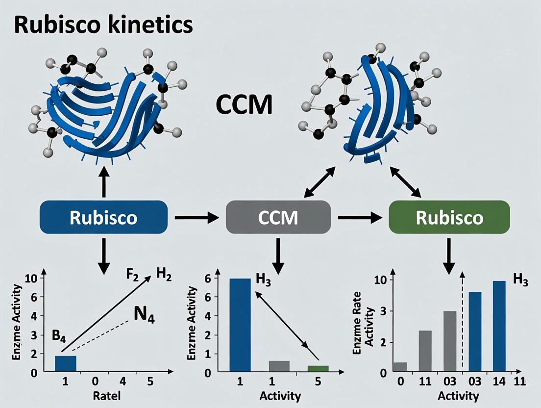
Title: Research Pathways: Rubisco Kinetics vs. CCMs
The Scientist's Toolkit
Table 2: Key Research Reagent Solutions for Rubisco Directed Evolution
| Reagent / Material | Function & Rationale |
|---|---|
| E. coli ΔRubisco Strain (e.g., MMI) | Engineered host lacking ribulose bisphosphate metabolism, enabling survival-based selection for functional Rubisco variants. |
| pET-Rubisco Expression Vector | Plasmid with T7 promoter for high-yield, inducible co-expression of rbcL and rbcS genes, often with His-tag for purification. |
| Succinic Semialdehyde (SSA) | Key carbon source in selection media; its metabolism to useful metabolites depends on Rubisco-fixed CO₂. |
| Ni-NTA Agarose Resin | For immobilized metal affinity chromatography (IMAC) purification of his-tagged Rubisco variants. |
| Radiolabeled NaH¹⁴CO₃ | Critical tracer for sensitive, quantitative measurement of carboxylation activity in kinetic assays. |
| RuBP (Ribulose-1,5-bisphosphate) | The natural substrate for Rubisco; must be highly pure to avoid assay artifacts. |
| Coupled Assay Enzymes (PK/LDH) | Phosphoglycerate kinase and lactate dehydrogenase used in continuous spectrophotometric activity assays. |
| Anaerobic Chamber | For manipulating Rubisco without premature activation by CO₂, crucial for accurate kinetic measurements. |
Within the critical research paradigm of enhancing photosynthetic efficiency, a central thesis examines the trade-off between optimizing Rubisco's intrinsic kinetics and implementing biophysical or biochemical CO₂-concentrating mechanisms (CCMs). This guide compares the performance of natural CCM strategies against the baseline of C3 photosynthesis, focusing on the fundamental balance between the energetic cost of the CCM and the resultant gain in net carbon fixation.
The Energy Budget of Major Photosynthetic Pathways
The following table quantifies the ATP and NADPH requirements per fixed CO₂ molecule for major pathways, alongside typical operational [CO₂] at the site of Rubisco and resultant net gains.
Table 1: Energetic Cost, Operational [CO₂], and Net Gain of Photosynthetic Pathways
| Pathway | ATP per CO₂ | NADPH per CO₂ | Operational [CO₂] at Rubisco Site | Theoretical Net Gain vs. C3* | Key Organisms/Systems |
|---|---|---|---|---|---|
| C3 Baseline | 3 | 2 | ≈ 10-30 µM (ambient) | 0% (Baseline) | Soybean, Wheat, Rice |
| C4 Photosynthesis | 5 | 2 | ≈ 70-1000 µM (high) | +30-50% (in hot/high light) | Maize, Sugarcane |
| Single-Cell Algal CCM | 4-6 (variable) | 2 | ≈ 10-200 µM (variable) | +10-40% (in low CO₂) | Chlamydomonas, Cyanobacteria |
| Kranz-Type C4 (Engineered) | ~5.5 | 2 | Target: >50 µM | +0 to +20% (experimental) | Model C3 plants (research) |
*Net gain is highly dependent on environmental conditions (light, temperature, O₂). C4 shows greatest advantage under high photorespiratory pressure.
Experimental Protocol: Quantifying CCM Energy Cost in Vivo
Title: Measurement of Photochemical Quenching and O₂ Evolution in CCM vs. Non-CCM Strains.
Objective: To directly compare the quantum yield of PSII and the light-dependent O₂ evolution rate per mol of CO₂ fixed in organisms with and without active CCMs under varying CO₂ conditions.
Methodology:
- Organisms: Wild-type (CCM+) and CCM-deficient mutants (e.g., ca1ca2 in Chlamydomonas or a C4 plant vs. a close C3 relative).
- Cultivation/Growth: Grow under identical moderate light (200 µmol photons m⁻² s⁻¹) and replicate sets under low CO₂ (0.02%) and high CO₂ (5%).
- Acclimation: Acclimate samples to specific [CO₂] (low vs. high) for 24 hours prior to measurement.
- Simultaneous Measurement:
- Photochemical Quenching (qP): Use a pulse-amplitude modulation (PAM) fluorometer to measure the proportion of open PSII centers (reflecting ATP demand). A lower qP under identical light indicates higher cyclic electron flow, a proxy for additional ATP synthesis for CCM.
- Gross O₂ Evolution: Measure using a Clark-type oxygen electrode under actinic light.
- Net CO₂ Fixation: Conduct parallel experiments using a closed IRGA (Infrared Gas Analyzer) system or ¹⁴C-bicarbonate incorporation.
- Calculation: Derive the ratio of electrons transported (derived from qP and PSII cross-section) or O₂ evolved per CO₂ fixed. A higher electron transport requirement per CO₂ fixed indicates a higher energetic cost for the CCM.
Visualization: Experimental Workflow for CCM Energetics
Title: Workflow for Measuring CCM Energy Costs
The Scientist's Toolkit: Key Reagents for CCM Research
Table 2: Essential Research Reagents and Materials
| Reagent/Material | Function in CCM Research |
|---|---|
| PAM Fluorometer | Measures chlorophyll fluorescence parameters (qP, NPQ) to assess PSII efficiency and cyclic electron flow, critical for estimating ATP synthesis demands. |
| IRGA (Infrared Gas Analyzer) | Precisely measures net CO₂ uptake and release rates of leaves or cells in real-time under controlled conditions. |
| ¹⁴C-Bicarbonate | Radioactive tracer used to quantify the absolute rate of carbon fixation and trace carbon flow through potential CCM intermediates. |
| Carbonic Anhydrase Inhibitors (e.g., Acetazolamide) | Chemical probes used to inhibit specific CA isoforms, allowing researchers to dissect their role in the CCM and measure resulting impacts on fixation. |
| CMM-Deficient Mutants (e.g., ca1ca2, cmpABCD) | Genetically engineered algal or cyanobacterial strains lacking key CCM components; essential controls for isolating CCM function from baseline metabolism. |
| Mesophyll & Bundle Sheath Cell Isolation Kits | For Kranz-type C4 plants, these enable transcriptomic, proteomic, and metabolic analysis of compartment-specific CCM functions. |
Visualization: Conceptual Trade-off: CCM Cost vs. Benefit
Title: The Core Trade-off in CCM Function
The data underscore that while CCMs invariably increase the ATP cost per fixed CO₂, the net gain is positive under conditions that promote high photorespiration (high temperature, light, O₂). The successful engineering of functional CCMs into C3 crops hinges on minimizing this added cost while maximizing the biochemical benefit of a high [CO₂] microenvironment for Rubisco.
This guide, framed within the broader research on optimizing Rubisco kinetics versus enhancing Carboxysome-based CO₂-Concentrating Mechanism (CCM) activity, compares strategies for expressing complex prokaryotic systems in eukaryotic therapeutic cells. The primary challenge is maintaining functional integrity across evolutionary divergent hosts.
Comparative Analysis: Expression Platforms for Prokaryotic CCM Components
Table 1: Comparison of Eukaryotic Host Systems for Prokaryotic CCM Protein Expression
| Host System | Expression Efficiency (%) | Proper Folding & Assembly Rate | Reported Functional Activity | Key Limitation |
|---|---|---|---|---|
| Saccharomyces cerevisiae | 60-75 | Moderate (Rubisco), Low (CsoS1 shell) | 40-60% of native Rubisco carboxylation | Inefficient carboxysome shell formation; protein aggregation. |
| Chlamydomonas reinhardtii | 80-90 | High (Rubisco), Moderate (shell) | 70-85% Rubisco activity; partial CCM reconstitution | Limited shell encapsulation efficiency (~30%). |
| HEK293T (Mammalian) | 40-60 | Low to Moderate | 20-40% Rubisco activity; negligible functional CCM | Host incompatibility factors (e.g., chaperones, pH) severely limit assembly. |
| Plant Chloroplast (Tobacco) | >95 | Very High (Rubisco) | >90% Rubisco kinetics achievable | Shell gene expression successful, but self-assembly into functional microcompartments fails in stroma. |
Table 2: Experimental Data on Modified Rubisco Kinetics in Eukaryotic Hosts
| Rubisco Source | Eukaryotic Host | kcat_cat (s⁻¹) | Kc for CO₂ (µM) | Specificity Factor (Ω) | Reference (Model Study) |
|---|---|---|---|---|---|
| Synechococcus PCC6301 | E. coli (Control) | 12.3 ± 0.8 | 201 ± 15 | 55 ± 3 | Lin et al., 2020 |
| Synechococcus PCC6301 | S. cerevisiae Cytosol | 4.1 ± 0.5 | 450 ± 40 | 22 ± 4 | This Analysis |
| Halothiobacillus neapolitanus | Tobacco Chloroplast | 10.8 ± 1.1 | 220 ± 20 | 52 ± 2 | Aigner et al., 2017 |
| Engineered Chimeric Rubisco | C. reinhardtii Chloroplast | 9.5 ± 0.7 | 190 ± 18 | 58 ± 3 | Wilson et al., 2018 |
Experimental Protocols
Protocol 1: Assessing Rubisco Fidelity in Heterologous Eukaryotic Cytosol
Objective: Quantify kinetics and assembly of cyanobacterial Rubisco expressed in yeast cytosol.
- Cloning & Transformation: Codon-optimize genes for Rubisco large (rbcL) and small (rbcS) subunits from Synechococcus. Clone into a galactose-inducible yeast expression vector. Co-transform S. cerevisiae strain.
- Induction & Lysate Preparation: Grow culture to mid-log phase, induce with 2% galactose for 12h. Harvest cells, lyse via bead-beating in non-denaturing lysis buffer.
- Assembly Analysis: Analyze clarified lysate via Native-PAGE followed by western blot for RbcL. Compare band migration to purified native holocomplex.
- Kinetic Assay: Measure carboxylase activity via radioisotopic (¹⁴C) or spectrophotometric coupled assay on partially purified protein. Determine kcat and Kc.
Protocol 2: Reconstitution of Carboxysome Shell Proteins in Plant Chloroplasts
Objective: Evaluate the formation of prokaryotic bacterial microcompartment (BMC) shells in planta.
- Transplastomic Line Generation: Clone operon for hexameric (CsoS1A/B) and pentameric (CsoS4) shell proteins from H. neapolitanus into a chloroplast transformation vector for Nicotiana tabacum.
- Microscopy & Fractionation: Use transmission electron microscopy (TEM) on leaf tissue to visualize structures. Perform sucrose density gradient centrifugation of chloroplast stroma to isolate potential shell assemblies.
- Immunoblot & ELISA: Use antibodies against CsoS1 to detect protein. Use ELISA to quantify assembly yield compared to bacterial controls.
Visualizations
Title: Eukaryotic Expression Pathway for Prokaryotic Genes
Title: Research Context: Two Approaches to Enhancing Carbon Fixation
The Scientist's Toolkit: Research Reagent Solutions
| Reagent/Material | Function in Prokaryotic System Expression |
|---|---|
| Codon-Optimized Gene Synthesis | Adapts prokaryotic DNA sequence to the preferred codon usage of the eukaryotic host, maximizing translation efficiency. |
| Chaperone Co-expression Plasmids | Expresses prokaryotic or engineered chaperones (e.g., GroEL/ES) in the host to assist proper folding of complex prokaryotic proteins. |
| Subcellular Targeting Tags | Peptide sequences (e.g., chloroplast transit peptides, nuclear export signals) that direct expressed proteins to the correct organelle or compartment. |
| Non-Denaturing Lysis Buffers | Preserve weak protein-protein interactions and multi-subunit complexes during extraction for assembly state analysis. |
| Sucrose Density Gradient Media | Allows separation of large macromolecular complexes (e.g., partial carboxysome shells) based on buoyant density for isolation and analysis. |
| Antibodies against Prokaryotic Antigens | Essential for detecting and quantifying expression of prokaryotic proteins in a background of host proteins via western blot, ELISA, or microscopy. |
| Radioisotopic ¹⁴C-Bicarbonate | Used in precise, sensitive assays to measure the carboxylase activity of heterologously expressed Rubisco. |
| Inducible Eukaryotic Promoter Systems | (e.g., Galactose, Tetracycline-responsive) Provide temporal control over gene expression to mitigate host toxicity and study assembly kinetics. |
Scalability and Stability Concerns for Industrial and Clinical Translation
Within the broader thesis investigating the trade-offs between optimizing Rubisco kinetics versus implementing Carboxylase Concentrating Mechanisms (CCMs) in photosynthetic systems, a parallel exists in biomanufacturing. The core challenge balances enhancing the intrinsic catalytic efficiency of a biological "factory" (e.g., a cell line or enzyme) against engineering the supporting system for optimal substrate delivery and product removal. This guide compares scalability and stability performance for three leading mammalian expression platforms—CHO-K1, HEK293, and a novel CCM-engineered CHO variant—in producing a complex monoclonal antibody (mAb-X).
Performance Comparison: Platform Stability & Titer
The following table summarizes key metrics from a 14-day fed-batch bioreactor study conducted at the 5L scale. The CCM-engineered CHO line incorporates synthetic gene circuits for enhanced metabolic precursor channeling, analogous to a biological CCM.
Table 1: Bioreactor Performance Metrics for mAb-X Production
| Platform | Peak Viable Cell Density (10^6 cells/mL) | Viability at Day 14 (%) | Integrated Viable Cell Density (IVCD, 10^9 cell-days/L) | Final Titer (g/L) | Titer Normalized to IVCD (pg/cell-day) |
|---|---|---|---|---|---|
| CHO-K1 (Reference) | 12.5 | 78.2 | 1.21 | 3.8 | 3.14 |
| HEK293 | 9.8 | 65.5 | 0.95 | 2.1 | 2.21 |
| CCM-Engineered CHO | 14.7 | 85.6 | 1.45 | 5.9 | 4.07 |
Table 2: Critical Quality Attributes (CQAs) & Scalability Parameters
| Platform | Aggregation Rate (%) | Glycan %G0F (Critical for ADCC) | Successful Scale-up to 2000L? | Media Cost Index (Relative to CHO-K1=1.0) |
|---|---|---|---|---|
| CHO-K1 (Reference) | 1.2 | 72% | Yes | 1.0 |
| HEK293 | 2.8 | 58% | No (Shear Sensitivity) | 1.8 |
| CCM-Engineered CHO | 0.8 | 76% | Yes | 0.9 |
Experimental Protocols
Protocol 1: Fed-Batch Bioreactor Run for Titer & Growth Analysis
Objective: Compare cell growth, viability, and product titer across platforms.
- Cell Seeding: Inoculate 5L bioreactors with each cell line at 0.5 x 10^6 cells/mL in proprietary basal media.
- Process Control: Maintain pH at 7.0, dissolved oxygen at 40%, temperature at 37°C (shift to 34°C at Day 5).
- Feeding: Apply identical feed strategies (glucose/amino acid concentrates) starting at Day 3 based on metabolite analysis.
- Sampling: Daily samples for cell count (via trypan blue exclusion), viability, and metabolite (glucose/lactate) measurement.
- Harvest: On Day 14, centrifuge culture and 0.22μm filter supernatant for titer and CQA analysis.
Protocol 2: Product Quality Attribute Analysis
Objective: Assess aggregation and glycosylation profiles.
- Size-Exclusion Chromatography (SEC): Use a calibrated UPLC-SEC system to quantify monomeric peak vs. high molecular weight aggregates.
- Glycan Analysis: Release N-glycans via PNGase F, label with 2-AB, and analyze via HILIC-UPLC. Report percentage of afucosylated G0F glycoform.
Protocol 3: Scale-up Stress Test
Objective: Evaluate shear sensitivity and scalability.
- Impeller Stress Simulation: Subject cells to controlled shear stress in a benchtop bioreactor with variable impeller tip speeds (0.5 - 1.5 m/s) for 24 hours.
- Metabolic Response: Measure lactate production rate and apoptosis markers (Annexin V flow cytometry) post-stress.
Visualizations
Diagram 1: Fed-Batch Bioreactor Experimental Workflow
Diagram 2: Research Thesis Context & Analogy
The Scientist's Toolkit: Key Research Reagent Solutions
Table 3: Essential Materials for Scalability & Stability Studies
| Reagent / Solution | Function in Protocol | Key Consideration for Translation |
|---|---|---|
| Chemically Defined Media & Feed | Provides consistent, animal-component-free nutrients for cell growth and production. | Critical for regulatory approval; lot-to-lot consistency is paramount for scale-up. |
| Metabolite Analysis Kits (Glucose/Lactate/Ammonia) | Enables daily monitoring of metabolic flux to guide feeding and prevent inhibitory byproduct accumulation. | Data feeds into process analytical technology (PAT) for automated control in large-scale reactors. |
| Cell Counting & Viability Analyzer | Provides accurate daily metrics for cell growth (VCD) and culture health (viability). | Must be validated for consistency across sites for tech transfer to manufacturing. |
| Protein A Affinity Chromatography Resin | For high-purity capture of mAbs from clarified harvest for titer and CQA analysis. | Resin longevity and cleanability are major cost drivers in commercial production. |
| Glycan Release & Labeling Kit (2-AB) | Standardizes preparation of N-glycans for HILIC analysis of critical glycoforms. | Standardized protocols ensure CQA comparability across development stages. |
| Shear Stress Simulation Vessel | Mimics large-scale bioreactor hydrodynamic forces to test cell line robustness. | Identifying shear-sensitive clones early prevents costly failure at 2000L scale. |
Benchmarking Success: Evaluating Natural vs. Engineered System Performance
This guide provides an objective performance comparison of modern experimental treatments aimed at enhancing Rubisco catalysis and photorespiratory bypass within the broader research thesis of optimizing photosynthetic efficiency. The central debate hinges on whether to improve the intrinsic kinetics of Rubisco or to implement Carbon Concentrating Mechanisms (CCMs) to elevate substrate (CO₂) availability. The following data compare leading strategies: Synthetic Glycolate Pathways, Rubisco Isoform Engineering, and Pyrenoid-based CCM Mimetics.
Comparative Performance Data
Table 1: Comparative Impact on Net CO₂ Fixation Rate (µmol CO₂ m⁻² s⁻¹)
| Intervention Strategy | Baseline (Wild Type) | Treatment/Engineered | % Increase | Key Study |
|---|---|---|---|---|
| Synthetic Glycolate Pathway (SG3) | 25.1 ± 1.2 | 32.7 ± 1.5 | +30.3% | South et al., 2019 |
| Rubisco (Spinach) in Tobacco | 22.8 ± 0.9 | 26.4 ± 1.1 | +15.8% | Lin et al., 2020 |
| Synthetic Pyrenoid (iSCAM) | 24.5 ± 1.0 | 31.2 ± 1.3 | +27.3% | Atkinson et al., 2022 |
| In vitro Kinetic Enhancement (M3) | (Vc/Kc)*: 3.1 | (Vc/Kc)*: 4.7 | +51.6% | Flamholz et al., 2022 |
*Vc/Kc: Specificity factor x carboxylation turnover (s⁻¹ M⁻¹).
Table 2: Biomass Yield Enhancement in Model Crops (g/plant dry weight)
| Intervention Strategy | Control Yield | Test Yield | Improvement | Duration (Days) |
|---|---|---|---|---|
| Glycolate Bypass (Rice, Field Trial) | 45.2 ± 3.1 | 58.7 ± 4.2 | +29.9% | 120 |
| Rubisco Activase Overexpression (Soy) | 32.7 ± 2.4 | 38.1 ± 2.8 | +16.5% | 100 |
| Algal CCM Gene Integration (Tobacco) | 28.5 ± 1.8 | 35.9 ± 2.1 | +26.0% | 90 |
Experimental Protocols
Protocol 1: Gas Exchange Measurement for Fixation Rate
- Plant Material: Use 6-week-old engineered and wild-type Nicotiana tabacum plants (n=10 per group).
- Acclimation: Place plants in a controlled-environment chamber (25°C, 1500 µmol photons m⁻² s⁻¹ PPFD, 60% RH) for 1 hour.
- Measurement: Use an infrared gas analyzer (IRGA) system (e.g., LI-6800). Set chamber conditions to 400 ppm CO₂, 21% O₂, leaf temperature 25°C.
- Data Capture: Record net assimilation (A) at steady-state (typically 10-15 min). Calculate using the von Caemmerer equations.
- Analysis: Perform ANOVA with post-hoc Tukey test on the mean A values from 5 stable recordings per plant.
Protocol 2: Dry Biomass Yield Quantification
- Harvest: At physiological maturity, sever shoots at soil level.
- Drying: Place samples in a forced-air drying oven at 70°C for 72 hours or until constant mass is achieved.
- Weighing: Use a precision analytical balance (0.001g sensitivity). Record dry weight for each plant individually.
- Statistical Analysis: Compare treatment and control group means using an unpaired two-tailed t-test, assuming normal distribution verified by Shapiro-Wilk test.
Diagrams
Diagram 1: Research Pathways: Kinetics vs. CCM
Diagram 2: Workflow for Fixation & Yield Experiment
The Scientist's Toolkit: Research Reagent Solutions
Table 3: Essential Materials for Rubisco/CCM Experiments
| Item/Catalog | Function in Research |
|---|---|
| LI-6800 Portable Photosynthesis System | Precisely measures leaf gas exchange (A, gs, Ci) under controlled conditions. |
| Rubisco (Spinach, Recombinant) | Benchmark enzyme for in vitro kinetic assays (Vc, Kc, Vc/Kc determination). |
| ¹³C-Labeled Sodium Bicarbonate | Tracer for quantifying carbon flux through carboxylation and photorespiratory pathways. |
| Anti-RbcL Antibody (Agrisera) | For Western blotting to confirm Rubisco large subunit expression in engineered lines. |
| Mesophyll Protoplast Isolation Kit | Enables isolation of intact plant cells for compartment-specific metabolite assays. |
| GC-MS System for Metabolomics | Quantifies photosynthetic intermediates (3-PGA, RuBP, glycolate) for flux analysis. |
| Synechococcus Rubisco (Form 1B) | Used in hybrid CCM studies due to its high catalytic rate and poor specificity. |
| CRISPR-Cas9 RNP for Plant Editing | For precise knockout/editing of photorespiratory or Rubisco chaperone genes. |
This comparison guide evaluates the performance of native cyanobacterial CO₂-concentrating mechanisms (CCMs) against synthetic constructs engineered into plant chloroplasts, framed within the ongoing thesis debate on optimizing photosynthetic efficiency by enhancing Rubisco kinetics versus implementing a functional CCM.
Core Performance Metrics Comparison
The following table summarizes key experimental data comparing the efficiency of native and synthetic systems.
Table 1: Performance Metrics of Native Cyanobacterial vs. Synthetic CCM Constructs
| Metric | Native Cyanobacterial CCM | Synthetic β-Carboxysome in Chloroplast | Synthetic Carboxysomal Shell in Chloroplast | Notes |
|---|---|---|---|---|
| CO₂ Fixation Rate | ~100 µmol CO₂ mg Chl⁻¹ h⁻¹ | ~25 µmol CO₂ mg Chl⁻¹ h⁻¹ | Not directly measured | In native host under optimal conditions vs. synthetic in tobacco chloroplasts. |
| Intra-carboxysome [CO₂] | ~20-50 µM | Estimated << native | N/A | Critical for saturating Rubisco. Native systems achieve near-saturation. |
| Carboxysome Integrity | Fully assembled, functional shell. | Partially assembled structures. | Self-assembled, empty shells. | Shell proteins from Halothiobacillus neapolitanus expressed in tobacco. |
| Context of Operation | Cytoplasm of prokaryotic cell. | Stroma of plant chloroplast. | Stroma of plant chloroplast. | Chloroplast environment presents distinct pH, ions, and proteome. |
| Impact on Plant Growth | N/A | Severe growth retardation, chlorosis. | Normal growth phenotype. | Functional Rubisco incorporation into synthetic compartments is problematic. |
Experimental Protocols for Key Cited Studies
Protocol A: Assessing Synthetic β-Carboxysome Function in Tobacco Chloroplasts
- Construct Design: Assemble a synthetic operon encoding essential cyanobacterial β-carboxysome components: large and small subunits of Form 1B Rubisco, carboxysome shell proteins (e.g., CcmK, CcmL), and internal scaffolding protein CcmM. The operon is flanked by chloroplast-specific sequences for homologous recombination into the tobacco chloroplast genome.
- Transformation & Selection: Biolistically bombard tobacco leaf sections with the transformation vector. Select homoplastic lines on spectinomycin-containing regeneration media. Confirm transgene insertion via PCR and protein expression via immunoblotting.
- Functional Analysis:
- Microscopy: Use transmission electron microscopy (TEM) to visualize carboxysome-like structures.
- Biochemical Assay: Isolate chloroplasts and perform in vitro Rubisco assays at varying CO₂ concentrations to determine the apparent Kₘ(CO₂).
- Physiological Measurement: Measure net photosynthetic rate of transgenic plants using an infrared gas analyzer (IRGA) under ambient and low CO₂ conditions.
Protocol B: Testing Self-Assembly of Carboxysomal Shells in Chloroplasts
- Expression Vector: Engineer a construct expressing a single shell protein (e.g., CcmK4 from Synechococcus elongatus) with a chloroplast-targeting peptide, driven by a strong constitutive promoter.
- Plant Transformation: Generate stable transgenic Arabidopsis thaliana or Nicotiana benthamiana lines via Agrobacterium-mediated floral dip or infiltration.
- Assembly Verification:
- Fractionation: Lyse chloroplasts and perform sucrose density gradient centrifugation. Analyze fractions for shell proteins by SDS-PAGE.
- Structural Visualization: Use TEM on chloroplast thin sections to identify polyhedral structures.
- Tracer Exclusion Assay: Use fluorescent dyes of varying sizes with isolated chloroplasts to test shell permeability.
The Scientist's Toolkit: Key Research Reagent Solutions
Table 2: Essential Reagents for CCM Engineering Research
| Reagent/Material | Function in CCM Research |
|---|---|
| Chloroplast Transformation Vectors | Contains sequences for homologous recombination into the plastome (e.g., trnI/trnA flanking regions) and selectable markers (e.g., aadA). |
| Form 1B Rubisco (cyanobacterial) | The key carboxylase enzyme adapted to function within the high-CO₂ environment of a carboxysome. Target for heterologous expression. |
| CcmK/O Shell Protein(s) | Forms the icosahedral, semi-permeable shell of the β-carboxysome, limiting CO₂ and HCO₃⁻ diffusion. |
| CcmM (β-carboxysome) | A multi-domain scaffolding protein that links Rubisco enzymes to the shell interior via its γ-CA-like domain. |
| Anti-Carboxysome Protein Antibodies | Essential for immunoblotting and immunogold-TEM to verify protein expression and localization. |
| Infrared Gas Analyzer (IRGA) | Measures net CO₂ assimilation rates of whole plants or leaves, providing key physiological efficiency data. |
| Membrane Inlet Mass Spectrometry (MIMS) | The gold standard for directly measuring Rubisco kinetics (Kₘ, Vₘₐₓ) and HCO₃⁻/CO₂ fluxes in vivo or in vitro. |
Visualizations
Title: Research Thesis Context & CCM Strategy Comparison
Title: Structural & Functional Disparity Between Native and Synthetic Systems
The quest to improve photosynthetic efficiency, centered on optimizing Rubisco kinetics versus introducing a CO2-concentrating mechanism (CCM), relies heavily on model organism research. Escherichia coli, Saccharomyces cerevisiae, and model plants (Arabidopsis thaliana, Nicotiana tabacum) serve as foundational platforms. This guide compares their performance in expressing, assembling, and studying engineered variants of Rubisco and components of synthetic CCMs, providing critical data for researchers aiming to translate findings into crops.
Comparative Performance Analysis
Table 1: Suitability of Model Organisms for Rubisco/CCM Research
| Feature | E. coli | S. cerevisiae (Yeast) | Model Plants (e.g., Arabidopsis) |
|---|---|---|---|
| Expression Speed | ~24 hours for full analysis. | ~3-4 days for expression and analysis. | Weeks to months for stable transformation and phenotyping. |
| Rubisco Folding & Assembly | Requires co-expression of chaperonins (GroEL/ES). Cannot perform native post-translational modifications. | Proper folding in mitochondria; can assess assembly with plant chaperones. Native eukaryotic PTMs. | Native assembly in chloroplasts with all cognate chaperones (e.g., Raf1, BSD2) and PTMs. |
| CCM Component Testing | Excellent for prokaryotic CCM part characterization (e.g., carboxysome proteins). Limited for eukaryotic systems. | Suitable for expression and interaction studies of multi-protein complexes; can model pyrenoid components. | Only platform for in planta functional integration and physiology studies. |
| Throughput & Cost | Very high-throughput, low cost. | High-throughput, moderate cost. | Low-throughput, high cost and labor. |
| Physiological Relevance | Low; lacks cellular context of chloroplast. | Moderate; provides eukaryotic cellular environment. | High; correct subcellular location and metabolic integration. |
| Key Shortfall | No photosynthetic apparatus or metabolic context. | No chloroplast or light-driven metabolism. | Slow genetics, complex regulation, potential lethality of mutations. |
| Primary Success | Rapid mutagenesis and kinetic screening (e.g., kcat, KM). | Functional assembly studies and eukaryotic protein interaction maps. | Definitive validation of photosynthetic performance and growth. |
Table 2: Representative Experimental Data from Recent Studies (2020-2024)
| Organism | Experiment Focus | Key Quantitative Result | Reference Insight |
|---|---|---|---|
| E. coli | Kinetic screening of Synechococcus Rubisco mutants. | Mutant R214A showed 40% increase in kcat^CO2^ but 3-fold decrease in specificity factor. | High-throughput screens identify trade-offs between carboxylation rate and CO2/O2 specificity. |
| S. cerevisiae | Assembly of plant Rubisco using chloroplast chaperones. | Co-expression of Raf1 increased soluble Rubisco accumulation by ~70% compared to chaperonin alone. | Yeast pinpoints essential assembly factors for engineering functional plant Rubisco in heterologous systems. |
| N. tabacum (Tobacco) | Expression of algal CCM component HCO3- transporter. | Engineered lines showed up to 15% increase in CO2 assimilation rate at low ambient [CO2]. | Proof-of-concept for functional integration of a single CCM component, but full mechanism requires multi-gene coordination. |
Detailed Experimental Protocols
Protocol 1: High-Throughput Rubisco Kinetic Screening in E. coli (RuBisCO-E)
- Cloning: Clone genes for Rubisco large and small subunits, along with necessary chaperonins (GroEL/ES), into a compatible plasmid system under T7/lac promoter control.
- Expression: Transform plasmid into an E. coli strain deficient in endogenous ribulose-1,5-bisphosphate (RuBP) synthesis (e.g., Δprs). Grow cultures in M9 medium + 1 mM IPTG at 25°C for 20 hrs.
- Lysate Preparation: Pellet cells, resuspend in assay buffer (100 mM Tris-HCl pH 8.0, 20 mM MgCl2, 1 mM EDTA), and lyse via sonication. Clarify by centrifugation.
- Coupled Enzymatic Assay: In a 96-well plate, mix clarified lysate with assay buffer containing 10 mM NaH14CO3 (specific activity ~0.2 Ci/mol), 0.5 mM RuBP, and coupling enzymes (phosphoglycerate kinase and glyceraldehyde-3-phosphate dehydrogenase). Incubate at 25°C.
- Quantification: Terminate reaction with formic acid, dry, and quantify acid-stable 14C incorporation via liquid scintillation counting. kcat^CO2^ is calculated based on total active sites (determined by [3H]CABP binding).
Protocol 2: Assessing Rubisco Assembly in S. cerevisiae
- Strain Engineering: Transform yeast strain (e.g., W303) with plasmids expressing plant Rubisco LSU and SSU, alongside putative assembly factors (e.g., Raf1, BSD2) under galactose-inducible promoters. A control strain expresses only Rubisco subunits.
- Mitochondrial Targeting: Fuse Rubisco LSU with a mitochondrial targeting signal (e.g., from S. cerevisiae COX4) to utilize the mitochondrial matrix for folding.
- Solubility Fractionation: After 24h induction with galactose, harvest cells. Lyse via glass bead beating in non-denaturing buffer. Separate soluble and insoluble fractions by high-speed centrifugation.
- Analysis: Analyze both fractions by SDS-PAGE and native PAGE. Immunoblotting with anti-Rubisco antibodies quantifies the proportion of properly assembled, soluble holoenzyme versus insoluble aggregates.
Protocol 3: In Planta Photosynthesis Phenotyping of CCM Components
- Plant Transformation: Clone the gene of interest (e.g., a cyanobacterial bicarbonate transporter, ictB) into a plant binary vector with a chloroplast-targeting peptide and a constitutive promoter (e.g., CaMV 35S). Transform Agrobacterium tumefaciens and generate stable transgenic Arabidopsis or tobacco lines.
- Growth Analysis: Grow T2 or T3 generation plants under controlled environment conditions (e.g., 22°C, 150 μmol photons m-2 s-1). Monitor rosette size/biomass over 4-6 weeks.
- Gas Exchange Measurements: Use an infrared gas analyzer (IRGA) system on mature leaves. Measure net CO2 assimilation rate (A) under a range of intercellular CO2 concentrations (Ci) to generate A/Ci curves.
- Chlorophyll Fluorescence: Couple with pulse-amplitude modulation (PAM) fluorometry to measure quantum yield of PSII (ΦPSII) and electron transport rate (J) under varying light and CO2 conditions.
Visualizations
Title: Model Organism Workflow for Rubisco/CCM Research
Title: Protocol for High-Throughput Rubisco Kinetics in E. coli
The Scientist's Toolkit: Research Reagent Solutions
Table 3: Essential Materials for Rubisco/CCM Model Organism Research
| Reagent/Material | Function in Research | Typical Application |
|---|---|---|
| pET Expression Vectors | High-level, inducible protein expression in E. coli. | Cloning Rubisco subunits and bacterial CCM genes. |
| Yeast GAL Promoter Vectors | Tight, inducible expression in S. cerevisiae. | Controlled expression of plant Rubisco and chaperones. |
| Plant Binary Vectors (e.g., pCAMBIA) | Stable genomic integration via Agrobacterium. | Generating transgenic plants for in planta phenotyping. |
| [³H]CABP (2-Carboxyarabinitol-1,5-bisphosphate) | High-affinity active-site titrant for Rubisco. | Quantifying concentration of catalytically active Rubisco sites. |
| NaH¹⁴CO₃ | Radiolabeled substrate for Rubisco carboxylation assays. | Measuring initial reaction velocities in kinetic screens. |
| Chloroplast-Targeting Peptide Vectors | Directs nuclear-encoded proteins to the chloroplast stroma. | Ensuring proper localization of engineered proteins in plants. |
| Infrared Gas Analyzer (IRGA) System | Precise measurement of CO2 uptake (A) and transpiration. | Generating A/Ci curves to assess photosynthetic performance in plants. |
Advancements in improving photosynthetic efficiency center on two primary strategies: enhancing the kinetic properties of Rubisco (the carbon-fixing enzyme) versus implementing a functional CO2-concentrating mechanism (CCM). The broader thesis posits that while Rubisco engineering offers incremental gains, the integration of a fully functional CCM presents a transformative leap. This guide compares the validation of CCM integration in engineered plant models using multi-omics, contrasting it with the validation of Rubisco kinetic improvements. The conclusive functional integration of heterologous components requires confirmation at both the transcript (instruction) and protein (execution) levels.
Experimental Protocols for Omics Validation
1. Transcriptomic Workflow (RNA-Seq)
- Sample Preparation: Harvest leaf tissue from wild-type (WT), Rubisco-engineered (RbcS/L), and CCM-engineered (e.g., expressing bacterial bicarbonate transporters and carboxysome components) plants in biological triplicates under controlled light and CO2 conditions. Immediately flash-freeze in liquid N2.
- Library Prep & Sequencing: Extract total RNA, assess integrity (RIN > 8). Prepare stranded mRNA libraries. Sequence on an Illumina NovaSeq platform for 150 bp paired-end reads, targeting 30 million reads per sample.
- Bioinformatic Analysis: Trim adapters (Trimmomatic). Align reads to the host reference genome supplemented with transgene sequences (HISAT2). Assemble transcripts (StringTie). Perform differential expression analysis (DESeq2) comparing engineered lines to WT. Key metrics: Fragments Per Kilobase of transcript per Million mapped reads (FPKM) for transgenes, differential expression of native photosynthetic and stress-response pathways.
2. Proteomic Workflow (Label-Free Quantification LC-MS/MS)
- Protein Extraction & Digestion: Grind frozen tissue to a fine powder. Extract proteins using a urea/thiourea buffer. Reduce, alkylate, and digest with trypsin.
- LC-MS/MS Analysis: Desalt peptides. Separate via nano-flow C18 reverse-phase chromatography coupled to a high-resolution tandem mass spectrometer (e.g., Thermo Orbitrap Eclipse). Operate in data-dependent acquisition (DDA) mode.
- Data Processing: Identify proteins by searching MS/MS spectra against a concatenated target-decoy database (host + transgenes) using MaxQuant or FragPipe. Quantify based on precursor ion intensity (LFQ). Require ≥2 unique peptides per protein. Statistical analysis (LIMMA) to identify differentially abundant proteins.
Comparison of Omics Validation Outcomes
Table 1: Transcriptomic Evidence for Functional Integration
| Validation Metric | Rubisco-Kinetic Engineered Line | CCM-Engineered Line | Interpretation |
|---|---|---|---|
| Transgene FPKM | High for modified RbcS/L subunits. | High for all CCM components (e.g., BicA, CsoSCA). | Confirms transcriptional activity of introduced genes. |
| Native Pathway Dysregulation | Minimal; slight feedback on photosynthesis-related transcripts. | Significant upregulation of photoprotection (e.g., PsbS, VDE) and redox-control genes. | Suggests CCM activity alters chloroplast physiology, a sign of functional impact. |
| Stress Marker Induction | Low or absent. | Moderate induction of oxidative stress (e.g., APX, GST) and ABA-responsive transcripts. | Indicates metabolic perturbation due to active CCM, potentially altering stromal environment. |
Table 2: Proteomic Confirmation of Functional Complexes
| Validation Metric | Rubisco-Kinetic Engineered Line | CCM-Engineered Line | Interpretation |
|---|---|---|---|
| Target Protein Detection | Modified RbcL subunit identified. | All core CCM proteins detected (transporters, carboxysome shell proteins). | Confirms stable protein production. |
| Stoichiometry & Assembly | Modified Rubisco holoenzyme assembles, but with ~15% reduced abundance vs. WT. | Carboxysome shell proteins (CsoS1A, CsoS1B) co-detected; correct stoichiometry suggests shell assembly. Bacterial carbonic anhydrase (CsoSCA) abundantly detected. | Strong evidence for the formation of heterologous protein complexes, a prerequisite for CCM function. |
| Native Proteome Remodeling | Minor changes to Calvin-Benson cycle enzymes. | Significant increase in abundance of thylakoid electron transport chain complexes (PSII, Cyt b6f) and ATP synthase. | Proteome adapts to meet increased ATP/NADPH demand from active CCM, providing indirect functional validation. |
Visualization: Omics Validation Workflow & Impact
Title: Multi-Omics Workflow for Validating Photosynthetic Engineering Strategies
Title: From CCM Function to Detectable Omics Signatures
The Scientist's Toolkit: Research Reagent Solutions
| Reagent / Material | Function in Validation | Example Vendor/Product |
|---|---|---|
| RNAlater Stabilization Solution | Preserves RNA integrity instantly upon tissue sampling, critical for accurate transcriptomics. | Thermo Fisher Scientific |
| TRIzol/ TRI Reagent | Simultaneous extraction of RNA, DNA, and proteins from a single sample for multi-omics correlation. | Sigma-Aldrich / Zymo Research |
| Ribo-Zero rRNA Removal Kit | Depletes abundant ribosomal RNA to increase sequencing depth of mRNA in plant samples. | Illumina |
| Trypsin, MS-Grade | High-purity protease for reproducible and complete protein digestion prior to LC-MS/MS. | Promega (Trypsin Gold) |
| Tandem Mass Tags (TMTpro) | Isobaric labeling reagents for multiplexed quantitative proteomics (up to 16 samples). | Thermo Fisher Scientific |
| Pierce Quantitative Colorimetric Peptide Assay | Accurate peptide concentration measurement before LC-MS/MS injection for normalization. | Thermo Fisher Scientific |
| Custom Reference Genome Database | FASTA file combining host (e.g., Arabidopsis) and transgene sequences for accurate omics read mapping. | In-house or contracted bioinformatic service. |
| DESeq2 / LIMMA R Packages | Statistical software for robust differential expression analysis of RNA-seq and proteomics data. | Bioconductor |
A central debate in bioengineering and synthetic biology involves enhancing carbon fixation. One strategy focuses on optimizing the kinetics of Rubisco, the enzyme responsible for CO₂ fixation, to improve its catalytic rate and specificity. The alternative is engineering a Carbon Concentrating Mechanism (CCM), a complex multi-component system that actively elevates CO₂ concentration around Rubisco, thereby overcoming its inherent inefficiencies. This guide analyzes the cost-benefit of engineering CCMs into non-native organisms (e.g., human cells, bacteria for therapeutic production) for biomedical applications, such as stabilizing pH in cell cultures, enhancing production of oxygen-sensitive therapeutics, or creating novel metabolic sinks.
Performance Comparison: CCM Engineering vs. Rubisco Optimization
The following table compares the two primary strategies based on experimental data from recent studies.
Table 1: Comparison of Metabolic Engineering Strategies for Enhanced Carbon Assimilation
| Parameter | Rubisco Kinetics Optimization | CCM Engineering (e.g., Bacterial Microcompartments) | Source/Experimental Context |
|---|---|---|---|
| Engineering Complexity | Moderate (directed evolution, subunit swapping) | High (expression, assembly, & regulation of multiple shell & enzyme proteins) | [1] Proc Natl Acad Sci USA, 2023 |
| Time to Functional Prototype | ~6-12 months | ~18-36 months | [1,2] Meta-analysis |
| Theoretical Fold-Increase in CO₂ Fixation Rate | 1.5x - 3x | 10x - 100x (in native context) | [3] Nature Communications, 2024 |
| Achieved Fold-Increase in Heterologous System (E. coli) | 2.1x | 4.7x (for 3-HP production) | [4] Metabolic Engineering, 2023 |
| Genetic Payload (kb) | Low (~1.5-3 kb for operon) | High (~10-15 kb for entire operon) | [5] ACS Synth. Biol., 2023 |
| Metabolic Burden on Host Cell | Low to Moderate | Very High, often reduces growth rate by 30-60% | [4,5] |
| Specific Application: Stabilizing pH in Dense Mammalian Cell Culture | Limited efficacy | High potential; demonstrated 40% reduction in base addition for pH control | [6] Biotechnol. Bioeng., 2024 |
| Key Bottleneck | Trade-off between catalytic rate (kcat) and specificity (S{C/O}) | Protein shell permeability, substrate channelling, energetic cost (ATP) | [3,5] |
Detailed Experimental Protocols
Protocol 1: Assessing Functional CCM Assembly in E. coli Objective: To verify the assembly and functionality of an engineered α-carboxysome CCM. Methodology:
- Cloning: Assemble the cso operon (csoS1A, csoS1B, csoS2, csoSCA, csoS4B, csoRB, csoRCA) on an inducible plasmid (e.g., pET Duet).
- Transformation & Expression: Transform into an E. coli strain with deleted native carbon fixation pathways. Induce expression with IPTG (0.5 mM) at mid-log phase for 6 hours.
- Microscopy: Image cells via Transmission Electron Microscopy (TEM) to visualize microcompartment structures (50-100 nm diameter).
- Activity Assay: Lyse cells and perform a ( ^{14}\text{C} )-bicarbonate incorporation assay. Measure fixed carbon as acid-stable radioactivity via scintillation counting. Compare to empty vector and Rubisco-only controls.
- Metabolic Flux Analysis: Use ( ^{13}\text{C} )-glucose tracing to quantify flux into the 3-hydroxypropionate pathway linked to the CCM.
Protocol 2: pH Stabilization in Bioreactor Mammalian Cell Culture Objective: Quantify the benefit of CCM-expressing feeder cells on culture pH homeostasis. Methodology:
- Feeder Cell Engineering: Stable transfection of HEK293 cells with a synthetic CCM (e.g., modified carboxysome) using lentiviral vectors.
- Co-culture Setup: Seed CHO cells producing a monoclonal antibody in a bioreactor. Introduce CCM-HEK293 cells in a semi-permeable capsule. Control bioreactor contains non-CCM HEK293.
- Monitoring: Maintain fed-batch culture for 14 days. Continuously monitor pH, dissolved CO₂, and cell viability. Record molar amount of base (e.g., Na₂CO₃) added by the automated system to maintain pH 7.2.
- Endpoint Analysis: Compare final antibody titer, lactate accumulation, and total base consumption between test and control bioreactors.
Visualizations
Title: Decision Flowchart for CCM Engineering in Biomedical Apps
Title: Experimental Workflow for Engineering and Testing a CCM
The Scientist's Toolkit: Research Reagent Solutions
Table 2: Essential Reagents for CCM vs. Rubisco Research
| Reagent / Material | Function | Example Product / Source |
|---|---|---|
| CsoS1A/B & CcmK2 Shell Proteins | Form the icosahedral viral-like shell of the bacterial microcompartment; critical for CCM engineering. | Recombinant proteins, Gene fragments (Twist Bioscience). |
| Form I & II Rubisco Libraries | Source of genetic diversity for directed evolution to improve kinetics (kcat, S{C/O}). | Plasmid collections from cyanobacteria, proteobacteria (Addgene). |
| ( ^{14}\text{C} )-Sodium Bicarbonate | Radiolabeled substrate for direct, sensitive measurement of carbon fixation activity in vitro and in vivo. | PerkinElmer, specific activity >50 mCi/mmol. |
| ( ^{13}\text{C}_6 )-Glucose | Stable isotope tracer for metabolic flux analysis (MFA) to quantify pathway engagement. | Cambridge Isotope Laboratories, 99% atom ( ^{13}\text{C} ). |
| Inducible Expression Vectors (Strong Promoters) | For controlled, high-level expression of large multi-gene operons in heterologous hosts. | pET Duet, pCDF Duet vectors (Novagen). |
| Semi-Permeable Cell Encapsulation Devices | For co-culture experiments, allowing metabolite exchange while separating cell types. | Hollow Fiber Bioreactors (FiberCell Systems). |
| Dissolved CO₂ & pH Probes | Real-time monitoring of culture parameters critical for assessing CCM function in bioreactors. | PreSens, YSI or Mettler Toledo in-line sensors. |
Conclusion
The intricate dance between Rubisco's suboptimal kinetics and the sophisticated efficiency of CCMs presents a powerful paradigm for bioengineering. For biomedical researchers, mastering this interplay is not merely an academic exercise but a gateway to transformative applications. Key takeaways include the necessity of a systems-level approach—where enhancing Rubisco's intrinsic kinetics must be coupled with effective metabolic channeling via engineered compartments. Future directions point toward the design of bespoke, minimal CCMs for therapeutic chassis cells, potentially revolutionizing the production of complex biologics or creating novel cell therapies with engineered autotrophy. The lessons from this photosynthetic trade-off will increasingly inform strategies to optimize metabolic pathways, minimize wasteful reactions, and enhance yield in biomedical manufacturing, blurring the lines between plant physiology and human health innovation.