The I-tip Method: A Comprehensive Guide for Cultivating Sponge-Associated Bacteria for Drug Discovery
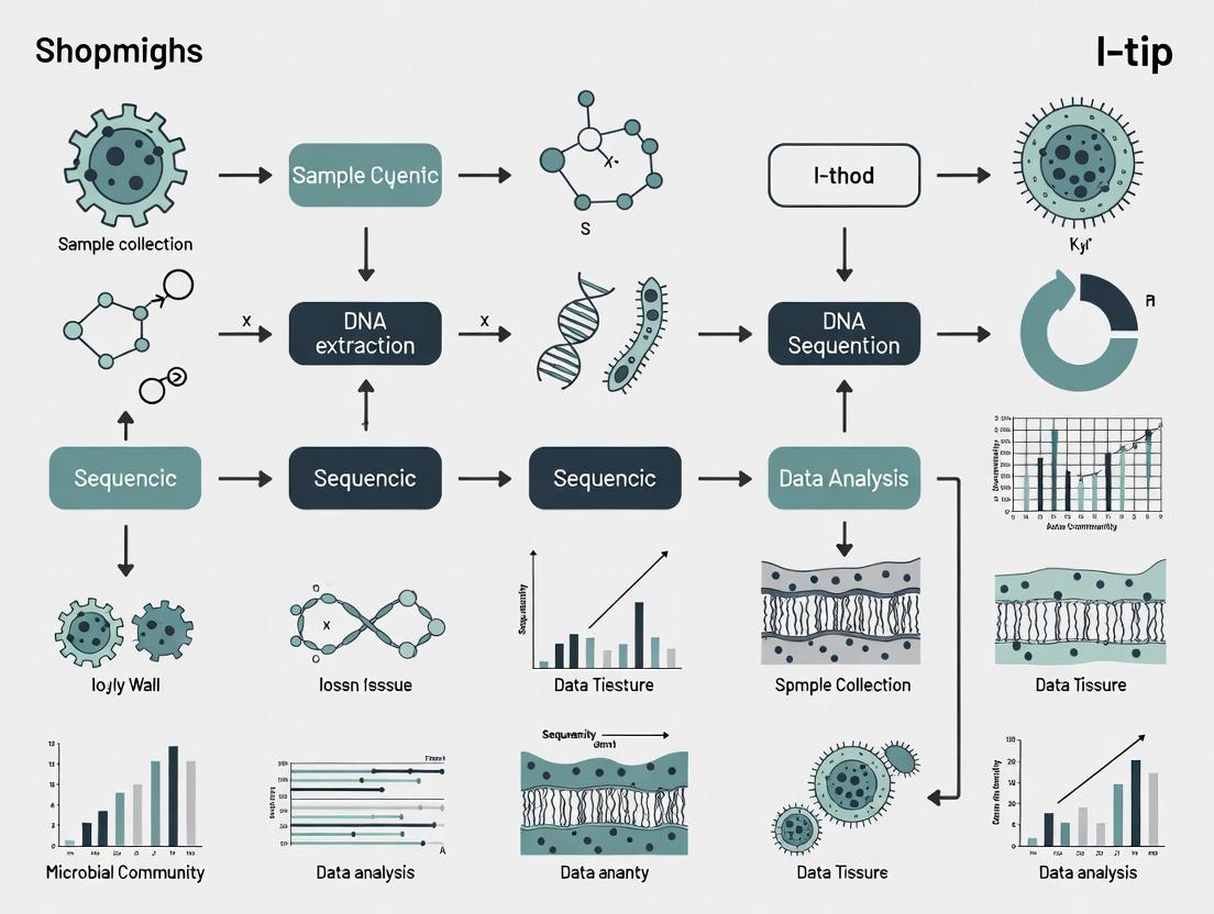
This article provides a detailed methodological framework for applying the I-tip inoculation technique to cultivate sponge-associated bacteria.
The I-tip Method: A Comprehensive Guide for Cultivating Sponge-Associated Bacteria for Drug Discovery
Abstract
This article provides a detailed methodological framework for applying the I-tip inoculation technique to cultivate sponge-associated bacteria. It covers the foundational rationale behind targeting this unique microbiome, a step-by-step protocol for I-tip application, troubleshooting common pitfalls, and a comparative analysis of the method's efficacy against traditional cultivation approaches. Designed for microbiologists, natural product researchers, and drug development professionals, this guide aims to enhance the recovery of novel bacterial taxa and bioactive compounds from marine sponge symbionts.
Why Sponge Microbiomes? Unlocking Marine Biodiversity for Novel Therapeutics
Application Notes: Integrating the I-tip Method into Sponge Microbiome Research
The study of sponge-associated microbial communities (the sponge holobiont) is critical for discovering novel bioactive compounds and understanding marine symbioses. The I-tip method (Intact-cell In situ Tip-digestion) provides a minimally disruptive technique for the selective enrichment and isolation of sponge-associated bacteria directly from sponge tissue, preserving their native physiological state. This protocol series details its application within a broader thesis framework focused on unlocking microbial diversity for drug discovery.
Table 1: Representative Diversity Metrics from Sponge Holobiont Studies
| Sponge Species | Estimated Bacterial Phyla | Unique OTUs* | Cultivable Fraction (Conventional vs. I-tip) | Reference Key Compound Classes |
|---|---|---|---|---|
| Theonella swinhoei | >20 | >25,000 | <1% vs. 3-5% | Polyketides, Nonribosomal Peptides |
| Cymbastela concentrica | 12-15 | ~10,000 | ~0.5% vs. ~4% | Brominated Alkaloids |
| Aplysina aerophoba | 8-12 | ~8,000 | <1% vs. 2-3% | Brominated Tyrosine Derivatives |
| Rhopaloeides odorabile | 10-14 | ~9,500 | ~1% vs. 5-8% | Cyclic Peptides |
*OTU: Operational Taxonomic Unit (16S rRNA gene similarity ≥97%)
Table 2: I-tip Method Performance Metrics vs. Conventional Homogenization
| Parameter | Conventional Tissue Homogenization | I-tip Method Enrichment |
|---|---|---|
| Host Eukaryotic DNA Co-isolation | High (≥60% of total sequences) | Low (≤15% of total sequences) |
| Bacterial Cell Viability Post-Processing | <10% | >75% |
| Novel OTUs Recovered (per sample) | 50-200 | 200-600 |
| Success Rate for Axenic Culture Initiation | 5-10% | 15-30% |
| Time to Initial Microbial Colony (avg.) | 14-21 days | 7-10 days |
Detailed Protocols
Protocol 1: I-tip Method for Enrichment of Sponge-Associated Bacteria
Objective: To gently dissociate and enrich intact, viable bacterial cells from sponge tissue with minimal host cell contamination.
Materials: See "Research Reagent Solutions" table.
Procedure:
- Sample Pre-processing: Under sterile conditions, rinse the sponge specimen (≈1 cm³) three times in filter-sterilized artificial seawater (F-ASW) to remove transient environmental microbes.
- Tissue Digestion: Place the sample in a sterile Petri dish. Using a sterile scalpel, make fine incisions. Apply 2-3 mL of the Enzymatic Dissociation Cocktail directly onto the tissue. Incubate at in situ temperature (e.g., 20°C) for 45-60 minutes without agitation.
- Selective Harvest: Mount a sterile, porous filter tip (I-tip) onto a micropipettor. Gently touch and depress the tip onto the digested tissue surfaces. The tip's pore size (5-10 µm) allows bacterial cells and small consortia to be drawn in while excluding most host sponge cells.
- Elution: Expel the contents of the I-tip into a tube containing 1 mL of Marine Broth Base diluted 1:10 with F-ASW. This forms the Primary Enrichment Inoculum.
- Immediate Processing: Use this inoculum for downstream plating (Protocol 2) or flow cytometry.
Diagram 1: I-tip Method Workflow
Protocol 2: Cultivation Using Diffusion Chambers and Co-culture Media
Objective: To cultivate previously uncultivated sponge bacteria using simulated natural conditions.
Procedure:
- Diffusion Chamber Setup: Prepare a sterile chamber (e.g., a 60mm petri dish with a 0.03 µm membrane bottom) filled with 1.5% low-melt Gellan Gum-based Marine Medium.
- Inoculation: Spread 100 µL of the Primary Enrichment Inoculum (from Protocol 1) onto the gellan gum surface.
- Conditioning: Surround the chamber with Sponge Tissue Homogenate Conditioning Broth (prepared from a separate, homogenized piece of the same sponge, filter-sterilized through 0.22 µm).
- Incubation: Seal the outer plate and incubate at in situ temperature for 4-12 weeks. Monitor weekly for microcolony formation.
- Sub-culturing: Using a micromanipulator, pick microcolonies and transfer to Cross-feeding Co-culture Plates seeded with helper strains (e.g., Ruegeria sp.).
Protocol 3: Metagenomic Analysis of I-tip Enriched Communities
Objective: To generate metagenome-assembled genomes (MAGs) from enriched communities.
Procedure:
- DNA Extraction: Concentrate cells from 10 mL of Primary Enrichment Inoculum by gentle centrifugation (6,000 x g, 10 min). Use a Mild Lysis Buffer followed by a commercial kit optimized for complex microbiomes.
- Sequencing Library Prep: Prepare libraries using a long-read (PacBio HiFi) or short-read paired-end (Illumina) platform, following manufacturer protocols. Pool 4-6 samples per lane.
- Bioinformatic Analysis: Process reads through a pipeline: quality filtering (FastP), assembly (metaSPAdes), binning (MaxBin2, MetaBat2), and dereplication (dRep). Annotate MAGs using Prokka.
Diagram 2: Metagenomics to Bioactivity Pipeline
The Scientist's Toolkit: Research Reagent Solutions
Table 3: Essential Materials for I-tip Based Sponge Microbiology
| Item Name | Function/Description | Key Consideration |
|---|---|---|
| Enzymatic Dissociation Cocktail | Blend of collagenase (1 mg/mL) and dispase II (0.5 mg/mL) in F-ASW. Gently degrades extracellular matrix to release microbial cells. | Must be prepared fresh; avoid proteases like trypsin that damage bacterial surfaces. |
| Porous Filter Tips (I-tips) | Sterile, polycarbonate micropipette tips with defined pore sizes (5µm, 10µm). Enable size-selective cell harvesting. | Pore size choice is critical: 5µm for single cells, 10µm for small consortia. |
| Gellan Gum-based Marine Medium | Solidifying agent for diffusion chambers. Provides a more natural, less rigid matrix than agar for slow-growing bacteria. | Use at low concentration (1-1.5%). Requires cations to gel. |
| Sponge Tissue Homogenate Conditioning Broth | Filter-sterilized homogenate of host tissue. Provides unknown growth factors and signaling molecules. | Use tissue from the same specimen/species. Filter through 0.22µm to remove cells. |
| Cross-feeding Co-culture Plates | Pre-conditioned media plates seeded with helper bacterial strains that provide essential metabolites. | Common helpers: Ruegeria sp., E. coli DH10B. Streak target strain close to helper. |
| Mild Lysis Buffer | Buffer containing lysozyme, proteinase K, and SDS at reduced concentrations for partial host cell lysis and preferential bacterial DNA release. | Incubation time (30-45 min at 37°C) must be optimized per sponge type. |
Historical Challenges in Cultivating Sponge-Associated Bacteria
The study of sponge-associated bacteria holds immense potential for novel drug discovery due to their prolific production of unique bioactive metabolites. However, research has been historically bottlenecked by cultivation limitations. This application note, framed within a thesis on the innovative In situ Trap for Incubated Prokaryotes (I-tip) method, details these challenges and provides refined protocols to overcome them.
A primary historical challenge is the extremely low recovery of sponge-associated bacteria using standard microbiological media.
Table 1: Historical Cultivation Yields from Marine Sponges
| Sponge Source | Standard Media Yield (%) | Specialized/Modified Media Yield (%) | Key Limitation Addressed |
|---|---|---|---|
| Diverse Marine Sponges | 0.01 - 1.0 | 5 - 15 | Nutrient Concentration (Oligotrophy) |
| Aplysina aerophoba | < 0.1 | ~8 | Quorum Sensing/ Signaling Molecules |
| Great Barrier Reef Sponges | ~0.3 | Up to 40 (in situ methods) | Simulation of Native Chemical/Physical Environment |
| Mediterranean Sponges | < 1.0 | 10 - 25 | Co-culture with Host Cells/Other Bacteria |
Protocol 1: Preparation of Diluted Nutrient Media for Oligotroph Enrichment
Purpose: To cultivate slow-growing, oligotrophic sponge bacteria inhibited by standard nutrient levels.
Materials:
- Autoclaved Natural Seawater (NSW)
- Basal Salt Mixture (e.g., Marine Agar, R2A Sea Salts)
- Heat-labile supplements (Vitamin solutions, trace elements)
- 0.22 µm Pore-Size Sterile Syringe Filters
Procedure:
- Prepare a 1x solution of basal salts in 800 mL of distilled water. Autoclave at 121°C for 15 minutes.
- Cool to room temperature. Aseptically add 200 mL of filter-sterilized (0.22 µm) NSW.
- Separately filter-sterilize stock solutions of vitamins (e.g., B12, biotin) and trace metals.
- Aseptically add vitamins and trace metals to the cooled basal salt-NSW mixture to achieve final concentrations typically 10-100x lower than standard recipes (e.g., 10 µg/L vitamin B12).
- For solid media, add purified agar to 1.0-1.5% w/v prior to autoclaving the basal salt/water component.
Protocol 2: Setup of a Transwell Co-culture System
Purpose: To facilitate growth of bacteria dependent on metabolites from sponge host cells or other microbial symbionts.
Materials:
- Sterile Transwell plate (e.g., 6-well, 0.4 µm pore membrane)
- Sponge cell suspension (primary or cell line)
- Bacterial inoculum from sponge homogenate
- Appropriate sponge cell culture medium
Procedure:
- In the lower chamber of the Transwell plate, seed and culture sponge cells until a monolayer or stable aggregate forms.
- In the upper chamber (insert), place a semi-permeable membrane coated with a thin layer of ultra-low-nutrient agar (e.g., 0.3% agarose in diluted medium).
- Gently resuspend the bacterial inoculum in a minimal volume of sterile NSW and spread onto the agar surface in the upper chamber.
- Assemble the insert into the well containing the sponge cells. The membrane allows for the diffusion of signaling molecules and metabolites while preventing physical contact.
- Incubate under conditions mimicking the sponge's native environment (temperature, low light).
The Scientist's Toolkit: Key Reagent Solutions
Table 2: Essential Research Reagents for Sponge-Associated Bacteriology
| Reagent Solution | Function & Rationale |
|---|---|
| Autoclaved Natural Seawater (NSW) | Provides trace elements, ionic balance, and unknown growth factors absent in synthetic salts. |
| Signal Molecule Cocktail (AHLs, AI-2) | Acyl-homoserine lactone and Autoinducer-2 solutions used to induce quorum sensing-dependent growth initiation. |
| Sponge Homogenate Supernatant (filtered) | Source of host-specific growth co-factors, vitamins, and chemical cues; used as a media supplement. |
| Gelrite (instead of Agar) | A gelling agent producing clearer plates and potentially lower concentrations of inhibitory impurities. |
| Cycloheximide Antifungal Solution | Selectively inhibits eukaryotic (fungal) contamination from sponge samples without affecting most bacteria. |
Visualization of Methodological Evolution and the I-tip Principle
Title: From Lab to Habitat: The I-tip Solution
Title: I-tip Chamber Function and Workflow
Application Notes
The I-tip (In-situ cultivation by tip) method represents a paradigm shift in microbial ecology, specifically targeting the "great plate count anomaly" in sponge-associated bacteria research. Conventional cultivation techniques fail to capture over 99% of microbial diversity, creating a critical bottleneck in natural product discovery. The I-tip method addresses this by mimicking the native chemical microenvironment.
Core Rationale and Advantages:
- Chemical Mimicry: Utilizes diffusion chambers to allow continuous, low-molecular-weight nutrient exchange between the encapsulated cells and their native environment, supplying unknown growth factors.
- In-situ Incubation: Maintains chemical and signaling gradients critical for growth initiation, which are lost in lab media.
- High-Throughput Potential: Enables parallel processing of数百 microbial cells directly from a homogenized environmental sample, significantly increasing cultivation yield.
Quantitative data from recent implementations highlights its efficacy:
Table 1: Comparative Cultivation Success Rates: I-tip vs. Conventional Methods
| Method | Avg. Colony Forming Units (CFUs) per mL Sample | Phylogenetic Diversity Recovered (%) | Novel Taxon Recovery Rate (%) | Avg. Incubation Time (Days) |
|---|---|---|---|---|
| I-tip (In-situ) | 5.2 x 10³ | 45-65 | 25-40 | 14-28 |
| Standard Agar Plates | 1.5 x 10² | 5-15 | 1-5 | 3-7 |
| Liquid Enrichment | 8.0 x 10¹ | <10 | <1 | 7-14 |
Table 2: Bioactivity Screening Yield from I-tip Isolates
| Screening Target | % of I-tip Isolates Showing Activity | % of Conventional Isolates Showing Activity | Most Promising Clades Identified |
|---|---|---|---|
| Antibacterial | 18.7 | 8.2 | Nitrospira, Poribacteria |
| Antifungal | 12.3 | 4.1 | Acidobacteria, Gemmatimonadetes |
| Cytotoxic | 9.8 | 3.5 | Chloroflexi, Entotheonellaeota |
Experimental Protocols
Protocol 1: I-tip Chip Preparation and Seeding
Objective: To fabricate and aseptically load diffusion chambers for in-situ cultivation.
Materials: See "Scientist's Toolkit" below. Procedure:
- Chip Fabrication: Cut a sterile silicone gasket (70 µm thick) to fit a standard microscope slide. Use a laser cutter to create an array of 400-600 microwells (diameter: 50 µm, depth: 60 µm) in the gasket.
- Membrane Assembly: Sandwich the gasket between two sterile, semi-permeable polycarbonate membranes (0.03 µm pore size). The pore size excludes environmental predators but allows diffusion of molecules <30 kDa.
- Sample Loading: Homogenize 1 cm³ of sponge tissue in 10 mL of filter-sterilized ambient seawater. Centrifuge gently (500 x g, 5 min) to remove large debris.
- Cell Seeding: Dilute the supernatant 1:100 in seawater. Pipette 100 µL onto the assembled I-tip chip. Use capillary action and gentle centrifugation (200 x g, 2 min) to drive single cells into the microwells.
- Sealing: Carefully place a cover glass over the top membrane and seal the edges with a biocompatible, UV-curing resin.
Protocol 2: In-situ Deployment and Recovery
Objective: To incubate the chip in its native habitat and retrieve grown microcolonies.
Procedure:
- Deployment Chamber: Place the sealed I-tip chip into a protective, perforated polypropylene cassette.
- In-situ Incubation: Secure the cassette at the original sponge collection site (e.g., attached to reef substrate near the host sponge) for 14-28 days.
- Recovery: Retrieve the cassette and immediately transport the I-tip chip to the lab in sterile seawater at in-situ temperature.
- Chip Scanning: Using an epifluorescence microscope with an automated stage, scan the entire chip. Identify microwells containing microcolonies (50-100 cells) via SYBR Gold nucleic acid staining.
- Colony Extraction: Use a micromanipulator equipped with a glass capillary (10 µm tip) to aspirate individual microcolonies from targeted wells.
Protocol 3: Lab Cultivation from Retrieved Microcolonies
Objective: To transition microcolonies to pure lab cultures.
Procedure:
- Transfer: Expel each retrieved microcolony into 50 µL of low-nutrient broth (e.g., 1:100 diluted Marine Broth 2216) in a 96-well microtiter plate.
- Conditioned Medium: For recalcitrant isolates, prepare medium using "spent" filtrate from a culture of the host sponge's homologous cells or other I-tip isolates to provide unknown growth factors.
- Incubation & Subculturing: Incubate plates at the environmental temperature with slow shaking (50 rpm). Monitor growth via optical density (OD600). Subculture 10 µL into fresh medium upon reaching stationary phase, gradually increasing nutrient concentration over 3-5 passages.
Visualizations
Diagram Title: I-tip Method Core Workflow
Diagram Title: Chemical Exchange in I-tip Cultivation
The Scientist's Toolkit
Table 3: Essential Research Reagent Solutions for I-tip Methodology
| Item | Function & Specification |
|---|---|
| Polycarbonate Membrane | Forms the semi-permeable barrier of the diffusion chamber. 0.03 µm pore size is critical for excluding predators while allowing nutrient exchange. |
| Silicone Gasket (70 µm) | Spacer material laser-cut to create microwell arrays, defining the isolation volume for single cells. |
| SYBR Gold Nucleic Acid Stain | A vital fluorescent dye for visualizing and identifying microcolonies within the I-tip chip post-incubation. |
| Filter-Sterilized Ambient Seawater | Used for sample dilution and chip priming. Maintains ionic balance and may contain essential, undefined co-factors. |
| Low-Nutrient Marine Broth | Transition medium, typically diluted 1:100 from standard recipe, to avoid "shocking" retrieved cells. |
| Conditioned Medium Filtrate | "Spent" medium from related cultures, used to supplement transition media with unknown growth-promoting factors. |
| UV-Curing Biocompatible Resin | For sealing the I-tip chip assembly, ensuring watertight integrity during in-situ deployment. |
| Glass Capillary (10 µm tip) | Attached to a micromanipulator for the precise, aseptic extraction of microcolonies from individual microwells. |
Key Microbial Phyla in Sponges and Their Bioactive Potential (e.g., Poribacteria, Chloroflexi)
Application Notes
Context within Thesis on the I-tip Method: The Integrated in situ cultivation, isolation, and processing (I-tip) method provides a targeted platform for accessing the uncultivated microbial majority within sponge tissues. This protocol focuses on applying the I-tip method to specifically enrich for and study key microbial phyla—notably Poribacteria and Chloroflexi—which are consistently dominant in sponge microbiomes and are hypothesized reservoirs of novel bioactive natural products. By integrating in situ cultivation with downstream molecular and chemical analysis, the I-tip method bridges the gap between phylogenetic identification and functional/biochemical validation.
Key Phyla and Their Bioactive Potential:
- Poribacteria: A candidate phylum almost exclusively found in marine sponges. Genomic analyses predict a facultatively anaerobic, heterotrophic lifestyle with gene clusters for polyketide synthase (PKS) and non-ribosomal peptide synthetase (NRPS), hinting at significant biosynthetic potential for antimicrobial and antitumor compounds.
- Chloroflexi (particularly the Chloroflexi sponge clades): Ubiquitous and abundant in sponges globally. Metagenomic studies suggest photoheterotrophic capabilities in some lineages and a rich arsenal of biosynthetic gene clusters (BGCs) for compounds like borrelidin and tambjamine analogs, with predicted antibiotic and cytotoxic activities.
- Acidobacteria, Actinobacteria, and Cyanobacteria are also prevalent and contribute to the sponge's chemical defense portfolio through diverse BGCs.
Table 1: Prevalence and Biosynthetic Potential of Key Sponge-Associated Microbial Phyla
| Microbial Phylum | Typical Relative Abundance in Sponge Microbiome (%) | Key Metabolite Gene Clusters Predicted (PKS/NRPS Hybrid) | Proposed Bioactive Potential |
|---|---|---|---|
| Poribacteria | 1–30% (host-dependent) | Type I PKS, NRPS | Antimicrobial, Anticancer |
| Chloroflexi | 5–65% (often dominant) | Type I & II PKS, NRPS, Terpene Synthases | Antibiotic, Cytotoxic, Antiviral |
| Acidobacteria | 1–20% | Type I & III PKS | Anti-inflammatory, Antimicrobial |
| Actinobacteria | 1–15% | Type I & II PKS, NRPS, Lantipeptides | Broad-spectrum Antibiotics |
| Cyanobacteria | 0.1–10% (variable) | NRPS, Hybrid PKS-NRPS (e.g., Microviridins) | Protease Inhibition, Cytotoxic |
Table 2: I-tip Method Yield for Targeted Phyla from Aplysina aerophoba
| Target Phylum | Pre-I-tip Abundance (16S rRNA amplicon %) | Post-I-tip Enrichment (16S rRNA amplicon %) | Cultivation Success (CFU/mL gel) |
|---|---|---|---|
| Chloroflexi | 28.5 | 41.7 | 1.2 x 10³ |
| Poribacteria | 8.2 | 15.3 | Not Cultivated (Enhanced detection) |
| Acidobacteria | 5.1 | 9.8 | 5.5 x 10² |
| Actinobacteria | 3.8 | 12.4 | 3.8 x 10⁴ |
Protocols
Protocol 1: I-tip Method for In Situ Enrichment of Sponge-Associated Bacteria
Objective: To cultivate and enrich sponge-associated bacteria, particularly elusive phyla like Poribacteria and Chloroflexi, within a semi-solid matrix placed directly in the sponge mesohyl.
Research Reagent Solutions & Materials:
| Item | Function/Explanation |
|---|---|
| Low-Melting-Point Agarose (0.8–1.2%) | Forms a diffusion-based cultivation gel within the I-tip capillary. |
| Marine Broth (MB) or R2A Marine Medium | Nutrient-rich base for the cultivation gel. |
| Filter-sterilized Sponge Homogenate (10% v/v) | Provides host-specific growth factors and signaling molecules. |
| Quorum Sensing Inducers (e.g., 5µM N-Acyl homoserine lactones) | Mimics microbial cross-talk to activate silent BGCs. |
| HD-PIT Tip (Hamilton-like syringe needle) | Precision tool for gel injection and sample aspiration. |
| Histoacryl Tissue Adhesive | Seals the injection point to prevent gel expulsion and contamination. |
| Cell Recovery Medium (CRM) | Enzymatic/chemi-mechanical solution for harvesting cells from the gel matrix post-incubation. |
Procedure:
- Gel Preparation: Prepare a sterile, low-melting-point semi-solid gel using diluted marine broth (e.g., 1/10 strength R2A seawater) supplemented with 10% filter-sterilized homogenate from the target sponge species. Add relevant signal molecules (e.g., AHLs). Maintain at 35°C to prevent solidification.
- Sponge Preparation: Aqueously acclimate the donor sponge (Aplysina aerophoba). Select a healthy osculum or branch for injection.
- I-tip Loading & Injection: Aspirate the warm gel into a sterile HD-PIT tip. Using a micromanipulator, carefully insert the tip ~2-3 mm into the sponge mesohyl. Deposit 5–10 µL of gel to form a micro-niche.
- Sealing & Incubation: Withdraw the tip and immediately seal the entry point with a drop of tissue adhesive. Return the sponge to its aquarium. Incubate for 2–4 weeks under ambient light/temperature conditions.
- Gel Retrieval & Processing: After incubation, surgically excise the gel plug and surrounding sponge tissue. Dissect the gel plug and place it in sterile Cell Recovery Medium. Gently vortex/incubate to liberate microbial cells for downstream analysis (DNA extraction, FACS, sub-cultivation).
Protocol 2: Targeted Metagenomic Analysis of I-tip Enriched Communities
Objective: To sequence and analyze the metagenome of the I-tip gel community to identify BGCs from enriched phyla.
Procedure:
- DNA Extraction: Extract high-molecular-weight genomic DNA from the harvested I-tip gel community using a kit optimized for complex matrices (e.g., MagAttract HMW DNA Kit).
- Sequencing Library Preparation: Prepare both short-read (Illumina 2x150bp, for high coverage) and long-read (Oxford Nanopore or PacBio, for scaffolding) libraries following manufacturer protocols.
- Bioinformatic Analysis:
- Assembly & Binning: Co-assemble reads using metaSPAdes. Recover metagenome-assembled genomes (MAGs) using binning tools (e.g., MaxBin2, MetaBat2). Assess MAG quality with CheckM.
- Phylogenetic Classification: Classify MAGs using GTDB-Tk.
- BGC Mining: Analyze MAGs for BGCs using antiSMASH. Prioritize BGCs from Poribacteria, Chloroflexi, and other target phyla.
Protocol 3: Heterologous Expression of Targeted BGCs
Objective: To express prioritized BGCs from uncultivated phyla in a culturable bacterial host (e.g., Pseudomonas putida).
Procedure:
- BGC Capture: Identify a target BGC (e.g., a large PKS cluster from a Chloroflexi MAG). Design primers to amplify the ~40-60 kb cluster using long-range PCR or capture it via transformation-associated recombination (TAR) in yeast.
- Vector Assembly: Clone the captured BGC into a broad-host-range expression vector (e.g., pSEVA series) containing an inducible promoter.
- Heterologous Expression: Electroporate the assembled vector into the expression host. Plate on selective media.
- Metabolite Induction & Extraction: Grow positive clones and induce BGC expression with IPTG. Extract metabolites from cell pellets and supernatant with ethyl acetate.
- Chemical Analysis: Analyze extracts via LC-HRMS/MS. Dereplicate using databases (GNPS). Isate novel compounds via HPLC for bioactivity testing (antimicrobial, cytotoxicity assays).
Visualizations
I-tip to Bioactive Compound Discovery Pipeline
Chloroflexi Biosynthetic Gene Cluster Logic
Within the broader thesis on the Integrated-Tip (I-tip) method for sponge-associated bacteria research, defining success for cultivation efforts is paramount. The I-tip method, which integrates sterile, porous polymer probes directly into the sponge mesohyl for in situ enrichment, aims to bridge the gap between environmental conditions and laboratory cultivation. This document outlines the target outcomes, application notes, and protocols for evaluating the success of cultivation initiatives derived from this innovative approach, focusing on yield, diversity, and biotechnological potential.
Success is multi-faceted. The following quantitative targets, informed by recent literature (2023-2024) on marine microbial cultivation, provide benchmarks.
Table 1: Primary Target Outcomes for Cultivation from I-tip Enrichment
| Outcome Metric | Definition & Measurement | Success Benchmark | Rationale |
|---|---|---|---|
| Cultivation Yield | Percentage of I-tip-enriched bacterial cells that form colonies on isolation media. | >15% of enriched population | Significantly higher than standard (<1%) methods; indicates effective in situ conditioning. |
| Phylogenetic Novelty | Percentage of isolated strains with 16S rRNA gene sequence similarity <98.7% to any described type strain. | >30% of isolate collection | Targets the "microbial dark matter" abundant in sponges. |
| Unique Isolate Recovery | Number of distinct strains (by genomic fingerprinting) recovered per sponge sample. | 50-100 unique strains | Demonstrates the method's ability to capture diversity. |
| Biosynthetic Gene Cluster (BGC) Richness | Average number of predicted BGCs per isolate genome (antiSMASH analysis). | >5 BGCs/genome | Indicates high drug discovery potential. |
| Bioactivity Hit Rate | Percentage of crude extracts showing antimicrobial or cytotoxic activity in primary screens. | >20% of extracts | Validates the cultivation of functionally relevant bacteria. |
Detailed Experimental Protocols
Protocol 3.1: I-tip Deployment and Retrieval
- Objective: To enrich sponge-associated bacteria in situ.
- Materials: Sterile I-tip probes (porous polymer matrix), diving/sampling gear, GPS, sterile scalpels, anaerobic jars.
- Procedure:
- Deployment: Underwater, gently insert multiple sterile I-tip probes into the mesohyl of target sponge species. Secure probes. Record location, depth, and sponge morphology.
- Incubation: Allow probes to remain integrated for 7-14 days.
- Retrieval: Carefully remove probes and immediately place them into individual sterile tubes containing anoxic transport medium. Store at in situ temperature during transport to the lab.
Protocol 3.2: Differential Cultivation from I-tip Eluate
- Objective: To maximize yield and diversity of isolates.
- Materials: Anaerobic chamber, dilution series tubes, diverse solid media (e.g., Marine Agar, R2A Marine, media supplemented with sponge extract).
- Procedure:
- Elution: In an anaerobic chamber, transfer each I-tip to a tube containing 10mL of sterile, reduced artificial seawater. Vortex gently for 2 minutes to dislodge cells.
- Plating: Perform serial dilutions (10⁻¹ to 10⁻⁶) of the eluate. Spread 100µL of each dilution onto a panel of 5-8 different cultivation media.
- Incubation: Incubate plates aerobically, microaerophilically (5% O₂), and anaerobically at 15°C, 25°C, and 30°C for up to 90 days.
- Picking: Visually distinguish morphologically distinct colonies. Subculture until pure.
Protocol 3.3: High-Throughput Screening for Bioactivity
- Objective: To rapidly assess the drug discovery potential of isolates.
- Materials: 96-well deep-well plates, liquid media, assay strains (E. coli, S. aureus, C. albicans, cancer cell lines*).
- Procedure:
- Fermentation: Inoculate each purified isolate into 1mL of medium in a 96-deep-well plate. Incubate with shaking for 5-7 days.
- Extraction: Add an equal volume of ethyl acetate to each well, shake for 1 hour. Centrifuge. Transfer organic (top) layer to a new plate. Evaporate solvent.
- Resuspension: Resuspend each extract in 100µL DMSO.
- Screening: Perform disk diffusion (antimicrobial) or MTT assay (cytotoxicity). Record zones of inhibition or IC₅₀ values.
Visualization: Workflow and Pathways
Title: I-tip Cultivation and Evaluation Workflow
Title: Quorum Sensing Pathway in Sponge Bacteria
The Scientist's Toolkit: Research Reagent Solutions
Table 2: Essential Materials for I-tip Cultivation & Analysis
| Item | Function & Rationale |
|---|---|
| Porous Polymer I-tip Probes | Sterile, inert matrix for in situ bacterial enrichment within sponge tissue. Mimics the natural porous architecture. |
| Anoxic Transport Medium | Preserves obligate anaerobic bacteria during sample retrieval and transport. Critical for diversity. |
| Marine Agar 2216, Modified | Standardized, nutrient-rich base medium for heterotrophic marine bacteria. |
| Sponge Extract Supplement | Autoclaved crude extract from host sponge homogenate. Provides specific growth factors. |
| Gifu Anaerobic Medium (GAM) Marine | Complex medium designed for demanding anaerobes. Essential for cultivating Poribacteria and related phyla. |
| AntiSMASH Software Suite | Bioinformatics platform for genome mining and identification of Biosynthetic Gene Clusters (BGCs). |
| Clinical & Industrial Assay Strains | Panel of Gram-positive, Gram-negative bacteria, fungi, and cell lines for primary bioactivity screening. |
| Reduced Artificial Seawater | Chemically defined, oxygen-scrubbed diluent and base for media, maintaining ionic balance. |
Step-by-Step Protocol: Implementing the I-tip Method for Sponge Samples
This document outlines critical pre-analytical protocols for research on sponge-associated bacteria, forming the foundational first pillar of a thesis utilizing the I-tip (Individual, Integrated Tip) method. The I-tip method is a novel micro-sampling and high-throughput sequencing platform designed for minimal-destructive, spatially resolved analysis of sponge microbiomes. The integrity of all downstream I-tip processing, genomic analysis, and bioprospecting outcomes is entirely contingent upon rigorous field collection, ethical sourcing, and immediate sample preservation as detailed herein.
Site Selection & Quantitative Environmental Data
Pre-deployment site surveys are mandatory. Data must be recorded using standardized instruments and compiled for meta-analysis.
Table 1: Essential Pre-Sampling Environmental Parameters
| Parameter | Measurement Instrument | Target Range/Notes | Relevance to Sponge Microbiology |
|---|---|---|---|
| Depth | Calibrated dive computer/CTD profiler | Record exact depth per specimen. | Light penetration, pressure, and thermal gradients influence microbiome composition. |
| Temperature | Seabird SBE 3plus or equivalent | ±0.001°C accuracy. Profile vs. depth. | Critical for metabolic rates; sharp changes indicate thermoclines. |
| Salinity | Conductivity sensor (CTD) | PSU (Practical Salinity Units). | Osmoregulatory stress on host and symbionts. |
| Dissolved Oxygen | SBE 43 or optode sensor | mg/L, % saturation. Record hypoxic thresholds (<2 mg/L). | Defines aerobic/anaerobic microbial niches within sponge mesohyl. |
| pH | SeaFET or spectrophotometric kit | Total scale, in situ. | Impacts microbial enzyme function and biogeochemical cycles (e.g., nitrification). |
| Turbidity/Nutrients | Niskin bottle + lab analysis (NO3-, PO4^3-, Si) | Filtered (0.2 µm), frozen. | Eutrophication indicators; affects filter-feeding and microbial autotrophy. |
Ethical Collection & Permitting Protocol
Objective: To obtain sponge specimens legally and sustainably, ensuring species protection and future reproducibility.
- Permitting: Secure prior informed consent and permits from relevant national and local authorities (e.g., CBD-Nagoya Protocol, CITES for endangered species).
- Collection Method:
- Using SCUBA or ROV, identify healthy, representative individuals (>5 specimens per species/site for statistical power).
- For the I-tip method, which is minimally invasive, the primary collection may still require a whole specimen for method validation. Use sterile titanium bone cutters or a scalpel to remove a small section (<10% of total biomass) from the margin, including outer and inner tissues.
- For whole specimens, sever at the base cleanly to allow regeneration. Avoid damaging surrounding fauna.
- Voucher Specimens: Preserve a tissue fragment in >95% ethanol or RNAlater for morphological and barcoding identification (CO1, 28S rRNA genes). A separate fragment should be deposited with a recognized marine biobank.
Sample Processing & Preservation Workflow for I-tip Analysis
Objective: To immediately stabilize nucleic acids and metabolites for accurate downstream I-tip micro-sampling and multi-omics.
Protocol 4.1: Tiered Preservation for Multi-Omics
- Materials: Sterile biopsy punches (5mm, 8mm), cryovials, liquid nitrogen dry shipper, sterile seawater (0.22 µm filtered), DNA/RNA shield buffer, -80°C freezer.
- Steps:
- Onboard Processing: Within <2 minutes of surfacing, rinse specimen briefly in sterile seawater to remove transient contaminants.
- Sub-sampling: Using sterile tools, excise three contiguous tissue cores per specimen.
- Core A (Metagenomics/Genomics): Place directly into DNA/RNA Shield (Zymo Research), incubate at room temp for 24h, then store at 4°C or -20°C.
- Core B (Metatranscriptomics): Submerge immediately in RNAlater (Invitrogen), incubate at 4°C overnight, then store at -80°C.
- Core C (Metabolomics & I-tip): Flash-freeze entire core in liquid nitrogen. Store at -80°C. This core is the source for subsequent I-tip micro-sampling under cryo-conditions.
Protocol 4.2: I-tip Cryo-Sampling Preparation
- Under a liquid nitrogen-cooled cryostat environment (-20°C to -30°C), mount frozen Core C.
- Using sterilized, cryo-cooled forceps, excise a microfragment (≈1 mm³) from specific sponge anatomical zones (e.g., ectosome, choanosome, osculum).
- This microfragment is immediately loaded into the sterile, integrated I-tip cartridge for subsequent in-tip lysis and library prep, per the core thesis methodology.
Title: Sponge Tissue Processing & I-tip Preparation Workflow
The Scientist's Toolkit: Key Reagent Solutions
Table 2: Essential Research Reagents for Pre-Sampling
| Reagent / Material | Supplier Example | Function & Rationale |
|---|---|---|
| DNA/RNA Shield | Zymo Research | Inactivates nucleases and preserves nucleic acid integrity at ambient temp for transport; critical for post-collection delay. |
| RNAlater Stabilization Solution | Invitrogen, Thermo Fisher | Penetrates tissue to stabilize and protect RNA profiles for transcriptomic studies. |
| Liquid Nitrogen (LN₂) & Dry Shipper | Taylor-Wharton, etc. | Provides instantaneous cryo-preservation of labile metabolites and halts all enzymatic activity. |
| Sterile Seawater (0.22 µm filtered) | Prepared in-lab | Removes loosely attached, non-symbiotic planktonic bacteria without osmotic shock. |
| Titanium Biopsy Punches & Scalpels | Fine Science Tools | Non-corrosive, sterilizable tools for clean tissue excision minimizing metal contamination. |
| Cryogenic Vials | Corning, Nunc | Withstand extreme temperatures of LN₂ and -80°C storage without cracking. |
| Permafrost or Similar Ethanol | Sigma-Aldrich | For fixation of voucher specimens for DNA barcoding and morphological reference. |
Title: Pre-Sampling Pillars Support Downstream I-tip Analysis
This application note details protocols for surface sterilization and tissue homogenization, critical steps in the preparation of marine sponge samples for microbial analysis. These procedures are foundational to a broader thesis employing the I-tip method for in-situ, spatially-resolved profiling of sponge-associated bacterial communities. Effective sterilization removes epibiotic contaminants, while optimal homogenization releases intracellular and tightly adherent microbiota for downstream genomic and culturomic applications in drug discovery pipelines.
Surface Sterilization Protocol
Principle: To eliminate transient and loosely attached surface microorganisms without affecting the endogenous symbiotic community within the sponge mesohyl.
Materials & Reagents
- Fresh or freshly frozen marine sponge specimen.
- Sterile artificial seawater (ASW) or phosphate-buffered saline (PBS, pH 7.4).
- Sterilization series: Ethanol (70%, v/v), Sodium hypochlorite (1-4% available chlorine), Betadine solution (1% povidone-iodine).
- Sterile antibiotics cocktail (e.g., 100 µg/mL ampicillin, 50 µg/mL nalidixic acid) in ASW.
- Sterile surgical blades, forceps, and dissection trays.
- Laminar flow hood.
Detailed Procedure
- Rinsing: Briefly rinse the intact sponge specimen (~1 cm³ piece) three times in sterile ASW to remove gross debris and seawater planktonic cells.
- Sterilization Bath Sequence: Immerse the specimen sequentially with gentle agitation: a. Sterile ASW wash: 2 minutes. b. Ethanol (70%): 30 seconds to 2 minutes (optimize per sponge type). c. Sodium hypochlorite (1-2%): 30 seconds to 1 minute. d. Betadine (1%): 1 minute. e. Antibiotics cocktail in ASW: 30 minutes at 4°C.
- Neutralization & Final Rinse: Rinse the specimen thoroughly five times in sterile ASW to quench sterilant activity.
- Validation: Plate the final rinse water on Marine Agar (MA) and Reasoner's 2A (R2A) agar. Incubate at relevant temperatures (e.g., 20°C, 30°C) for 48-72 hours. Successful sterilization yields no colony-forming units (CFUs).
Table 1: Efficacy of Common Sterilants on Sponge Surface Microbiota
| Sterilant | Concentration | Exposure Time (s) | Log Reduction CFU/cm²* | Notes |
|---|---|---|---|---|
| Ethanol | 70% (v/v) | 60-120 | 2.5 - 3.8 | Quick, evaporates; may not kill spores. |
| Sodium Hypochlorite | 1-2% Av. Cl | 30-60 | 4.0 - >6.0 | Strong oxidizer; can damage tissue if overused. |
| Povidone-Iodine | 1% (w/v) | 60 | 3.0 - 4.5 | Broad-spectrum; must be thoroughly rinsed. |
| Antibiotic Cocktail | Variable | 1800 | 1.0 - 2.5* | Targets specific groups; used as a final step. |
*Representative data from recent sponge microbiome studies; actual reduction depends on sponge porosity and initial biofilm load.
Tissue Homogenization for Microbial Release
Principle: To physically disrupt the sponge matrix to liberate bacterial cells while minimizing lysis and genomic DNA shear.
Methods Comparison
Three primary methods are evaluated for integration with the I-tip micro-sampling workflow.
A. Mechanical Blender Homogenization (Bulk)
- Protocol: Transfer sterile tissue (0.5g) to a sterile bag or tube with 5mL sterile ASW/PBS. Homogenize using a paddle blender (e.g., BagMixer) at high speed for 2 x 60s with a 30s rest on ice.
- Application: Bulk community DNA/RNA extraction.
B. Bead Beating (Micro-scale)
- Protocol: Place 0.1-0.2g tissue in a 2mL tube with 1mL ASW and a mix of zirconia/silica beads (0.1, 0.5 mm). Process in a bead beater for 3 x 45s cycles, with 2-minute ice incubations between cycles.
- Application: Effective for tough sponges; compatible with multi-well formats for high-throughput processing.
C. Gentle Potter-Elvehjem (Tissue Grinder)
- Protocol: Use a loose-fitting glass/Teflon grinder. Manually homogenize tissue on ice with 5-10 gentle strokes in 2mL buffer.
- Application: Preferred for delicate sponges or when preserving cell viability for cultivation (e.g., I-tip inoculation).
Table 2: Homogenization Method Impact on Microbial Yield and Integrity
| Method | Relative Cell Yield (%)* | DNA Shearing (Fragment Size) | Viability Post-Homogenization | Suitability for I-tip |
|---|---|---|---|---|
| Mechanical Blender | 100% (Baseline) | Moderate (5-20 kb) | Very Low | Low (bulk sample only) |
| Bead Beating | 110-130% | High (Severe, 1-5 kb) | Low | Medium (lysate analysis) |
| Potter-Elvehjem | 70-90% | Low (>30 kb) | High | High (viable cells) |
*Yield compared to a standardized baseline; data from comparative analyses.
Integrated Workflow for I-tip Method
This protocol connects sample prep to the thesis's core I-tip micro-sampling technique, which involves using fine, sterile capillary tips to collect and deposit microscopic quantities of homogenate for cultivation or molecular analysis.
- Perform surface sterilization as in Section 1.
- Aseptically dissect to obtain a fragment from the inner mesohyl (avoiding the cortex).
- Homogenize this fragment using the Gentle Potter-Elvehjem method in 1mL of specialized marine cell preservation medium.
- Filter homogenate through a sterile 100 µm nylon mesh to remove large debris.
- The resulting microbial suspension is ready for I-tip micro-sampling:
- The I-tip is immersed in the suspension.
- A nanoliter-volume aliquot is captured via capillary action.
- This aliquot is precisely deposited onto an ultra-low nutrient agar plate or into a microfluidic growth chip for cultivation of "uncultivable" symbionts.
The Scientist's Toolkit
Table 3: Essential Research Reagent Solutions
| Item | Function in Protocol | Example/Composition |
|---|---|---|
| Sterile Artificial Seawater (ASW) | Physiological rinsing and dilution medium; maintains osmolarity to prevent cell lysis. | 3.2% NaCl, 0.07% KCl, 0.53% MgCl₂·6H₂O, 0.1% CaCl₂, pH adjusted to 7.4-7.6. |
| Antioxidant & Chelator Buffer | Added to homogenization buffer to inhibit host-derived nucleases and reactive oxygen species. | 10-20 mM Sodium ascorbate, 1-5 mM EDTA in ASW. |
| Marine Cell Preservation Medium | Protects viability of fastidious symbionts post-homogenization for I-tip culturing. | ASW supplemented with 0.1% thioglycolate (oxygen scavenger), 0.01% glutathione (antioxidant). |
| Differential Filtration Membranes | Size-based separation of sponge eukaryotic cells from smaller bacterial cells. | Sequential filtration through 100 µm (debris), 20 µm (host cells), and 5 µm (collect bacteria). |
| Percoll Density Gradient Medium | Enriches for bacterial cells by separating them from lighter sponge cell debris based on buoyancy. | Iso-osmotic Percoll solution prepared in ASW, centrifuged at low speed (800 x g). |
Visualizations
Title: Integrated Sample Prep and I-tip Workflow
Title: Sequential Surface Sterilization Steps
Within the broader thesis investigating sponge-associated bacteria for novel bioactive compound discovery, the I-tip inoculation method is established as the foundational, reproducible technique for establishing axenic and defined co-culture models. This protocol details the core physical setup and aseptic technique required to transfer bacterial isolates from sponge homogenate or stock cultures onto and into solid and liquid media, minimizing contamination and physiological stress, which is critical for subsequent compound extraction and bioactivity screening.
Core Equipment Setup and Configuration
The I-tip inoculation station is a dedicated, organized workspace for aseptic sample handling.
Table 1: Core Equipment for I-tip Inoculation Setup
| Equipment | Specification/Model Example | Primary Function in Protocol |
|---|---|---|
| Class II Biosafety Cabinet (BSC) | Nuaire NU-425-400S | Provides sterile work area, protects user and sample from airborne contaminants. |
| Inoculation Tool (I-tip) | Fine-gauge hypodermic needle (22-27G) or micro-scalpel, mounted on handle. | Precision tool for excising tiny tissue fragments or picking bacterial colonies. |
| Bunsen Burner or Bacti-Cinerator | Lab Gas Burner or Microincinerator 230V | Creates convective updraft for sterile field; sterilizes I-tip via heating to red-hot. |
| Microscope (Dissecting/Stereo) | Leica S9E with 10x-40x magnification | Visualizes sponge matrix or colonies for precise I-tip targeting. |
| Precision Micromanipulator (Optional) | Narishige MN-153 | Provides ultra-fine, vibration-damped control for I-tip under high magnification. |
| Vacuum System with Filter | Portable HEPA-filtered aspirator | Removes excess moisture or media from sample area during manipulation. |
| Temperature-Controlled Stage | Linkam PE120 | Maintains sample at in situ sponge temperature (e.g., 10°C for deep-sea isolates) during manipulation. |
Detailed Application Notes & Protocols
Protocol 3.1: Aseptic Setup of I-tip Workstation
- Decontamination: Wipe down BSC interior surfaces with 70% ethanol followed by a sporicidal agent (e.g., 1% peracetic acid). Place all pre-sterilized tools (Petri dishes, media, collection tubes) inside.
- Equipment Arrangement: Arrange tools in order of use from left to right (clean to dirty side). Position microscope inside BSC if possible, or at immediate access port. Place burner/microincinerator to the dominant-hand side.
- Field Sterilization: Turn on BSC and allow 10-minute purge. Flame the interior metal surfaces of the BSC where tools will be placed. Create a defined "sterile field" near the flame updraft.
Protocol 3.2: I-tip Inoculation of Sponge Fragment onto Agar
Objective: Transfer a bacteria-laden sponge fragment to complex marine agar (e.g., Marine Agar 2216) for initial cultivation. Materials: Sterile glass Petri dish containing sponge fragment (<1 mm³) in artificial seawater (ASW), target agar plates, I-tip tool.
Workflow:
- Tool Sterilization: Flame the I-tip until red-hot, then cool for 10-15 seconds in the sterile air flow of the BSC.
- Fragment Selection: Under dissecting microscope, identify a target fragment. Use vacuum aspirator to remove excess ASW if needed.
- Fragment Engagement: Gently spear or scoop the fragment with the tip of the I-tip.
- Inoculum Transfer: Swiftly move the I-tip to the target agar plate. Use a "dabbing and rolling" motion to deposit the fragment onto the agar surface.
- Streaking (Optional): If aiming for isolation, gently streak the fragment away from the inoculation point in 2-3 successive streaks using a sterile loop.
- Sealing & Incubation: Seal plate with parafilm and incubate under conditions mimicking sponge habitat (temperature, gas atmosphere).
Diagram 1: Sponge Fragment Inoculation Workflow
Protocol 3.3: I-tip Colony Picking for Sub-culturing or Assay Setup
Objective: Isolate a single bacterial colony from a primary sponge culture for genetic analysis or bioactivity screening. Materials: Primary culture plate, target media (agar deeps, 96-well assay plates, fresh agar), I-tip.
Workflow:
- Colony Identification: Visually or microscopically identify a well-isolated, morphologically distinct colony.
- Tool Sterilization: Flame and cool I-tip as in 3.2.
- Colony Picking: Gently touch the top of the target colony with the I-tip, collecting a minuscule amount of biomass.
- Inoculation: For agar deeps (oxygen gradient studies): stab I-tip vertically into the center of the agar column. For 96-well plates: dip I-tip into broth medium and agitate gently. For streak plates: proceed with standard quadrant streaking.
- Disposal: Dispose of used I-tip into sharps container. Never re-flame a used tip without prior decontamination.
Table 2: Key Research Reagent Solutions
| Reagent/Material | Composition/Example | Function in I-tip Protocols |
|---|---|---|
| Artificial Seawater (ASW) | 3.1% NaCl, 0.1% KCl, 0.05% NaHCO₃, Mg/Ca salts. | Maintenance medium for sponge fragments; prevents osmotic shock. |
| Marine Agar 2216 | Peptone, Yeast Extract, Ferric Citrate in aged seawater. | General non-selective medium for initial cultivation of heterotrophic marine bacteria. |
| Sponge Homogenate Supplement | 0.22µm-filtered homogenate of host sponge. | Adds species-specific growth factors for fastidious sponge symbionts. |
| Cycloheximide Solution | 100 µg/mL in ethanol, filter-sterilized. | Selective agent added to media to inhibit eukaryotic (fungal/sponge) cell growth. |
| Anoxic Medium | Marine broth/agar supplemented with reducing agents (Cysteine, Na₂S). | For cultivating obligate anaerobic sponge-associated bacteria. |
Advanced Protocol: I-tip Inoculation for Micro-Colony Single-Cell Genomics
Objective: Physically pick a micro-colony (<100 µm) derived from a single cell for whole genome amplification.
Protocol:
- Preparation: Coat the I-tip (30G needle) with 1 µL of sterile molecular grade glycerol using a micro-pipette.
- Micro-colony Visualization: Use an inverted microscope at 400x magnification within an anaerobic chamber if required.
- Targeted Pick: Under direct visualization, gently touch the glycerol-coated tip to the target micro-colony.
- Transfer: Immediately transfer the tip into a PCR tube containing lysis buffer. Rinse by pipetting up and down.
- Downstream Processing: Proceed with MDA (Multiple Displacement Amplification) for WGA.
Diagram 2: Micro-Colony Picking for Single-Cell Genomics
A core challenge in the I-tip method for isolating and cultivating sponge-associated bacteria is the transition from in situ sampling to in vitro cultivation. The "great plate count anomaly" is acute in sponge microbiology, as <1% of microbial diversity is culturable on standard media. This application note details media formulation strategies designed to mimic the chemical and physical microenvironment of sponge tissue, thereby increasing cultivation success within the I-tip workflow. By recreating critical aspects of the sponge milieu—including nutrient gradients, signaling molecules, and surface topography—researchers can access novel bacterial taxa for downstream drug discovery pipelines.
Key Components of Sponge Microenvironment Media
The sponge microenvironment is characterized by specific chemical cues and physical constraints. Media formulations must move beyond rich, homogeneous broths to incorporate these elements.
Table 1: Quantitative Analysis of Representative Sponge Interstitial Fluid Components
| Component Category | Example Molecules | Typical Concentration Range in Sponges | Proposed Media Concentration | Function in Media |
|---|---|---|---|---|
| Dissolved Organic Matter | Amino acids, Nucleosides | 10-500 µM (variable) | 1-100 µM (gradient) | Low-nutrient conditioning; mimics in situ flux. |
| Inorganic Ions | Silicate (Si), Germanium (Ge) | [Si]: 10-70 µM (in demosponges) | 5-50 µM | Essential for silicifying bacteria; metabolic cofactors. |
| Secondary Metabolites | Brominated compounds, Alkaloids | Nano- to micromolar (highly variable) | Sub-inhibitory (nM-µM) | Quorum sensing mimics; stress inducers for bioactive compound production. |
| Gels & Polymers | Collagen, Mycalolides | Not easily quantified | 0.01-0.1% w/v | Creates hydrogel matrix; simulates physical architecture. |
Detailed Protocols
Protocol 1: Preparation of Sponge Homogenate-Enriched Seawater (SHES) Base
This protocol creates a nutrient base reflecting the complex dissolved organic pool of the sponge mesohyl.
Materials:
- Fresh or frozen sponge tissue (1g, from I-tip biopsy).
- Filter-sterilized (0.22 µm) natural seawater (NSW), 100 ml.
- Centrifuge and ultracentrifuge equipment.
- 3 kDa molecular weight cut-off (MWCO) centrifugal filters.
Procedure:
- Homogenize 1g of sponge tissue in 10ml of cold NSW using a sterile pestle and mortar or gentle bead-beating.
- Centrifuge the homogenate at 4°C, 10,000 x g for 20 minutes to remove eukaryotic cells and debris.
- Filter the supernatant through a 5 µm syringe filter.
- Use a 3 kDa MWCO centrifugal filter to concentrate the filtrate. The retentate (>3 kDa) contains sponge-specific polymers.
- Filter the flow-through (<3 kDa) through a 0.22 µm PES filter. This is the Low-Molecular-Weight (LMW) fraction.
- Autoclave the retentate (>3 kDa) separately. This is the High-Molecular-Weight (HMW) fraction.
- To prepare SHES Base, combine 90 ml sterile NSW with 10 ml of sterile LMW fraction and 0.5 ml of sterile HMW fraction.
Protocol 2: Establishing a Nutrient Gradient in Solid Media Using the I-Tip
This protocol leverages the I-tip's design to create a diffusion-based nutrient gradient on an agar plate.
Materials:
- I-tip device containing a fresh sponge biopsy.
- Low-nutrient agar plate (e.g., 0.1x Marine Agar 2216 with 1.5% purified agar).
- SHES Base (from Protocol 1).
- Small, sterile filter disc (5 mm diameter).
Procedure:
- Aseptically prepare a low-nutrient agar plate. Allow surface to dry completely.
- Place a sterile filter disc in the center of the plate.
- Using the I-tip, gently expel a small volume (10-20 µL) of the sponge interstitial fluid or a concentrated SHES Base onto the filter disc.
- Immediately seal the plate and incubate under appropriate conditions. Nutrients will diffuse from the disc, creating a concentration gradient. Bacterial cells extruded from the I-tip onto the agar surface will be exposed to varying nutrient levels, simulating the heterogeneous sponge matrix.
Visualizations
Diagram 1: I-tip media formulation workflow.
Diagram 2: Signaling and response in mimetic media.
The Scientist's Toolkit: Research Reagent Solutions
| Item | Function in Mimetic Media Formulation |
|---|---|
| Marine Broth 2216 (Diluted) | Base nutrient source; used at 0.1-0.5x strength to avoid nutrient shock. |
| Natural Seawater (0.22 µm filtered) | Essential ionic and trace element base; superior to artificial seawater for many fastidious isolates. |
| GeO₂ (Germanium Dioxide) | Selective inhibitor of diatom growth in sponge-associated bacterial cultures. Use at 1-10 µM. |
| Cycloheximide / Nystatin | Eukaryotic inhibitors to suppress fungal growth from sponge tissue. |
| Agarose / Gellan Gum | Alternative gelling agents that reduce polysaccharide content which may inhibit some bacteria. |
| 3kDa MWCO Centrifugal Filters | Critical for fractionating sponge homogenate into LMW (nutrients) and HMW (polymer) fractions. |
| N-Acyl Homoserine Lactone (AHL) Mix | Synthetic quorum sensing molecules to add at nM concentrations to induce cooperative behaviors. |
| Marine Collagen / Alginate | Polymers to create hydrogel overlays or solid media, mimicking the sponge mesohyl texture. |
Within the broader thesis on the I-tip method for sponge-associated bacteria research, precise control of incubation parameters is critical for mimicking the native sponge microenvironment and successfully cultivating previously unculturable symbionts. The I-tip method, which involves the in situ inoculation of individual bacterial cells into enclosed, nutrient-supplemented environments, relies heavily on optimizing temperature, atmospheric composition, and incubation duration to reduce physiological shock and promote growth. This document outlines application notes and standardized protocols for determining these key parameters.
Table 1: Optimized Incubation Parameters for Sponge-Associated Bacterial Classes
| Bacterial Phylogenetic Group | Optimal Temperature Range (°C) | Recommended Atmosphere | Typical Incubation Duration (Days) | Key Rationale |
|---|---|---|---|---|
| Marine Actinobacteria | 20 - 25 | Microaerophilic (2-5% O₂) | 14 - 28 | Mimics oxygen gradients within sponge mesohyl. |
| Proteobacteria (e.g., Rhodobacteraceae) | 15 - 22 | Anaerobic or Microaerophilic | 7 - 21 | Many are facultative anaerobes in symbiotic state. |
| Marine Bacteroidetes | 20 - 28 | Aerobic to Microaerophilic | 10 - 21 | Sensitive to rapid oxygen shifts; require gradual adaptation. |
| Candidate Phyla Radiation (CPR) | 15 - 20 | Anaerobic (with H₂/CO₂ supplement) | 30 - 60+ | Ultra-small, slow-growing; often episymbionts. |
| Nitrate-Reducing Bacteria | 18 - 24 | Anaerobic (with NO₃⁻) | 14 - 35 | Supports respiration in anoxic sponge niches. |
Table 2: Impact of Temperature on Growth Yield in I-tip Assays
| Test Temperature (°C) | Mean Colony Formation Units (CFU) per 100 I-tips | Standard Deviation | Notes |
|---|---|---|---|
| 4 (Cold adaptation) | 5 | ± 2 | Psychrophilic isolates only. |
| 15 | 18 | ± 5 | Maximum diversity recovery for temperate sponges. |
| 22 | 25 | ± 6 | Optimal for many sponge core microbiomes. |
| 28 | 15 | ± 4 | Increased growth but reduced diversity. |
| 37 | 3 | ± 1 | Largely inhibitory for marine symbionts. |
Experimental Protocols
Protocol 3.1: Determining Optimal Incubation Temperature Gradient
Objective: To empirically determine the temperature yielding maximum cultivability from a sponge homogenate using the I-tip method. Materials: I-tip array pre-inoculated with single cells from disaggregated sponge tissue; marine broth supplements; thermal gradient incubator. Procedure:
- Prepare I-tip arrays as per standard I-tip methodology (see core thesis).
- Place identical arrays into separate, controlled atmosphere chambers.
- Incubate chambers at temperatures: 4°C, 10°C, 15°C, 20°C, 25°C, 30°C.
- Maintain a constant microaerophilic atmosphere (5% O₂, 10% CO₂, balance N₂) across all temperatures.
- Monitor weekly for micro-colony formation via low-magnification microscopy (20x) for 60 days.
- Terminate incubation at each timepoint (7, 14, 21, 28, 60 days) for a subset of tips. Stain with LIVE/DEAD BacLight and count viable micro-colonies.
- Plot CFU vs. Temperature and vs. Time to identify optima.
Protocol 3.2: Establishing Controlled Atmospheres for Anaerobic-Microaerophilic Transitions
Objective: To cultivate bacteria requiring a shift from anaerobic to microaerophilic conditions, simulating host interface gradients. Materials: Anaerobic chamber (Coy Laboratory type), gas mixing system (O₂, CO₂, N₂), oxygen microsensor, pre-inoculated I-tip arrays. Procedure:
- Place all I-tip arrays inside the anaerobic chamber (<0.1% O₂) for initial incubation (14 days).
- After 14 days, gradually introduce oxygen using the gas mixing system.
- Increase O₂ concentration by 1% increments every 48 hours until the target level (e.g., 5%) is reached.
- Continuously monitor chamber O₂ with a calibrated microsensor.
- Incubate at the target O₂ level for an additional 14-28 days, monitoring for growth.
- Compare colony formation to control arrays maintained at constant atmospheres.
Visualization: Experimental Workflow and Parameter Decision Logic
Title: I-tip Incubation Parameter Decision Workflow
Title: Parameter Influence on Cultivation Outcomes
The Scientist's Toolkit: Research Reagent Solutions
Table 3: Essential Materials for I-tip Incubation Parameter Studies
| Item | Function/Application in I-tip Protocols | Example Product/Note |
|---|---|---|
| Controlled Atmosphere Chamber | Precise regulation of O₂, CO₂, N₂ for mimicking in situ sponge gradients. | Coy Laboratory Vinyl Anaerobic Chamber; or microbiological workstations with gas mixing. |
| Thermal Gradient Incubator | Simultaneous testing of multiple temperatures for optimization. | Grant (or similar) gradient block incubator for 6-12 parallel conditions. |
| Oxygen Microsensor | Real-time, non-destructive measurement of O₂ within I-tip array environments. | Unisense OX-MR microsensor; tip diameter < 50 µm. |
| Marine Broth Base (Modified) | Low-nutrient, seawater-based medium to reduce physiological shock. | Use 1/10 R2A sea water agar, supplemented with sponge homogenate filtrate (0.22 µm). |
| LIVE/DEAD BacLight Viability Kit | Staining for viability counts of micro-colonies within opaque I-tips. | Thermo Fisher Scientific L7005; use with long-working-distance fluorescence microscopy. |
| Gas-Permeable Membrane Seal | Allows for gradual atmospheric exchange while maintaining sterility for I-tip arrays. | Breathe-Easy sealing membrane or PTFE film. |
| Reducing Agent (for Anaerobic) | Maintains a low redox potential critical for anaerobic symbiont growth. | Cysteine-HCl (0.05% w/v) or sodium thioglycolate, added to medium pre-inoculation. |
| Sponge-Derived Signal Molecules | Quorum sensing or growth factors to induce cultivability. | Filter-sterilized aqueous extract of host sponge added at 1% (v/v). |
This protocol details the critical post-incubation phase following the application of the I-tip ("Inoculation-tip") method for isolating sponge-associated bacteria. The I-tip method, which involves the direct mechanical inoculation of sponge tissue micro-fragments onto solid media using a sterile pipette tip, yields a diverse array of microbial colonies. The subsequent, meticulous process of colony picking and pure culture isolation is paramount for obtaining axenic strains suitable for phylogenetic identification, bioactivity screening, and downstream drug discovery pipelines. Failure to ensure purity can lead to erroneous genomic data and misattribution of metabolic functions.
Recent studies employing direct inoculation methods similar to the I-tip approach highlight key metrics for success.
Table 1: Post-Incubation Outcomes from Marine Invertebrate-Associated Bacteria Isolation Studies
| Study Parameter | Typical Range / Value | Notes & Context |
|---|---|---|
| Initial Colony Forming Units (CFUs) per inoculum | 10 - 200+ | Highly variable based on sponge species, tissue type, and medium selectivity. |
| Morphologically Distinct Colonies | 15 - 50% of total CFUs | Visual pre-screening for diversity (color, form, elevation, margin). |
| Successful Sub-culturing to Purity | 70 - 90% of picked colonies | Contamination by fast-swarming or ubiquitous microbes is a common cause of failure. |
| Time to Visible Colony | 3 days - 8 weeks | Many marine bacteria, especially oligotrophs, exhibit slow growth. |
| Average Colonies Picked per Sponge Sample | 50 - 200 | Required to capture a representative fraction of culturable diversity. |
Detailed Experimental Protocols
Protocol: Visual Screening and Primary Colony Selection
Objective: To identify and select morphologically unique bacterial colonies for further purification from I-tip inoculation plates.
Materials:
- Primary isolation plates (e.g., Marine Agar, R2A Sea Water Agar, media with sponge extracts).
- Sterile, fine-tipped marking pens (ethanol-stable).
- Sterile inoculation loops (1µL, disposable preferred).
- Sterile phosphate-buffered saline (PBS) or artificial seawater.
- Binocular dissecting microscope or high-magnification plate viewer.
Procedure:
- Examination: After incubation (typically 7-28 days at relevant temperatures, e.g., 20-25°C), examine plates under a microscope at 10-40x magnification.
- Mapping: Using a sterile marking pen, assign a unique identifier (e.g., SampleIDPlate#Colony#) on the underside of the plate near each selected colony.
- Selection Criteria: Prioritize colonies based on:
- Form: Circular, filamentous, rhizoid, punctiform.
- Elevation: Raised, convex, umbonate, crateriform.
- Margin: Entire, undulate, filamentous, lobate.
- Surface: Smooth, wrinkled, rough, glistening, dry.
- Pigmentation: Note any distinctive colors.
- Documentation: Photograph each selected colony with its identifier. Maintain a log linking the ID to morphological descriptors.
Protocol: Streak-for-Isolation to Obtain Pure Cultures
Objective: To separate individual bacterial cells from a picked colony to obtain a genetically homogeneous, axenic culture.
Materials:
- Fresh, appropriate solid medium plates (same as primary or less nutrient-rich).
- Sterile inoculating loops or sterile toothpicks.
- Incubator.
Procedure:
- Initial Pick: Using a sterile loop or toothpick, lightly touch the top of the target colony. Avoid digging into the agar to prevent transferring contaminants from underlying layers.
- Quadrant Streak:
- Area 1: Smear the inoculum over a small area (~25%) at one edge of a fresh plate.
- Sterilize & Cool: Flame and cool the loop.
- Area 2: Drag the loop 3-4 times through Area 1, then streak into a new, adjacent quadrant in a tight, back-and-forth pattern without touching Area 1 again.
- Repeat: Sterilize, cool, and repeat the process for Areas 3 and 4, each time streaking from the previous area to achieve dilution.
- Incubation: Invert and incubate the plate under appropriate conditions until isolated, well-separated colonies appear in the later streak areas (Area 3 or 4).
- Purity Check: Perform Gram staining and observe under 100x oil immersion from a single, isolated colony. Homogeneity in cell morphology is an initial indicator. Confirm purity by re-streaking a single colony and/or by 16S rRNA gene sequencing of multiple picks.
Visualization: Colony Picking and Purity Verification Workflow
Title: Pure Culture Isolation Workflow from I-tip Plates
The Scientist's Toolkit: Key Reagent Solutions & Materials
Table 2: Essential Research Reagents for Post-Incubation Processing
| Item | Function & Rationale |
|---|---|
| Marine Agar 2216 (or variations) | Standard non-selective medium for heterotrophic marine bacteria; baseline for morphology observation. |
| Dilute Nutrient Media (e.g., R2A + SW) | Low-nutrient media promote growth of slow-growing, oligotrophic sponge symbionts. |
| Selective Media Supplements | Cycloheximide (anti-fungal), Nalidixic Acid (inhibits some G- bacteria) used to target specific groups. |
| Sterile Artificial Seawater (ASW) | Used for preparing dilution blanks and re-suspending cells; maintains osmotic balance. |
| Cryopreservation Solution | 20-25% Glycerol in ASW or growth medium for long-term storage at -80°C of pure isolates. |
| Disposable Inoculating Loops (1µL/10µL) | Ensure sterility and consistency during picking and streaking; prevent cross-contamination. |
| Gram Stain Kit | Initial, rapid phenotypic assessment of pure culture cell wall structure and homogeneity. |
| PCR Reagents for 16S rRNA Gene | For definitive phylogenetic identification and confirmation of culture purity (single sequence). |
Overcoming Cultivation Hurdles: Troubleshooting the I-tip Method for Sponge Bacteria
1. Introduction and Thesis Context The I-tip micro-cultivation method represents a significant advancement in accessing the "dark matter" of sponge-associated microbiomes for drug discovery. This protocol, which employs diffusion chambers incubated in situ or in simulated conditions, aims to mimic the natural chemical environment to trigger the growth of previously uncultivable bacteria. A core challenge within this thesis work is the frequent occurrence of low colony yield or no growth from I-tip chambers, negating the potential for subsequent isolation and bioactivity screening. This application note provides a structured diagnostic framework and targeted solutions, synthesizing current best practices.
2. Diagnostic Framework and Quantitative Data Summary
Table 1: Primary Causes and Diagnostic Indicators for Low Yield in I-tip Cultivation
| Cause Category | Specific Factor | Diagnostic Evidence | Typical Yield Impact (Reported Range) |
|---|---|---|---|
| Sample Quality & Processing | Sponge tissue necrosis / improper washing | High 16S rRNA copy number but zero CFUs; dominance of non-sponge-specific taxa in molecular assays. | 0-5% of expected diversity |
| Excessive homogenization | Microscopy shows predominantly broken cells; low viability staining. | Colony count reduction >70% | |
| Chamber & Diffusion | Pore size (≤0.03 µm) too small | Limited diffusion of growth factors; chambers clear while surrounding medium shows turbidity. | Viability drops ~60-80% vs. 0.2 µm |
| Membrane fouling / blockage | Visible debris on membrane; inconsistent growth between replicate chambers. | Unpredictable, often 100% failure | |
| Nutrient & Signaling | Nutrient concentration too high | "Poisoned" wells; lawn of very few fast-growing contaminants. | Target colonies: 0 |
| Lack of essential growth factors | No growth in chambers despite healthy inoculum. | Target colonies: 0 | |
| Incubation Conditions | Incorrect in situ placement / lab simulation | No growth in experimental chambers, positive controls grow. | Context-dependent, can be 100% failure |
| Temperature / pH mismatch | Species-specific failure; mismatched vs. native environment metrics. | Diversity reduction 30-90% | |
| Quorum Sensing Disruption | Absence of autoinducer signals (AHLs, AI-2) | Isolated cells fail to initiate division; growth only at very high inoculum density. | Critical for <10^3 cells/chamber |
3. Experimental Protocols for Diagnosis and Optimization
Protocol 3.1: Inoculum Viability and Purity Check Objective: Confirm that the initial sponge homogenate contains viable, intact bacterial cells. Materials: SYBR Green I, Propidium Iodide (PI), phosphate-buffered saline (PBS), 0.22 µm black polycarbonate membrane filter, epifluorescence microscope. Steps:
- Dilute sponge homogenate 1:1000 in sterile PBS.
- Stain with SYBR Green I (1X final) and PI (5 µg/mL final) for 15 min in the dark.
- Filter onto membrane. Rinse gently.
- Mount and image. Calculate viability ratio: (SYBR+/PI- cells) / (Total SYBR+ cells). Interpretation: Viability <5% indicates processing damage. Proceed to gentler homogenization (e.g., manual dissection with sterile scalpel).
Protocol 3.2: Diffusion Chamber Pore Size and Nutrient Optimization Assay Objective: Empirically determine the optimal pore size and nutrient concentration. Materials: I-tip chambers with 0.03 µm, 0.1 µm, and 0.2 µm pore membranes; R2A marine agar at 1x, 0.1x, and 0.01x strength; sponge extract (1% w/v). Steps:
- Prepare a standardized viable inoculum.
- Load identical inoculum into chamber types (n=3 per condition).
- Place chambers on agar plates of varying nutrient strength, all supplemented with sponge extract.
- Incubate in situ or in a simulated tank for 4 weeks.
- Weekly, count colony-forming units (CFUs) per chamber under a stereomicroscope. Interpretation: Compare CFUs and diversity (morphotypes) across the 3x3 matrix to identify the combination yielding the highest yield and morphological diversity.
Protocol 3.3: Cross-Feeding and Signaling Factor Supplementation Objective: Introduce missing quorum-sensing molecules or metabolic intermediates. Materials: Synthetic autoinducers (e.g., C4-HSL, C12-HSL, AI-2); spent medium from a mature sponge microbiome batch culture; sterile diffusion chambers. Steps:
- Prepare a low-nutrient agar (0.01x Marine Broth) as base.
- Supplement experimental plates with: a) 10 µM autoinducer mix, b) 10% v/v filtered spent medium, c) both, d) none (control).
- Deploy I-tip chambers loaded with low-yield inoculum.
- Incubate and monitor as in Protocol 3.2. Interpretation: Increased CFU counts in supplemented conditions indicates a lack of essential signaling molecules in the baseline protocol.
4. Visualization of Workflows and Pathways
Title: I-tip Cultivation Yield Diagnostic & Optimization Workflow
Title: Quorum Sensing Failure and Intervention Pathway
5. The Scientist's Toolkit: Research Reagent Solutions
Table 2: Essential Reagents for I-tip Yield Optimization
| Item | Function in Context | Example/Note |
|---|---|---|
| Polycarbonate Membranes (0.03, 0.1, 0.2 µm) | Forms the diffusion barrier of the I-tip. Smaller pores retain cells but may block signal molecules. | Must be autoclaved or gas-sterilized. |
| R2A Marine Medium (powder) | Low-nutrient base medium; reduces the growth speed of fast-growing competitors, favoring slow-growers. | Prepare at 0.01x to 1x strength for optimization assays. |
| Sponge Extract | Source of unknown growth factors specific to the host environment. Critical for triggering growth. | Prepare from sterile, healthy sponge tissue (1% w/v in seawater, filter sterilize). |
| Synthetic Autoinducers (C4-HSL, C12-HSL, AI-2) | Defined quorum-sensing molecules to compensate for low cell density and initiate cooperative behaviors. | Use at 1-10 µM in agar base. Store in aliquots at -20°C. |
| SYBR Green I / Propidium Iodide (PI) | Dual viability stain for Protocol 3.1. SYBR stains all DNA; PI stains only membrane-compromised cells. | Use fresh working solution; avoid light exposure. |
| Gelrite / Gellan Gum | Alternative solidifying agent for oligotrophic media. Allows clearer visualization of micro-colonies vs. agar. | Requires divalent cations (Mg²⁺, Ca²⁺) to solidify. |
| Filter-Sterilized Spent Medium | Contains a natural cocktail of metabolites and signals produced by a cultivating microbiome. | Harvest from late-log phase cultures of diverse sponge bacteria. |
Within the broader thesis on the I-tip (Individual colony tip) method for isolating and cultivating sponge-associated bacteria, media optimization is a critical step. The I-tip method enables the physical transfer of individual microbial cells from complex sponge matrices to cultivation media. To replicate the native chemical microenvironment and unlock uncultivated diversity, media must be supplemented with sponge-derived nutrients and chemical signals. This application note details protocols for preparing sponge extract, incorporating key bacterial signaling molecules like acyl-homoserine lactones (AHLs), and using selective inhibitors to suppress fast-growing opportunists, thereby promoting the growth of slow-growing, symbiont-like bacteria.
Preparation of Sponge Crude Extract
Protocol 1.1: Aseptic Extraction of Water-Soluble Compounds
Objective: To prepare a sterile, aqueous sponge extract rich in host-derived nutrients and signaling factors.
Materials:
- Fresh or frozen sponge tissue (e.g., Aplysina aerophoba, Crambe crambe)
- Artificial Seawater (ASW)
- Blender or mortar and pestle (pre-chilled)
- Sonicator with probe
- Centrifuge and rotors for 50mL tubes
- 0.22 μm PES membrane vacuum filtration units
- Lyophilizer
- -80°C freezer
Procedure:
- Homogenization: Weigh 100 g of sponge tissue. Add 200 mL of chilled, sterile ASW. Homogenize on ice for 3 x 1-minute bursts.
- Sonication: Sonicate the homogenate on ice (50% amplitude, 30 sec pulse, 30 sec rest) for a total of 5 minutes to disrupt cells.
- Clarification: Centrifuge the lysate at 10,000 x g for 30 minutes at 4°C. Retain the supernatant.
- Sterile Filtration: Filter the supernatant sequentially through 5.0 μm and 0.22 μm PES filters.
- Concentration & Storage: Aliquot the sterile filtrate for immediate use. For long-term storage and standardization, lyophilize 50 mL aliquots. The resulting powder can be reconstituted in ASW to a standardized concentration (e.g., 10 g/L) for media supplementation.
Incorporating Signaling Molecules
Background
Many sponge-associated bacteria use quorum sensing (QS) for communication. AHL-based QS regulates behaviors like biofilm formation and secondary metabolite production, which are often linked to culturability.
Protocol 2.1: Preparing AHL Supplemented Media
Objective: To create a gradient of AHLs to stimulate QS-dependent growth in I-tip cultures.
Stock Solutions:
- Prepare 100 mM stock solutions of common AHLs in dimethyl sulfoxide (DMSO). Filter-sterilize (0.22 μm).
- N-(3-Oxododecanoyl)-L-homoserine lactone (3-oxo-C12-HSL)
- N-Butyryl-L-homoserine lactone (C4-HSL)
- N-Hexanoyl-L-homoserine lactone (C6-HSL)
Supplementation Protocol:
- Prepare a base marine medium (e.g., Marine Agar 2216, R2A-Sea).
- After autoclaving and cooling to ~50°C, add sterile sponge extract to a final concentration of 5-10% (v/v).
- Add AHL stock solutions to achieve final concentrations ranging from 1 nM to 10 μM. A DMSO control (0.01% v/v) is mandatory.
- Pour plates and use immediately for I-tip inoculation.
Table 1: Common AHLs and Their Typical Working Concentrations
| AHL Molecule | Primary Receptor Type | Typical Working Concentration Range | Proposed Ecological Role |
|---|---|---|---|
| C4-HSL | LuxR-type | 1 nM - 1 μM | Intra-species signaling, biofilm maturation |
| 3-oxo-C12-HSL | LuxR-type (e.g., LasR) | 100 nM - 10 μM | Virulence, interspecies communication |
| C6-HSL | LuxR-type | 10 nM - 5 μM | Secondary metabolite regulation |
Application of Selective Inhibitors
Background
To counter the overgrowth of fast-growing, often non-target bacteria, selective growth inhibitors can be incorporated. This shifts the cultivable population toward slow-growing, potentially novel taxa.
Protocol 3.1: Using Cyclic Di-GMP and Antibiotic Cocktails
Objective: To suppress common, fast-growing marine heterotrophs while permitting growth of slow-growing bacteria.
Inhibitor Stocks:
- Cyclic Di-GMP: Prepare a 10 mM stock in sterile ASW. Filter sterilize.
- Antibiotic Cocktail: Prepare individual 10 mg/mL stocks in appropriate solvent (H₂O or DMSO). Filter sterilize.
- Kanamycin (targets Gram-negatives & positives)
- Nalidixic Acid (targets DNA gyrase in fast-growers)
- Cycloheximide (targets eukaryotes, e.g., fungi)
Media Formulation:
- Prepare the base medium with sponge extract (as in Protocol 2.1).
- Add inhibitors from sterile stocks to the cooled medium to final concentrations listed in Table 2.
- Pour plates. Conduct control plates without inhibitors.
Table 2: Inhibitor Cocktail for Selective Cultivation
| Inhibitor | Target Group / Mechanism | Final Concentration in Medium | Purpose in I-tip Context |
|---|---|---|---|
| Cyclic Di-GMP | Modulates bacterial lifestyle (motility vs. biofilm) | 50 - 200 μM | Encourages biofilm, sessile growth state |
| Nalidixic Acid | DNA gyrase inhibitor (fast-growing bacteria) | 10 - 25 μg/mL | Suppresses rapidly dividing opportunists |
| Kanamycin | Protein synthesis (broad-spectrum) | 5 - 20 μg/mL | Selects for resistant, often symbiotic bacteria |
| Cycloheximide | Eukaryotic protein synthesis | 50 - 100 μg/mL | Inhibits fungal contamination from sponge |
Integrated Experimental Workflow for I-tip Media Optimization
Protocol 4.1: Combined Media Optimization and Screening
Workflow:
- Media Matrix Preparation: Prepare plates with combinations of: a) Sponge Extract (0%, 5%, 10%), b) AHLs (None, C4, 3-oxo-C12), c) Inhibitors (None, Cocktail from Table 2).
- I-tip Inoculation: Using the I-tip micromanipulation system, transfer individual bacterial cells/particles from disaggregated sponge slurry to each plate variant.
- Incubation & Monitoring: Incubate plates at in situ sponge temperature (e.g., 16°C or 22°C) for 4-12 weeks. Monitor colony appearance weekly.
- Analysis: Compare colony counts, morphology, and time-to-appearance across conditions. Identify optimal combinations for target taxa (e.g., Acidobacteria, Chloroflexi) via 16S rRNA gene sequencing.
Visualizations
Title: I-tip Media Optimization Workflow
Title: AHL Quorum Sensing Pathway & Inhibition
The Scientist's Toolkit: Key Reagent Solutions
| Reagent / Material | Function in Media Optimization | Key Consideration |
|---|---|---|
| Sponge Aqueous Extract | Provides host-specific nutrients, cofactors, and unknown growth factors critical for symbionts. | Source sponge species and preservation method (fresh vs. frozen) significantly impact composition. |
| AHL Quorum Sensing Molecules | Mimic bacterial intercellular signaling to induce a "culturable" physiological state (e.g., biofilm). | Hydrolysis in aqueous media; use fresh plates. DMSO vehicle control is essential. |
| Cyclic Di-GMP | A ubiquitous secondary messenger that promotes a sessile, biofilm lifestyle over motility. | High concentrations can be inhibitory; a gradient (50-200 μM) must be tested. |
| Selective Antibiotic Cocktail | Suppresses fast-growing, common marine planktonic bacteria (e.g., some Alphaproteobacteria). | Cocktail composition should be tailored based on sponge microbiota pre-screening. |
| Dimethyl Sulfoxide (DMSO) | Universal solvent for hydrophobic signaling molecules and inhibitors. | Final concentration in media should not exceed 0.1% (v/v) to avoid cytotoxicity. |
| 0.22 μm PES Membrane Filters | For sterile filtration of heat-labile supplements (extract, AHLs, antibiotics). | Low protein binding is crucial to avoid sequestering signaling molecules. |
Addressing Contamination from Fast-Growing Opportunists
Within the broader thesis investigating the In-tip (I-tip) method for cultivating and studying sponge-associated bacteria, a significant technical challenge is the overgrowth of fast-growing opportunistic contaminants. These organisms, often environmental Gammaproteobacteria or Bacilli, can rapidly dominate culture-independent enrichment protocols, obscuring the slow-growing, novel symbionts of interest. This application note details protocols to mitigate such contamination, thereby enhancing the fidelity of the I-tip method for targeted drug discovery.
Key Contaminant Identities & Quantitative Data
Current literature and internal I-tip studies identify common fast-growing opportunists. Their growth rates critically impact protocol success.
Table 1: Common Fast-Growing Opportunists in Marine Enrichment Cultures
| Genus / Group | Typical Division Time (Hours) | Common Source | Resistance Profile |
|---|---|---|---|
| Pseudomonas spp. | 0.5 - 1.5 | Seawater, labware | Intrinsic multidrug efflux pumps |
| Vibrio spp. | 0.3 - 1.0 | Marine sample, cross-contamination | Often ampicillin-resistant |
| Alteromonas spp. | 0.8 - 1.8 | Sterile seawater supply | Broad nutrient utilization |
| Bacillus spp. | 0.7 - 1.4 | Airborne spores, reagents | Spore-forming, heat-resistant |
| Fast-growing Actinomycetes | 2.0 - 4.0 | Sediment particles | Often nalidixic acid resistant |
Table 2: Impact of Contaminants on I-tip Recovery of Target Bacteria
| Contamination Mitigation Level | % of Tips with Target Growth (n=100) | Avg. Novel Species Isolated per 100 Tips | Time to Contaminant Overgrowth (Days) |
|---|---|---|---|
| No Mitigation (Standard Enrichment) | 12% | 1.2 | 2-3 |
| Physical Separation Only | 31% | 3.5 | 5-7 |
| Physical + Selective Chemical | 65% | 8.7 | >14 |
| Physical + Chemical + Metabolic | 78% | 12.4 | >21 |
Experimental Protocols
Protocol 3.1: I-tip Fabrication with Size-Exclusion Barriers
Purpose: To physically separate sponge tissue fragments from free-swimming opportunistic bacteria during initial inoculation. Materials: 200µL sterile pipette tips, sterile 0.4µm polycarbonate membrane discs, sterile surgical glue (e.g., Vetbond), micromanipulator. Procedure:
- Under a sterile laminar flow hood, use a micromanipulator to place a 0.4µm membrane disc 5mm from the tip opening of a 200µL pipette tip.
- Apply a minimal amount of surgical glue circumferentially to secure the membrane, ensuring no gaps. UV-sterilize for 15 minutes per side.
- The membrane allows diffusion of nutrients and signaling molecules but blocks cells >0.4µm.
- Inoculate the tip's upper chamber (above membrane) with a single, minimally macerated sponge fragment (1mm³) in 20µL of sterile seawater.
- Lower the I-tip into a well containing 1mL of enriched marine broth. Contaminants from the sponge sample are confined with the fragment, while environmental contaminants cannot swim up into the fragment chamber.
Protocol 3.2: Preparation of Selective Supplement Cocktails
Purpose: To supplement media with sub-inhibitory concentrations of compounds selective against common contaminants while permitting growth of many slow-growing symbionts. Materials: Stock solutions of antibiotics (filter-sterilized), cycloheximide, sodium azide, sterile artificial seawater (ASW). Procedure:
- Prepare a "Vibrio-Suppressive Cocktail" in ASW: 10µg/mL ampicillin, 0.5% NaCl above standard marine medium.
- Prepare a "Gram-Negative Opportunist Suppressive Cocktail": A combination of 2µg/mL erythromycin (many novel Gram-negatives are resistant) and 0.001% sodium azide (inhibits cytochrome oxidases).
- Critical: Pre-test all cocktails on a panel of known sponge isolates (targets) and contaminants. Use in I-tip wells at 1:100 (v/v) dilution into primary enrichment medium.
Protocol 3.3: Metabolic Starvation Pre-Incubation
Purpose: To exploit the metabolic versatility of many symbionts and the fast-growth dependency of opportunists. Procedure:
- Prior to I-tip setup, suspend the washed sponge fragment in 100µL of Carbon/Nitrogen-Limited Basal Salts Medium (C/N-LBSM) for 48 hours at in-situ temperature.
- C/N-LBSM contains only 0.001% peptone and 0.001% glucose as C/N sources.
- This step reduces the viable count of fast-growing opportunists dependent on rich substrates. Many sponge symbionts enter a state of dormancy or slow metabolism.
- After pre-incubation, wash fragment twice in sterile ASW and proceed to I-tip inoculation (Protocol 3.1).
Visualizations
Diagram Title: I-tip Workflow with Contamination Mitigation
Diagram Title: Contrasting Metabolic Pathways of Targets vs. Contaminants
The Scientist's Toolkit: Research Reagent Solutions
Table 3: Essential Materials for Contamination Mitigation in I-tip Studies
| Item / Reagent | Function in Protocol | Key Consideration |
|---|---|---|
| 0.4µm Polycarbonate Membrane Discs | Creates physical size-exclusion barrier in I-tip. | Must be non-cytotoxic and compatible with surgical glue. |
| Cycloheximide (100mg/mL stock) | Eukaryotic inhibitor; suppresses fungal/protozoan contaminants in enrichments. | Use at 50-100µg/mL final concentration. Do not autoclave. |
| Nalidixic Acid (10mg/mL stock) | Selective for some Gram-negatives; can inhibit certain Vibrio. | Used at low conc. (5µg/mL) in combination with other agents. |
| Artificial Seawater (ASW) Base | Provides consistent ionic background without organic nutrients. | Formulate without carbon/nitrogen for starvation steps. |
| Sponge Homogenization Buffer (SHB) | Gentle, isotonic buffer for minimal tissue disruption. | Contains Mg²⁺ and Ca²⁺ to maintain symbiont integrity. |
| Viable-but-Non-Culturable (VBNC) Dyes (e.g., SYBR Green + PI) | Distinguishes live target cells from dead cells/contaminants via flow cytometry of I-tip effluent. | Staining must be optimized for marine bacteria. |
| Taxon-Specific 16S qPCR Primers | Quantitative monitoring of specific contaminant vs. target load during enrichment. | Design primers against common contaminant genera and target sponge-specific clades. |
This document provides application notes and protocols for manipulating two critical parameters—hydrostatic pressure and quorum sensing (QS) signaling—within the framework of the I-tip (In-situ cultivation by Tip) method for sponge-associated bacteria research. The broader thesis posits that simulating a bacterium’s native physicochemical niche is paramount for cultivating the "uncultivable" majority and unlocking their biosynthetic potential for drug discovery. While the I-tip method physically isolates cells in their native chemical environment, this supplement details how to systematically adjust the physical (pressure) and chemical (QS mimics) dimensions to further enhance cultivation success and secondary metabolite induction.
Research Reagent Solutions Toolkit
| Item/Category | Function/Explanation in Context |
|---|---|
| High-Pressure Bioreactors (e.g., stainless steel vessels with O₂ control) | Apply precise, stable hydrostatic pressure to simulate benthic depths (10-300 atm). Essential for recreating the piezophilic niche of deep-sea sponge symbionts. |
| Quorum Sensing Mimics (QSM) | Synthetic autoinducer analogs (e.g., AHL, AI-2 analogs) or antagonists that modulate bacterial QS pathways without supporting microbial growth themselves, used to induce silent biosynthetic gene clusters (BGCs). |
| N-Acyl Homoserine Lactone (AHL) Library | A panel of synthetic AHL molecules with varying acyl chain lengths (C4-C18) to probe and activate specific LuxI/LuxR-type QS systems prevalent in Gram-negative sponge symbionts. |
| Furanosyl Borate Diester (AI-2) | The universal autoinducer-2 molecule, used to induce interspecies QS responses in mixed I-tip cultures. |
| QS Antagonists (e.g., halogenated furanones) | Compounds that disrupt autoinducer-receptor binding, used as negative controls to confirm QS-dependent phenotypes. |
| Pressure-Tolerant Growth Media | Pre-reduced, anaerobic media prepared with degassed buffers to prevent cavitation and oxidative shock during pressurization/depressurization cycles. |
| LuxR-based Biosensor Strains (e.g., E. coli pSB401, A. tumefaciens A136) | Reporter strains used to detect and quantify AHL production from I-tip isolates before and after QSM supplementation. |
Protocol: Cultivation Under Elevated Hydrostatic Pressure
Objective: To cultivate I-tip-isolated sponge bacteria under hydrostatic pressure mimicking their native benthic environment.
Materials:
- High-pressure bioreactor system with temperature control
- Pressure-tolerant culture vessels (serum bottles)
- Pre-reduced, anaerobic marine broth or defined medium
- Inert gas (N₂ or He) supply
- I-tip inoculum (bacterial cells in native sponge extract matrix)
Method:
- Inoculum Preparation: Following I-tip retrieval and microscopic confirmation of cell viability, elute the captured microorganisms into 5 mL of pre-cooled (4°C), pre-reduced medium.
- Loading: Aseptically transfer 2 mL of inoculum into sterile, pressure-tolerant serum bottles. Flush headspace with inert gas for 5 minutes and seal.
- Pressurization: Place vessels inside the high-pressure reactor. Fill the reactor with pre-cooled water as the pressure-transmitting fluid.
- Parameter Setting: Set the target pressure (see Table 1) and temperature. Ramp pressure gradually at a rate of ~10 atm/min to the target.
- Incubation: Incubate cultures under constant pressure for 14-28 days. Monitor for optical density changes in situ if possible.
- Depressurization: After incubation, decompress slowly (rate ≤5 atm/min) to prevent cell lysis.
- Downstream Processing: Plate decompressed cultures on conventional agar for colony isolation. Compare diversity and colony counts with ambient-pressure controls.
Protocol: Induction with Quorum Sensing Mimics (QSMs)
Objective: To activate silent BGCs in I-tip isolates or co-cultures using synthetic QS molecules.
Materials:
- Pure I-tip isolate or defined co-culture in mid-log phase
- QSM stock solutions (e.g., C8-AHL, C12-AHL, AI-2 in DMSO or acidified ethyl acetate)
- QS antagonist stock solution (negative control)
- Appropriate sterile growth medium
- Biosensor agar plates for AHL detection
Method:
- Baseline QS Activity: Assess native autoinducer production of the isolate. Spread 100 μL of cell-free supernatant onto an agar lawn of a LuxR-based biosensor strain. Incubate and observe bioluminescence or colorimetric change.
- Culture Setup: Inoculate test cultures at low density (10³ CFU/mL) to minimize endogenous QS signal.
- QSM Supplementation: At the time of inoculation, add QSMs to treatment groups at a final concentration of 1-10 μM. Include a vehicle control (e.g., 0.1% DMSO) and a QS antagonist control (10 μM).
- Incubation & Harvest: Incubate cultures under optimal conditions. Harvest samples at 24-hour intervals over 5-7 days: one aliquot for growth measurement (OD₆₀₀), one for chemical extraction (metabolomics), and one for RNA extraction (transcriptomics).
- Analysis: Compare metabolite profiles (via LC-MS) and gene expression (e.g., of NRPS/PKS genes) between QSM-treated and control groups. Link activation of specific BGCs to the added QSM signal.
Data Tables
Table 1: Simulated Pressure Parameters for Sponge Habitats
| Sponge Source Habitat Depth (m) | Calculated Hydrostatic Pressure (atm)* | Recommended Experimental Pressure Range (atm) | Target Microbial Type |
|---|---|---|---|
| Shallow Reef (<30 m) | 1 - 4 | 1 - 5 | Piezosensitive |
| Continental Slope (200-1000 m) | 21 - 101 | 20 - 100 | Piezotolerant |
| Deep Sea / Vents (>2000 m) | >200 | 200 - 300 | Piezophilic |
*Pressure increase: ~1 atm per 10 m depth.
Table 2: Common QS Mimics and Their Applications
| QS Mimic Type | Example Compound | Typical Working Concentration | Expected Effect on Sponge Bacteria |
|---|---|---|---|
| Short-Chain AHL Agonist | N-Butyryl-L-homoserine lactone (C4-AHL) | 1-10 μM | Induce LuxR-type systems in many Gram-negative bacteria; early biofilm formation. |
| Long-Chain AHL Agonist | N-(3-Oxododecanoyl)-L-HSL (3-oxo-C12-AHL) | 1-10 μM | Often associated with virulence and secondary metabolism in Pseudomonas and related genera. |
| Broad-Spectrum Autoinducer | (S)-4,5-Dihydroxy-2,3-pentanedione (DPD, AI-2) | 10-100 μM | Modulate interspecies behavior in mixed communities; potentially induces cryptic BGCs. |
| AHL Antagonist | N-(Heptylsulfanylacetyl)-L-homoserine lactone | 10-50 μM | Competitively inhibit AHL binding to LuxR, used to confirm QS-dependent phenotypes. |
Visualization Diagrams
Advanced Co-cultivation and Diffusion Chamber Setups using I-tip
Within the broader thesis on the I-tip method for sponge-associated bacteria research, this document details advanced cultivation strategies. The intrinsic limitations of axenic culture are a primary bottleneck in drug discovery from marine microbiomes. The I-tip, a capillary-based microsampling and isolation tool, enables precise, minimally disruptive collection of single microbial cells or consortia directly from sponge tissue. This application note expands its utility into sophisticated co-cultivation and diffusion-dependent setups, designed to mimic the chemical and physical gradients of the natural sponge microenvironment, thereby activating cryptic biosynthetic pathways for novel therapeutic compound production.
The success of these methods hinges on recreating key ecological parameters. The following table summarizes targeted environmental factors and corresponding cultivation setup parameters.
Table 1: Key Ecological Parameters & Cultivation Design Targets
| Ecological Factor in Sponge | Cultivation Design Target | Typical Parameter Range in Setup |
|---|---|---|
| Spatial Organization | Physical proximity of bacteria & host cells | 50-500 µm distance in co-culture; Diffusion chamber membrane pore size: 0.1-0.4 µm |
| Nutrient Gradient | Controlled, slow nutrient flux | Low-nutrient media (e.g., 1/10 R2A, Marine Broth); Feed rate in microfluidics: 0.1-1 µL/h |
| Quorum Sensing | Critical cell density for signaling | Seeding density: 10³ - 10⁵ cells per compartment; Co-culture partner ratio: 1:1 to 1:10 |
| Chemical Exchange | Permeable, non-contact interaction | Diffusion chamber membrane molecular weight cutoff: 1-12 kDa |
| Oxygen Gradient | Microaerobic zones | Oxygen concentration gradient: 0.1-10% O₂ in designated zones |
Experimental Protocols
Protocol 1: I-tip-Mediated Microdroplet Co-cultivation
Objective: To initiate direct co-culture of a target sponge bacterium with a triggering partner (e.g., another bacterium, fungal spore, or sponge cell) in a nanoliter-scale droplet.
Materials:
- I-tip capillary (≤ 50 µm orifice)
- Micromanipulator with microinjector system
- Sterile, filtered (0.22 µm) marine-based low-nutrient medium (e.g., diluted R2A seawater)
- Agarose-coated microcultivation dish (1.5% low-melt agarose in medium)
- Source cultures (pure isolates or sponge tissue homogenate)
Method:
- Preparation: Coat a 60 mm Petri dish with a thin layer of sterile, low-melt agarose medium. Allow to solidify.
- Primary Cell Harvest: Using the I-tip under microscopic guidance, gently penetrate a sponge tissue section or a pure colony of the primary bacterium of interest. Apply slight suction to draw approximately 10-50 picoliters of material.
- Partner Cell Harvest: Without expelling, move the I-tip to a source of the intended co-culture partner (a different colony or cell suspension). Draw a similar volume.
- Microdroplet Deposition: Navigate the I-tip to the agarose-coated dish. Expel the combined cellular contents as a single, nanoliter-scale droplet onto a predefined location on the agarose surface.
- Sealing & Incubation: Immediately cover the droplet with a sterile, thin layer of mineral oil or a gas-permeable membrane to prevent evaporation. Incubate the dish at in situ sponge temperature (e.g., 15-22°C) for 14-28 days.
- Monitoring: Observe regularly under an inverted microscope for microcolony formation.
- Recovery: Use a fresh I-tip to harvest from the microcolony and transfer to a larger scale or directly for analysis.
Protocol 2: Diffusion Chamber (I-Chip) Assembly using I-tip Inoculation
Objective: To cultivate sponge bacteria in a device that allows chemical exchange with the native environment (sandwiched between semi-permeable membranes) while preventing physical contact.
Materials:
- Sterile diffusion chambers (e.g., commercial "I-Chip" or custom-made polycarbonate washers)
- Semi-permeable polycarbonate membranes (0.03 µm pore size, 12 mm diameter)
- Syringe filters (0.22 µm)
- Low-gelling-temperature agarose (1% in seawater)
- Sterile forceps
- I-tip capillary
Method:
- Chamber Assembly (Sterile): Aseptically place one membrane on a flat surface. Position the diffusion chamber (washer) on top.
- Agarose Inoculation: Prepare 1% low-melt agarose in the target medium, cool to ~30°C. Using the I-tip, draw up a concentrated suspension of the target sponge bacterium (harvested directly from tissue or a primary culture). Expel this suspension into the warm agarose and mix gently by pipetting.
- Loading: Quickly pipette the inoculated agarose into the chamber well, filling it completely.
- Sealing: Place the second membrane on top, creating a sandwich. Secure the assembly within a larger, sterile Petri dish.
- Environmental Incubation: Add a small amount of sterile seawater to the Petri dish to maintain humidity. For in situ activation, the entire assembly can be returned to the sponge's habitat in a protective cage. For lab-based activation, incubate in a chamber with sponge tissue extracts or co-culture partner media in the outer compartment.
- Retrieval & Recovery: After 2-6 weeks, disassemble the chamber. Excise the agarose plug, and use an I-tip to isolate colonies formed within, or dissolve the agarose with appropriate enzymes (e.g., agarase) to recover cells for subculturing.
Signaling Pathways in Microbial Cross-Talk
Co-cultivation often activates pathways silenced in monoculture. Key pathways involved in sponge-bacteria interactions are summarized below.
Diagram Title: Bacterial Signaling Pathways Activated in Co-culture
The Scientist's Toolkit: Key Research Reagent Solutions
Table 2: Essential Materials for I-tip Co-cultivation Experiments
| Item | Function/Description | Example Product/Type |
|---|---|---|
| I-tip Capillaries | Precision microsampling with minimal disturbance. Orifice size is critical. | Custom-drawn borosilicate glass capillaries (1B100F-4, WPI) or equivalent. Orifice: 10-50 µm. |
| Low-Nutrient Marine Media | Simulates oligotrophic sponge mesohyl conditions to prevent overgrowth. | 1/10 Strength R2A with 3% NaCl; M1 Medium; Marine Agar (diluted). |
| Semi-Permeable Membranes | Enables chemical diffusion while containing cells in chambers. | Polycarbonate membranes, 0.03-0.4 µm pore, 12-25 mm diameter. |
| Low-Melt Agarose | Solidifies at low temp for embedding cells without thermal shock. | SeaPlaque GTG or equivalent (1-2% in medium). |
| Microcultivation Dish | Provides a stable, evaporation-controlled surface for microdroplets. | Glass-bottom dishes coated with agarose or commercially available microfluidic plates. |
| Quorum Sensing Inhibitors/Promoters | Chemical tools to probe the role of cell-density-dependent signaling. | Synthetic AHLs; Furanoes (e.g., C-30); Antibiotics at sub-inhibitory concentrations. |
| Cell Recovery Solution | Dissolves agarose plugs for cell retrieval without mechanical shearing. | Agarase enzyme in appropriate buffer (e.g., from Pseudomonas atlantica). |
| Gas-Permeable Seals | Allows O₂/CO₂ exchange while preventing droplet evaporation in microplates. | Breathable sealing membranes (e.g., Breathe-Easy seals). |
Experimental Workflow for Advanced Setups
The following diagram outlines the logical progression from sample to compound discovery using these advanced I-tip methods.
Diagram Title: I-tip Advanced Cultivation Workflow
Assessing Efficacy: How the I-tip Method Compares to Traditional Techniques
Application Notes
This document provides detailed application notes and protocols for the evaluation of cultivation success within the context of the I-tip (In situ diffusion cultivation) method for sponge-associated bacteria research. The effective recovery of microbial diversity is critical for accessing novel biosynthetic gene clusters (BGCs) and bioactive metabolites. The following three metrics serve as the primary quantitative framework for comparing the I-tip method against conventional cultivation techniques.
- Diversity Recovery (DR): This metric quantifies the ability of a cultivation method to capture the phylogenetic breadth of the original environmental sample. It is calculated as the percentage of Operational Taxonomic Units (OTUs) or Amplicon Sequence Variants (ASVs) recovered in culture relative to those identified in the source sponge microbiome via high-throughput 16S rRNA gene sequencing.
- Novelty Rate (NR): This metric assesses the potential for discovering novel taxa. It is defined as the percentage of cultivated bacterial isolates that show less than 98.65% 16S rRNA gene sequence similarity to any described type strain in a curated database (e.g., EzBioCloud). A high NR indicates success in accessing uncharted phylogenetic space.
- Cultivation Efficiency (CE): This operational metric evaluates the practical yield of the method. It is expressed as the number of distinct, pure isolates obtained per unit of input material (e.g., per gram of sponge tissue) or per cultivation device (e.g., per I-tip chamber), normalized by the processing time.
The table below summarizes hypothetical quantitative data from a comparative study between the I-tip method and standard agar plate cultivation for a marine sponge sample (Aplysina aerophoba).
Table 1: Comparative Performance Metrics for Sponge-Associated Bacteria Cultivation
| Metric | Formula / Definition | I-tip Method (Mean ± SD) | Conventional Plating (Mean ± SD) | Notes / Interpretation |
|---|---|---|---|---|
| Diversity Recovery (DR) | (No. of Cultured OTUs / No. of Source Sample OTUs) x 100 | 18.5% ± 2.1% | 3.2% ± 0.8% | I-tip recovers a significantly broader subset of the native community. |
| Novelty Rate (NR) | (No. of Isolates with <98.65% 16S similarity / Total Isolates) x 100 | 41.3% ± 5.6% | 12.7% ± 3.1% | I-tip yields a >3x higher proportion of putatively novel taxa. |
| Cultivation Efficiency (CE) | No. of Pure Isolates / (g tissue * days) | 8.4 ± 1.5 isolates g⁻¹ day⁻¹ | 1.2 ± 0.4 isolates g⁻¹ day⁻¹ | I-tip provides higher isolate throughput relative to effort and material. |
| Phyla Recovered | Count of bacterial phyla represented among isolates | 7 (e.g., Poribacteria, Chloroflexi, Acidobacteriota) | 3 (Predominantly Proteobacteria, Actinobacteriota) | I-tip accesses "microbial dark matter" phyla rarely cultured. |
Experimental Protocols
Protocol: I-tip Device Deployment and Harvest for Sponge-Associated Bacteria
Objective: To cultivate bacteria from a sponge microbiome using in situ diffusion-based substrate exchange.
Materials: Sterile I-tip devices (semi-permeable chambers prefilled with low-nutrient marine agar), sterile surgical blade, marine sponge specimen, sterile artificial seawater (ASW), sterile forceps and scalpel, anaerobic chamber (if targeting anaerobes), 50 mL conical tubes containing 10 mL sterile ASW for transport.
Procedure:
- Sponge Sample Collection: Collect a healthy sponge specimen by SCUBA diving. Using a sterile blade, detach a ~5 cm³ piece from the interior mesohyl to avoid surface contaminants. Immediately place in a sterile container with seawater from the collection site.
- I-tip Implantation: In a laminar flow hood, rinse the sponge piece three times with sterile ASW. Using sterile forceps and scalpel, make a small incision (~5 mm deep). Insert the I-tip device into the incision, ensuring the semi-permeable membrane is in full contact with sponge tissue. Seal the incision gently around the I-tip stem with sterile marine agar.
- In Situ Incubation: Return the sponge with the implanted I-tip to a flow-through seawater aquarium at the in situ collection temperature (e.g., 22°C) for 14-21 days. Alternatively, incubate in a laboratory aquarium mimicking natural conditions.
- Device Harvest and Primary Processing: Aseptically remove the I-tip from the sponge. In a laminar flow hood, open the I-tip chamber using a sterile cutter. Add 5 mL of sterile ASW to the chamber and gently homogenize the gel and any microbial colonies using a sterile loop.
- Isolation and Purification: Serially dilute the homogenate in sterile ASW. Spread 100 µL of appropriate dilutions (e.g., 10⁻¹ to 10⁻⁴) onto Marine Agar 2216 and low-nutrient media (e.g., R2A prepared with seawater). Incubate plates at 22°C for 4-8 weeks. Periodically inspect for slow-growing colonies. Purify colonies by repeated streaking.
Protocol: Quantification of Diversity Recovery and Novelty Rate
Objective: To calculate DR and NR metrics for the cultivated isolate collection.
Materials: Purified bacterial genomic DNA, primers for 16S rRNA gene amplification (27F/1492R), PCR reagents, sequencing facility access, bioinformatics workstation (QIIME 2, USEARCH, EzBioCloud web platform).
Procedure:
- 16S rRNA Gene Sequencing: Amplify and Sanger sequence the near-full-length 16S rRNA gene from each purified isolate. For the source sponge sample, perform environmental DNA extraction and Illumina-based 16S rRNA amplicon sequencing (V3-V4 region) of the microbiome.
- Bioinformatic Processing (Source Sample): Process raw Illumina reads through QIIME 2 pipeline: demultiplex, quality filter (q-score >30), denoise (DADA2), and cluster into ASVs. Assign taxonomy using a trained classifier against the SILVA 138 database.
- Bioinformatic Processing (Isolates): Trim and quality-check Sanger sequences. Perform a BLASTn search against the EzBioCloud 16S database. Record the percent similarity to the closest type strain.
- Metric Calculation:
- DR: Align isolate full-length 16S sequences against the source sample ASV sequences (using USEARCH global alignment at 97% similarity). Count the number of source ASVs that have a cultured representative. DR = (This count / Total source ASVs) x 100.
- NR: Count the number of isolates with <98.65% 16S similarity to a type strain. NR = (This count / Total isolates sequenced) x 100.
Visualizations
I-tip Cultivation and Analysis Workflow
Three Key Metrics for Cultivation Success
The Scientist's Toolkit
Table 2: Key Research Reagent Solutions for I-tip Cultivation Experiments
| Item / Reagent | Function / Rationale | Example Product / Formulation |
|---|---|---|
| I-tip Cultivation Device | Semi-permeable chamber allowing diffusion of quorum signals and nutrients from the host tissue while containing developing microcolonies. | Custom fabricated polycarbonate chamber with 0.1 µm pore-size polycarbonate membrane. |
| Low-Nutrient Marine Media | Mimics the oligotrophic conditions of the sponge mesohyl, preventing overgrowth of fastidious organisms. | M1 medium (0.1% peptone, 0.1% yeast extract in sterile-filtered seawater) or 10% strength Marine Agar 2216. |
| Artificial Seawater (ASW) | Provides consistent ionic background for media preparation and sample rinsing, free of organic contaminants. | Commercial synthetic sea salts (e.g., Tropic Marin) dissolved in Milli-Q water to 35 ppt salinity. |
| DNA/RNA Shield for Tissue | Preserves the in vivo microbial community structure and gene expression profile immediately upon sampling for baseline analysis. | Zymo Research DNA/RNA Shield, added to sponge tissue at collection. |
| Sponge Homogenization Buffer | Sterile buffer with mild detergents (e.g., Tween 80) for gentle dissociation of bacterial cells from sponge matrix for source community analysis. | Phosphate-buffered saline (PBS) with 0.01% Tween 80. |
| PCR Inhibitor Removal Kit | Critical for clean DNA extraction from sponge tissue, which is high in polyphenols and polysaccharides that inhibit downstream PCR. | Zymo Research OneStep PCR Inhibitor Removal Kit or Qiagen PowerClean Pro. |
| Long-read Sequencing Service | For obtaining complete 16S sequences from isolates and for metagenomic assembly of BGCs from cultured consortia. | PacBio HiFi or Oxford Nanopore Technologies sequencing. |
Application Notes
This document provides a direct experimental comparison between the Innovative Tip (I-tip) isolation method and standard serial dilution plating for the cultivation of sponge-associated bacteria. Conducted within the broader thesis investigating the I-tip method as a superior tool for accessing the hidden microbial diversity of marine sponges, this study quantifies key performance metrics including culturability, diversity recovery, and procedural efficiency.
The I-tip method utilizes a sterile, porous matrix at the tip of a handheld device to physically extract and micro-disperse microbial cells from sponge tissue, aiming to mimic natural microenvironments and reduce oxidative stress during processing. Standard plating relies on maceration and serial dilution in an artificial medium.
Table 1: Quantitative Comparison of Cultivation Outcomes
| Metric | Standard Plating Method | I-tip Method | Notes |
|---|---|---|---|
| Average Colony Forming Units (CFU) / g tissue | 2.1 x 10^4 ± 3.2 x 10^3 | 8.7 x 10^4 ± 9.1 x 10^3 | Mean ± SD; n=6 sponge replicates |
| Observed OTUs (97% similarity) | 45 ± 6 | 112 ± 14 | From 16S rRNA gene sequencing of cultivated isolates |
| Shannon Diversity Index (H') | 2.55 ± 0.31 | 3.82 ± 0.28 | Calculated from isolate genotypes |
| Time to First Colony Appearance (hrs) | 72.5 ± 12.1 | 42.3 ± 8.7 | On Marine Agar, 25°C |
| Procedural Time (min per sample) | 35 ± 5 | 18 ± 3 | From tissue processing to incubation setup |
| Cross-Contamination Events | 0 | 0 | Verified by negative controls and sample-specific genotype screening |
Table 2: Taxonomic Profile of Recovered Isolates (%)
| Phylum/Class | Standard Plating | I-tip Method |
|---|---|---|
| Gammaproteobacteria | 68% | 42% |
| Alphaproteobacteria | 22% | 28% |
| Bacteroidota | 7% | 12% |
| Actinomycetota | 2% | 11% |
| Firmicutes | 1% | 5% |
| Others | <1% | 2% |
Experimental Protocols
Protocol 1: I-tip Method for Sponge-Associated Bacteria
Purpose: To isolate bacteria from marine sponge tissue using minimal mechanical and chemical stress. Materials: See "The Scientist's Toolkit" below. Procedure:
- Sponge Processing: Aseptically collect a ~1 cm^3 subsample from the inner mesohyl of a freshly collected sponge. Rinse briefly in sterile artificial seawater (ASW) to remove loosely associated debris.
- I-tip Preparation: Aseptically attach a sterile, porous polymer I-tip (pore size 50 µm) to the handheld applicator.
- Direct Extraction: Gently press and rotate the I-tip onto the freshly cut sponge surface for 10 seconds, allowing the tip to absorb tissue fluids and associated microbial cells via capillary action.
- Micro-Dispersion: Touch the loaded I-tip directly onto the surface of pre-poured agar plates (e.g., Marine Agar, R2A-SeaWater, M1) in a zig-zag pattern. Use one plate per medium type.
- Incubation: Invert plates and incubate at temperatures matching the sponge's habitat (e.g., 22-25°C for temperate sponges) for up to 8 weeks, monitoring weekly.
Protocol 2: Standard Serial Dilution Plating
Purpose: To isolate sponge-associated bacteria using conventional maceration and dilution. Procedure:
- Homogenization: Aseptically weigh 1 g of rinsed sponge tissue. Macerate in 9 mL of sterile ASW using a sterile pestle and mortar or tissue grinder on ice for 2 minutes.
- Serial Dilution: Prepare a 10-fold serial dilution series up to 10^-6 in sterile ASW.
- Plating: Spread-plate 100 µL aliquots from the 10^-3, 10^-4, and 10^-5 dilutions onto duplicate agar plates of the same media used for the I-tip method.
- Incubation: Inculate as per Protocol 1, Step 5.
Visualizations
Experimental Workflow Comparison
Stress Pathways in Each Method
The Scientist's Toolkit
Table 3: Key Research Reagent Solutions & Materials
| Item | Function in Experiment | Specific Example/Note |
|---|---|---|
| Sterile Artificial Seawater (ASW) | Washing sponge samples; dilution medium for standard plating. | Per 1L: 26.5 g NaCl, 0.75 g KCl, 5.3 g MgSO₄·7H₂O, 7.0 g MgCl₂·6H₂O, 1.4 g CaCl₂·2H₂O, pH 7.4-7.6. |
| Marine Agar | General-purpose medium for heterotrophic marine bacteria. | Commercial preparation or lab-made with peptone, yeast extract in ASW base. |
| R2A Agar + 75% Seawater | Low-nutrient medium to recover oligotrophic bacteria. | Standard R2A formula prepared with 75% (v/v) sterile ASW. |
| M1 Agar | Medium designed for selective cultivation of Actinomycetota. | Contains starch, casein, nitrate; prepared with ASW. |
| I-tip Applicator & Tips | Core device for micro-extraction and direct plating. | Sterile, single-use porous polymer tips (50µm pores). |
| Anaerobic Chamber / Bags | For creating reduced-oxygen incubation conditions post-plating. | Optional, to mimic sponge interior anoxic micro-niches. |
| Tissue Grinder (on ice) | For homogenization in standard plating protocol. | Kept ice-cold to minimize heat stress during maceration. |
| PCR Reagents for 16S rRNA Gene | Genotypic identification and diversity analysis of isolates. | Primers 27F/1492R; used for Sanger sequencing of pure colonies. |
This application note provides detailed protocols for validating the I-tip (Individual-tip) cultivation method for sponge-associated bacteria. The broader thesis posits that the I-tip method, which isolates bacteria in micro-droplets directly from sponge tissue homogenate, captures a more representative and viable fraction of the sponge microbiome compared to conventional bulk cultivation. Validation is achieved by comparing the 16S rRNA gene profiles from I-tip cultures against the metagenomic profile of the source sponge tissue. This direct comparison assesses cultivation bias and identifies which phylogenetic groups are successfully recruited into culture.
Table 1: Comparison of 16S rRNA Gene Analysis Approaches
| Parameter | I-tip Cultured Isolates (Amplicon Seq) | Sponge Tissue Metagenome (Shotgun) | Conventional Bulk Cultivation (Amplicon Seq) |
|---|---|---|---|
| Primary Objective | Profile diversity of viable, cultivated bacteria from the I-tip method. | Profile total bacterial community structure & functional potential. | Profile cultivable diversity on selected agar media. |
| DNA Source | Pooled genomic DNA from pure I-tip isolates. | Total DNA extracted directly from sponge tissue. | Pooled genomic DNA from colonies on plates. |
| Sequencing Target | 16S rRNA gene (V3-V4 region, ~460 bp). | Whole-genome shotgun sequencing. | 16S rRNA gene (V3-V4 region). |
| Key Metric | Relative abundance of bacterial taxa that grew. | Relative abundance of all bacterial taxa present. | Relative abundance of bacteria that grew on specific media. |
| Typical Yield (Phylum Level) | 4-8 major phyla (e.g., Proteobacteria, Bacteroidota, Actinobacteriota). | 10-20+ bacterial phyla. | 2-4 phyla (often dominated by Proteobacteria). |
| % Community Coverage | 15-40% of metagenomic relative abundance. | 100% reference. | 5-15% of metagenomic relative abundance. |
| Major Advantage | Links taxonomy to viable, isolate genomes for downstream drug screening. | Culture-independent, comprehensive community snapshot. | Simple, low-cost. |
| Major Limitation | Inherent cultivation bias persists, though reduced. | Does not yield live isolates for functional assays. | High bias; misses most microbial diversity. |
Table 2: Example Validation Results from a Tedania sp. Sponge
| Bacterial Phylum | Relative Abundance in Metagenome (%) | Relative Abundance in I-tip Cultures (%) | Detection in Conventional Cultures |
|---|---|---|---|
| Proteobacteria | 45.2 | 55.1 | Yes (Dominant) |
| Bacteroidota | 28.7 | 32.4 | Yes (Low) |
| Acidobacteriota | 12.5 | 8.1 | No |
| Chloroflexota | 7.3 | 2.5 | No |
| Verrucomicrobiota | 3.1 | 1.9 | No |
| Others | 3.2 | 0.0 | No |
| Cultured Diversity (ASVs) | - | 127 | 41 |
Detailed Experimental Protocols
Protocol 3.1: I-tip Cultivation and DNA Extraction from Isolates
Objective: To generate pure bacterial cultures from a sponge sample using the I-tip method and prepare genomic DNA for 16S rRNA gene sequencing.
Materials: See Scientist's Toolkit (Section 5.0).
Procedure:
- Sponge Processing: Aseptically dissect ~1 cm³ of sponge tissue. Rinse in sterile artificial seawater (ASW) to remove transient cells. Homogenize gently in 10 mL sterile ASW.
- I-tip Dispensing: Serially dilute the homogenate (10⁻¹ to 10⁻³) in marine broth. Using an electronic multichannel pipette, dispense 2 µL micro-droplets of each dilution into the wells of a 384-well microtiter plate prefilled with 50 µL of low-nutrient marine agar per well.
- Incubation & Picking: Incubate plates at 20-22°C for 4-8 weeks. Monitor weekly for microbial growth (turbidity). Using a sterile tip, pick each positive well and streak for purity on marine agar plates. Confirm purity by colony PCR (Protocol 3.2, Step 1).
- Culture & DNA Extraction: Inoculate each pure isolate into 5 mL of appropriate marine broth. Incubate with shaking until late-log phase. Harvest cells by centrifugation (8,000 x g, 10 min).
- Genomic DNA (gDNA) Isolation: Use a commercial microbial DNA isolation kit (e.g., DNeasy PowerLyzer). Include bead-beating step (5 min at 30 Hz) for thorough cell lysis. Elute DNA in 50 µL TE buffer. Quantify DNA using a fluorometric assay (e.g., Qubit dsDNA HS Assay). Pool equal masses of gDNA from all isolates (e.g., 10 ng per isolate) to create a composite "I-tip Cultured" sample for amplicon sequencing.
Protocol 3.2: 16S rRNA Gene Amplification & Sequencing (I-tip Pool)
Objective: To generate amplicon libraries from the pooled I-tip isolate DNA for sequencing.
Procedure:
- Colony PCR (for purity check): For each isolate, perform PCR directly from a colony using universal 16S primers 27F (5'-AGAGTTTGATCMTGGCTCAG-3') and 1492R (5'-GGTTACCTTGTTACGACTT-3'). Analyze products by agarose gel electrophoresis. A single band ~1500 bp indicates purity.
- Amplicon Library Prep (for pooled DNA): Amplify the V3-V4 hypervariable region using primers 341F (5'-CCTAYGGGRBGCASCAG-3') and 806R (5'-GGACTACNNGGGTATCTAAT-3') with overhang adapters.
- Reaction Mix (50 µL): 2X KAPA HiFi HotStart ReadyMix (25 µL), 10 µM each primer (5 µL), template DNA (5 ng of pooled gDNA), nuclease-free water to volume.
- Cycling Conditions: 95°C for 3 min; 25 cycles of: 95°C for 30s, 55°C for 30s, 72°C for 30s; final extension 72°C for 5 min.
- Library Indexing & Clean-up: Index each sample with unique dual indices (Nextera XT Index Kit) in a limited-cycle PCR (8 cycles). Clean the final library using magnetic beads (e.g., AMPure XP). Validate library size (~550 bp) on a bioanalyzer and quantify by qPCR.
- Sequencing: Pool libraries at equimolar concentration. Sequence on an Illumina MiSeq or iSeq platform using a 2x250 bp paired-end v2 kit.
Protocol 3.3: Metagenomic DNA Extraction & Sequencing (Sponge Tissue)
Objective: To obtain a comprehensive genetic profile of the total bacterial community within the source sponge tissue.
Procedure:
- Total DNA Extraction: Using a separate portion of the same sponge tissue (~0.5 g), perform total DNA extraction with a kit designed for complex environmental samples (e.g., DNeasy PowerSoil Pro Kit).
- Critical: Include negative extraction controls.
- Homogenization: Use vigorous bead-beating (5-10 min) to lyse tough bacterial and sponge cells.
- DNA QC & Quantification: Assess DNA integrity by agarose gel electrophoresis (should be high molecular weight). Quantify using a fluorometric assay. Acceptable A260/A280 ratio is 1.8-2.0.
- Shotgun Library Preparation: Use 1 ng of input DNA with a library prep kit suitable for low-input metagenomes (e.g., Illumina DNA Prep). Perform fragmentation, adapter ligation, and PCR amplification (12-14 cycles) according to manufacturer instructions.
- Sequencing: Pool and sequence on an Illumina NextSeq 2000 or NovaSeq 6000 platform to generate a minimum of 10-20 million 2x150 bp paired-end reads per sample.
Protocol 3.4: Bioinformatic Analysis & Validation
Objective: To process sequencing data and compare community profiles.
Procedure:
- 16S Amplicon Data (I-tip pool):
- Use DADA2 (in QIIME 2) for denoising, paired-end merging, chimera removal, and Amplicon Sequence Variant (ASV) calling.
- Assign taxonomy against the SILVA 138 database.
- Generate an ASV table and compute relative abundances.
- Metagenomic Data (Sponge tissue):
- Perform quality trimming (Fastp) and host read removal (by mapping to sponge transcriptome, if available, or using Kraken2 with a eukaryotic database).
- For taxonomic profiling, use Kraken2/Bracken with the GTDB database to estimate bacterial relative abundances from the shotgun reads.
- Comparative Analysis:
- Overlap Calculation: Determine the proportion of ASVs/taxa from the I-tip pool that are also detected in the metagenome.
- Bias Assessment: Calculate the fold-change difference in relative abundance for each taxon between the I-tip profile and the metagenome profile.
- Visualization: Generate stacked bar charts (phylum level), Venn diagrams (genus/ASV level), and correlation plots.
Diagrams
Diagram 1: Experimental Workflow for Validation
Diagram 2: Assessing Cultivation Bias and Coverage
The Scientist's Toolkit: Research Reagent Solutions
Table 3: Essential Materials for I-tip Validation Experiments
| Item | Function in Protocol | Example Product/ Specification |
|---|---|---|
| Artificial Seawater (ASW) | Washing and homogenizing sponge tissue; base for media. | 0.22 µm filtered, sterile, based on Aquil recipe (salinity ~35 ppt). |
| Low-Nutrient Marine Agar/Broth | I-tip cultivation medium; mimics natural nutrient conditions to stimulate growth of oligotrophs. | 1-10% strength of standard Marine Broth 2216 (Difco) in ASW, solidified with 0.8-1.2% agar for plates. |
| 384-Well Microtiter Plates | Platform for I-tip micro-droplet cultivation. | U-bottom, sterile, non-treated polystyrene plates. |
| Electronic Multichannel Pipette | Precise, reproducible dispensing of 2 µL micro-droplets. | 8- or 12-channel, volume range 0.5-10 µL. |
| Microbial DNA Isolation Kit | Extraction of high-quality gDNA from pure bacterial isolates. | Includes bead-beating for Gram-positive cells (e.g., Qiagen DNeasy PowerLyzer). |
| Metagenomic DNA Isolation Kit | Extraction of total DNA from complex sponge tissue, inhibiting humic acids. | Optimized for soil/marine environmental samples (e.g., Qiagen DNeasy PowerSoil Pro). |
| 16S rRNA Gene Primers (with Adapters) | Amplification of the V3-V4 region for Illumina amplicon sequencing. | 341F (5'-CCTAYGGGRBGCASCAG-3') / 806R (5'-GGACTACNNGGGTATCTAAT-3') with Illumina overhangs. |
| High-Fidelity PCR Master Mix | Accurate, low-bias amplification of 16S targets from pooled DNA. | KAPA HiFi HotStart ReadyMix or equivalent. |
| Dual Index Kit (Illumina) | Unique indexing of samples for multiplexed sequencing. | Nextera XT Index Kit v2. |
| SPRI Magnetic Beads | Size-selective purification and clean-up of PCR products and sequencing libraries. | AMPure XP Beads. |
| Fluorometric DNA Quant Kit | Accurate quantification of low-concentration DNA for library prep. | Invitrogen Qubit dsDNA HS Assay Kit. |
Application Notes
This application note details the successful cultivation of a previously uncultured Pseudovibrio sp. strain S-B1-1, isolated from the marine sponge Haliclona simulans, and the subsequent discovery of a novel polyketide synthase (PKS)-derived bioactive compound, designated as Pseudovibrolide A. The study was conducted as a core validation of the I-tip in situ cultivation method within a broader thesis investigating its efficacy for accessing the pharmacologically relevant metabolome of sponge-associated bacteria.
Traditional laboratory conditions fail to support the growth of an estimated >99% of microbial species. The I-tip method bridges this gap by simulating a chemical diffusion-based microenvironment, allowing for the in situ cultivation of bacteria in their native habitat before transfer to the lab. In this case, the I-tip device, containing low-nutrient marine agar, was embedded within the sponge matrix for a four-week in situ incubation period. This facilitated the growth of strain S-B1-1, which was subsequently purified on artificial seawater nutrient (ASN) agar.
Bioactivity screening of ethyl acetate extracts from lab-grown cultures revealed potent anti-methicillin-resistant Staphylococcus aureus (MRSA) activity. Bioassay-guided fractionation, employing HPLC-MS and NMR spectroscopy, led to the isolation of Pseudovibrolide A. Genomic analysis confirmed the presence of a novel, silent biosynthetic gene cluster (BGC) responsible for its production, which was activated under specific I-tip-induced physiological conditions.
Key Quantitative Data Summary
Table 1: Cultivation Efficiency Comparison: I-tip vs. Direct Plating
| Method | Total CFUs per cm³ sponge | Unique OTUs Recovered | Cultivation Efficiency (%)* |
|---|---|---|---|
| I-tip in situ Cultivation | 5.2 x 10⁴ ± 3.1 x 10³ | 18 | 14.5 |
| Conventional Direct Plating | 8.7 x 10² ± 1.5 x 10² | 6 | 1.2 |
*Estimated as (Cultured OTUs / Total OTUs from sponge amplicon sequencing) x 100.
Table 2: Bioactivity Profile of Purified Pseudovibrolide A
| Test Organism | MIC (µg/mL) | IC₅₀ (HeLa Cells, µg/mL) | Selectivity Index (IC₅₀/MIC) |
|---|---|---|---|
| S. aureus (MRSA) | 1.95 | 62.5 | 32.0 |
| E. faecalis (VRE) | 3.91 | 62.5 | 16.0 |
| E. coli | >125 | 62.5 | <0.5 |
| P. aeruginosa | >125 | 62.5 | <0.5 |
Protocols
Protocol 1: I-tip Device Preparation and In Situ Deployment Objective: To construct and deploy I-tip devices for the in situ cultivation of sponge-associated bacteria. Materials: Sterile 200 µL pipette tips, 1.5% Agarose in Filtered Natural Seawater (FNS), 0.22 µm syringe filters, sterile 50 mL conical tubes, scalpel, sterile gloves, buoyant marker. Procedure:
- Prepare 1.5% low-melt agarose in FNS, autoclave, and cool to ~40°C.
- Using a sterile pipette, fill ~150 µL of the agarose medium into each sterile 200 µL pipette tip. Allow to solidify at 4°C.
- During a scientific dive, collect a healthy Haliclona simulans specimen using a sterile scalpel.
- Immediately on the support vessel, make a small incision (~2 cm deep) into the sponge mesohyl using a sterile scalpel.
- Insert the agar-filled I-tip into the incision. Trim the tip edge if necessary for a snug fit.
- Return the sponge with the embedded I-tip to its original habitat, securing it near the marker.
- Allow for in situ incubation for 28 days.
Protocol 2: Recovery and Laboratory Cultivation of I-tip Isolates Objective: To recover bacteria from the I-tip and establish axenic cultures. Materials: Sterile forceps, mortar and pestle, 1x PBS (pH 7.4), ASN-III agar plates, marine broth (MB). Procedure:
- After 28 days, retrieve the sponge and aseptically remove the I-tip device.
- Place the entire I-tip in a sterile mortar. Gently homogenize with a pestle in 1 mL of sterile 1x PBS.
- Serially dilute the homogenate (10⁻¹ to 10⁻⁴) in PBS.
- Spread 100 µL of each dilution onto ASN-III agar plates. Incubate at 22°C for 4-8 weeks.
- Periodically inspect plates for microcolony formation.
- Pick distinct colonies and streak for purity on fresh ASN-III plates. Maintain pure isolates on MB slants at 4°C and as glycerol stocks (20% v/v) at -80°C.
Protocol 3: Bioassay-Guided Fractionation of Active Metabolites Objective: To isolate and purify the bioactive compound from a bacterial fermentation. Materials: Fermentation broth (MB, 7 days, 22°C, shaking), Amberlite XAD-16N resin, ethyl acetate, HPLC-MS system, C18 semi-preparative column, MRSA ATCC 43300 culture, Mueller-Hinton Broth (MHB). Procedure:
- Extract the whole fermentation broth (20 L) with XAD-16N resin (1% w/v). Elute organics with ethyl acetate.
- Concentrate the extract in vacuo to yield a crude gum.
- Conduct an initial bioautography assay (TLC plate vs. MRSA) to confirm antimicrobial activity.
- Fractionate the crude extract via vacuum liquid chromatography (VLC) using a stepwise gradient of hexane/ethyl acetate/methanol.
- Test all fractions for anti-MRSA activity using a standard broth microdilution MIC assay in 96-well plates.
- Subject the active fraction to reverse-phase HPLC-MS (C18 column, gradient: 20-100% MeCN in H₂O + 0.1% formic acid over 30 min).
- Collect UV-active peaks (210-400 nm) and screen each for bioactivity.
- Repeat HPLC purification of the active peak until purity >95% (by analytical HPLC). Characterize via HR-MS and 1D/2D NMR.
Visualizations
Title: I-tip Cultivation Workflow for Sponge Bacteria
Title: Bioassay-Guided Fractionation Workflow
The Scientist's Toolkit: Key Research Reagent Solutions
Table 3: Essential Materials for I-tip Cultivation & Discovery
| Item | Function/Benefit |
|---|---|
| Low-Melt Agarose in FNS | Provides a solid, diffusion-permeable, low-nutrient matrix mimicking the sponge environment. |
| Amberlite XAD-16N Resin | Hydrophobic resin for efficient adsorption of secondary metabolites from large volumes of fermentation broth. |
| ASN-III Agar (Artificial Seawater Based) | Low-nutrient, high-ionic-strength medium ideal for cultivating fastidious marine bacteria post I-tip recovery. |
| C18 Reverse-Phase HPLC Columns | For high-resolution separation and purification of medium-polarity bioactive natural products. |
| 96-well Microtiter Plates | Enables high-throughput broth microdilution assays for antimicrobial activity screening (MIC determination). |
Application Notes: Enhancing I-tip Capabilities Through Strategic Integration
The I-tip method, a core focus of this thesis, has proven invaluable for the direct, in situ sampling of bacterial cells from marine sponge matrices. Its primary strength lies in minimally disturbing the native spatial structure and chemical microenvironment of the symbiont community. However, our research delineates clear limitations where integration with complementary platforms, notably microfluidics, becomes essential to address complex questions in sponge-microbe symbiosis and drug discovery.
Table 1: Quantitative Limitations of Standalone I-tip Method and Benefits of Integration
| Aspect | Standalone I-tip Limitation | Integrated Approach (I-tip + Microfluidics) | Quantitative/Functional Benefit |
|---|---|---|---|
| Throughput & Screening | Low-throughput; manual sampling limits candidate number. | I-tip samples inoculated into droplet microfluidics for encapsulation. | Enables high-throughput screening (>10⁴ droplets/sec) of bacterial metabolites or growth conditions. |
| Downstream Analysis | Bulk processing of sample loses single-cell resolution. | I-tip eluate fed into microfluidic chip for single-cell trapping and analysis. | Enables genomic or phenotypic analysis at true single-cell resolution (100% isolation efficiency possible). |
| Chemical Gradient Studies | Cannot replicate complex in hospite chemical gradients. | I-tip-isolated bacteria cultured in microfluidic gradient generators. | Allows precise, dynamic control of microenvironment (e.g., [O₂], [pH], [sponge-derived metabolite]). |
| Temporal Dynamics | Provides a single temporal snapshot. | I-tip samples taken at time points, with cells introduced into microfluidic chemostats or observation chambers. | Enables continuous, real-time monitoring of community dynamics (data points every few seconds/minutes). |
| Metabolite Exchange | Difficult to assess metabolite transfer between partners. | Co-cultivation of I-tip-isolated bacteria and sponge cells in microfluidic compartments. | Permits real-time imaging and quantification of metabolite exchange via designed channels (µm-scale). |
Protocol 1: I-tip Sampling for Downstream Microfluidic Single-Cell Analysis
Objective: To obtain a viable, non-homogenized bacterial suspension from a sponge specimen for loading into a microfluidic single-cell encapsulation or trapping device.
Materials (Research Reagent Solutions Toolkit):
- I-tip Probes: Sterile, end-rounded capillary tubes (OD 2-5 µm).
- Sterile Artificial Seawater (ASW) Buffer: 0.22 µm-filtered, with 30 mM HEPES, pH 7.5. Function: Physiological immersion and elution medium.
- Microfluidic Device: PDMS-based droplet generator or microwell array chip.
- Droplet Generation Oil: Fluorinated oil with 2-5% biocompatible surfactant. Function: Continuous phase for water-in-oil droplet formation.
- Lysis/Direct PCR Mix: (If for single-cell genomics). Function: Cell lysis and amplification within micro-confined volumes.
- Cell Staining Mix: SYBR Green I & Propidium Iodide in ASW. Function: Viability assessment pre-loading.
Procedure:
- Gently rinse the sponge fragment in sterile ASW to remove loose debris.
- Under a dissecting microscope, carefully penetrate the sponge mesohyl surface with a sterile I-tip probe using a micromanipulator.
- Allow capillary action to draw tissue fluid and cells into the probe for 60 seconds.
- Withdraw the probe and expel its contents into a 0.5 mL microcentrifuge tube containing 20 µL of sterile ASW buffer. This is the I-tip Eluate.
- (Optional) Assess cell concentration and viability via microscopy with staining (2 µL stain + 8 µL eluate).
- Dilute the I-tip eluate in ASW to a target concentration of ~10⁵ cells/mL for microfluidic loading.
- Load the cell suspension into the aqueous inlet of a droplet generator chip. Co-flow with droplet generation oil at optimized flow rates (e.g., aqueous: 3000 µL/hr, oil: 10000 µL/hr) to generate monodisperse, cell-containing droplets (~100 µm diameter).
- Collect droplets in a PCR tube for incubation and downstream assay (e.g., fluorescent metabolite screening, single-cell PCR).
Protocol 2: Microfluidic Gradient Cultivation of I-tip-Derived Isolates
Objective: To subject bacteria sampled via I-tip to precise, sponge-mimicking chemical gradients to study growth and metabolic responses.
Procedure:
- Prepare a pure culture of a bacterial isolate previously obtained via I-tip sampling and cultivation.
- Prepare two media reservoirs: Reservoir A (High Inducer): ASW with rich carbon source; Reservoir B (Low Inducer): ASW with minimal carbon source but potential sponge-derived signal molecule (e.g., 10 µM homoserine lactone).
- Load the bacterial suspension (OD600 ~0.05) into the cell inlet of a three-inlet linear gradient generator microfluidic chip.
- Infuse Reservoir A and Reservoir B into the two side inlets using syringe pumps at equal flow rates (e.g., 10 µL/min each).
- The chip generates a stable, linear concentration gradient of the signal molecule across the central observation channel, where cells are immobilized in a trapping array or allowed to adhere.
- Mount the chip on a time-lapse microscope inside an environmental chamber. Monitor bacterial growth (e.g., phase contrast) and reporter gene expression (e.g., GFP) every 30 minutes for 24-48 hours.
- Quantify growth rates and expression profiles as a function of the local concentration gradient.
Visualizations
Title: Decision Flowchart for I-tip & Microfluidics Integration
Title: I-tip to Microfluidic Gradient Assay Workflow
Conclusion
The I-tip method represents a significant, practical advancement in the cultivation toolbox for accessing the untapped pharmaceutical potential of sponge-associated bacteria. By addressing the unique physiological and ecological needs of these symbionts—through targeted inoculation and optimized environmental mimicry—it enables a higher throughput and diversity of isolates compared to conventional plating. This directly translates to expanded libraries for drug screening pipelines. Future directions should focus on high-throughput automation of the I-tip process, integration with genomic data for targeted cultivation, and its application to other challenging host-associated microbiomes. Ultimately, mastering such methodological nuances is crucial for translating microbial diversity from marine sponges into tangible leads for new antibiotics, anticancer agents, and other therapeutic compounds.